Application
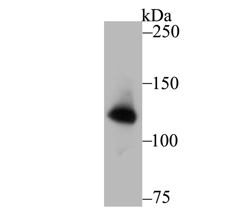
Fig1: Western blot analysis of hUPF1 on PC-3M cell lysate using anti-hUPF1 antibody at 1/1,000 dilution.Application
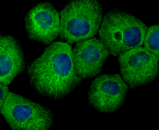
Fig2: ICC staining hUPF1 in A431 cells (green). The nuclear counter stain is DAPI (blue). Cells were fixed in paraformaldehyde, permeabilised with 0.25% Triton X100/PBS.Application
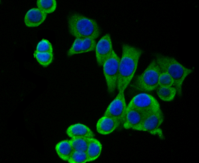
Fig3: ICC staining hUPF1 in LOVO cells (green). The nuclear counter stain is DAPI (blue). Cells were fixed in paraformaldehyde, permeabilised with 0.25% Triton X100/PBS.Application
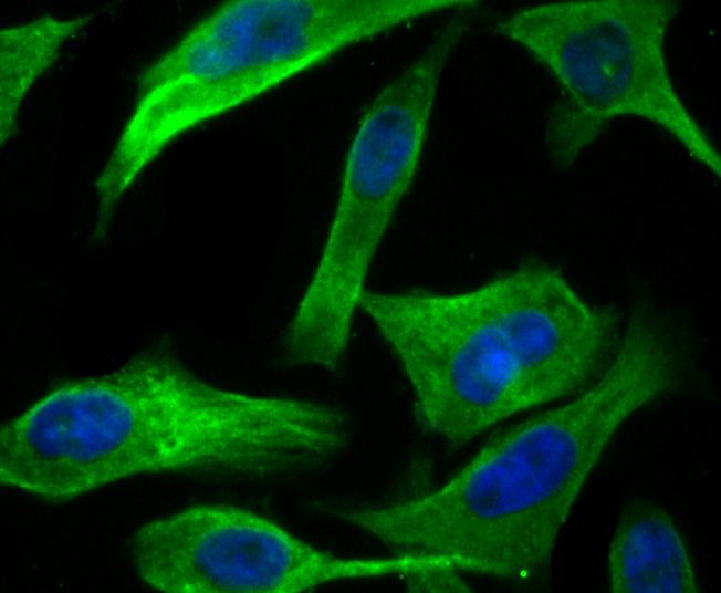
Fig4: ICC staining hUPF1 in PC-3M cells (green). The nuclear counter stain is DAPI (blue). Cells were fixed in paraformaldehyde, permeabilised with 0.25% Triton X100/PBS.Application
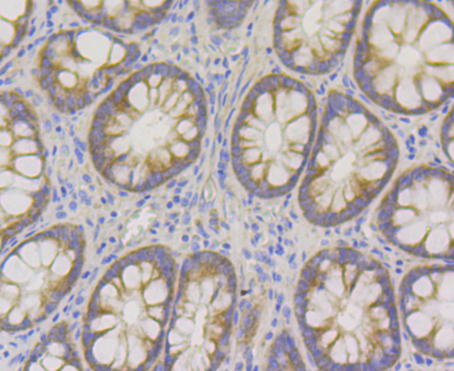
Fig5: Immunohistochemical analysis of paraffin-embedded human colon tissue using anti- hUPF1 antibody. Counter stained with hematoxylin.Application
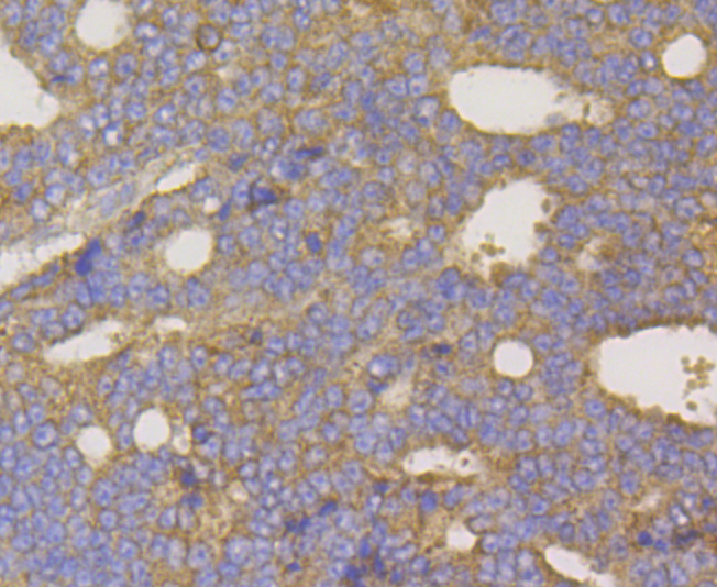
Fig6: Immunohistochemical analysis of paraffin-embedded human prostate cancer tissue using anti-hUPF1 antibody. Counter stained with hematoxylin.Application
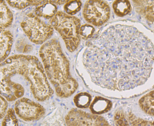
Fig7: Immunohistochemical analysis of paraffin-embedded human kidney tissue using anti- hUPF1 antibody. Counter stained with hematoxylin.Application
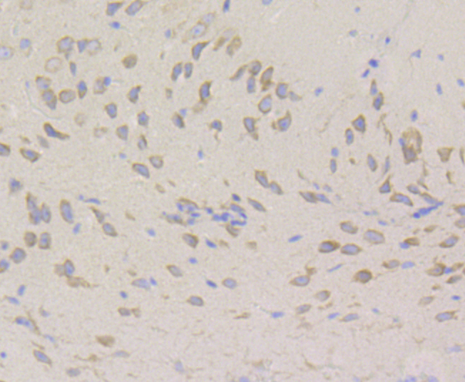
Fig8: Immunohistochemical analysis of paraffin-embedded mouse brain tissue using anti- hUPF1 antibody. Counter stained with hematoxylin.Application
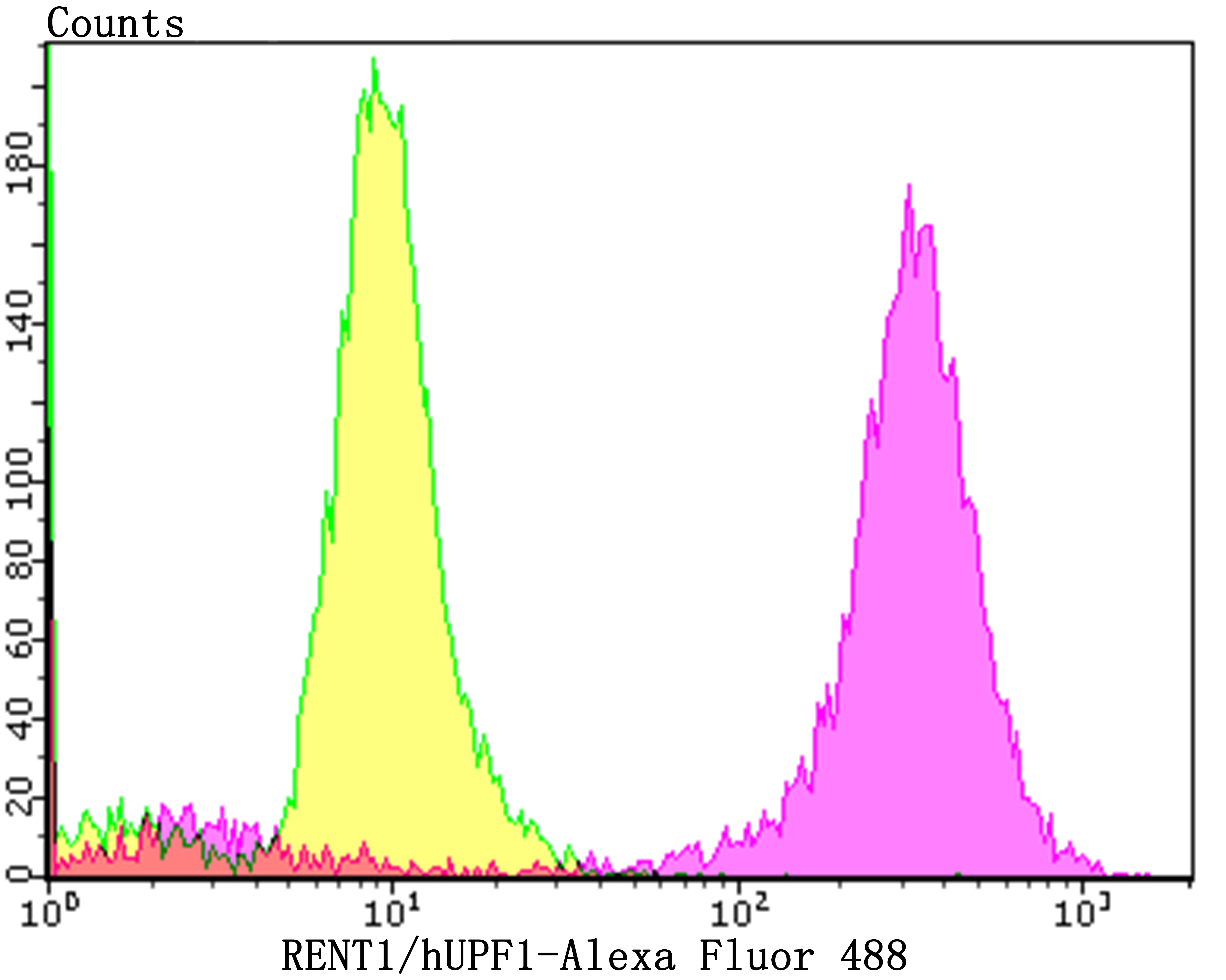
Fig9: Flow cytometric analysis of PC-3M cells with hUPF1 antibody at 1/100 dilution (purple) compared with an unlabelled control (cells without incubation with primary antibody; yellow). Alexa Fluor 488-conjugated goat anti-rabbit IgG was used as the secondary antibody.| Product Name | hUPF1 [JG38-44] |
|---|---|
| Antibody Type | Primary Antibodies |
| Antigen Alias | ATP dependent helicase RENT1 antibody ATP-dependent helicase RENT1 antibody Delta helicase antibody FLJ43809 antibody FLJ46894 antibody HUPF 1 antibody hUpf1 antibody KIAA0221 antibody Nonsense mRNA reducing factor 1 antibody NORF 1 antibody NORF1 antibody pNORF 1 antibody pNORF1 antibody Regulator of nonsense transcripts 1 antibody RENT 1 antibody RENT1 antibody RENT1_HUMAN antibody Smg 2 antibody Smg 2 homolog nonsense mediated mRNA decay factor antibody UP Frameshift 1 antibody Up frameshift mutation 1 homolog (S. cerevisiae) antibody Up frameshift mutation 1 homolog antibody Up frameshift suppressor 1 homolog antibody Up-frameshift suppressor 1 homolog antibody UPF 1 antibody UPF 1 regulator of nonsense transcripts homolog antibody upf1 antibody UPF1 regulator of nonsense transcripts homolog antibody UPF1 RNA helicase and ATPase antibody Yeast Upf1p homolog antibody |
| Product description | In eukaryotes, it is essential to have the ability to detect and degrade transcripts that lack full coding potential. Nonsense-mediated RNA decay (NMD) protects the organism by avoiding the translation of truncated peptides with dominant negative or deleterious gain-of-function potential. Rent1, a mammalian ortholog of Upflp, is essential for embryonic viability. Rent1 (also designated regulator of nonsense transcripts and HUpf1) contains an N-terminal zinc finger-like domain, NTPase domains and a region comprised of domains that define Rent1 as a superfamily group I helicase. Rent1 protein has nucleic-acid-dependent ATPase activity and 5' to 3' helicase activity. In addition, Rent1 is an RNA-binding protein whose activity is modulated by ATP and directly interacts with Rent2, which is a mammalian homolog of Upf2p. Two mammalian orthologs to Upf3p, Rent3a and Rent3b, are encoded by two separate genes. Rent3b (also known as Rent3X) is encoded by a X-linked gene and localizes primarily to the nucleus, while Rent 1 and Rent 2 localize primarily in the cytoplasm. Specific Rent3 protein interactions with Y14 and spliced mRNA suggest Rent3a and Rent3b serve as a link between splicing and NMD in the cytoplasm. |
| Immunogen | Recombinant protein within human hUPF1 aa 1-200. |
| Clonality | Monoclonal |
|---|---|
| Isotype | IgG |
| Host Species | Recombinant rabbit |
| Tested Applications | WBICCIFIPIHCFC |
| WB:1:500-1:2,000 ICC/IF:1:50-1:200 IHC:1:50-1:200 FC:1:50-1:100 IP:1:10-1:50 |
|
| Species Reactivity | HumanMouse |
| Concentration | 1mg/ml |
| Alternative Names | ATP dependent helicase RENT1 antibody ATP-dependent helicase RENT1 antibody Delta helicase antibody FLJ43809 antibody FLJ46894 antibody HUPF 1 antibody hUpf1 antibody KIAA0221 antibody Nonsense mRNA reducing factor 1 antibody NORF 1 antibody NORF1 antibody pNORF 1 antibody pNORF1 antibody Regulator of nonsense transcripts 1 antibody RENT 1 antibody RENT1 antibody RENT1_HUMAN antibody Smg 2 antibody Smg 2 homolog nonsense mediated mRNA decay factor antibody UP Frameshift 1 antibody Up frameshift mutation 1 homolog (S. cerevisiae) antibody Up frameshift mutation 1 homolog antibody Up frameshift suppressor 1 homolog antibody Up-frameshift suppressor 1 homolog antibody UPF 1 antibody UPF 1 regulator of nonsense transcripts homolog antibody upf1 antibody UPF1 regulator of nonsense transcripts homolog antibody UPF1 RNA helicase and ATPase antibody Yeast Upf1p homolog antibody |
|---|---|
| Molecular Weight(MW) | 124 kDa |
| Cellular Localization | Cytoplasm. Nucleus. |
Application
Fig1: Western blot analysis of hUPF1 on PC-3M cell lysate using anti-hUPF1 antibody at 1/1,000 dilution.
Application
Fig2: ICC staining hUPF1 in A431 cells (green). The nuclear counter stain is DAPI (blue). Cells were fixed in paraformaldehyde, permeabilised with 0.25% Triton X100/PBS.
Application
Fig3: ICC staining hUPF1 in LOVO cells (green). The nuclear counter stain is DAPI (blue). Cells were fixed in paraformaldehyde, permeabilised with 0.25% Triton X100/PBS.
Application
Fig4: ICC staining hUPF1 in PC-3M cells (green). The nuclear counter stain is DAPI (blue). Cells were fixed in paraformaldehyde, permeabilised with 0.25% Triton X100/PBS.
Application
Fig5: Immunohistochemical analysis of paraffin-embedded human colon tissue using anti- hUPF1 antibody. Counter stained with hematoxylin.
Application
Fig6: Immunohistochemical analysis of paraffin-embedded human prostate cancer tissue using anti-hUPF1 antibody. Counter stained with hematoxylin.
Application
Fig7: Immunohistochemical analysis of paraffin-embedded human kidney tissue using anti- hUPF1 antibody. Counter stained with hematoxylin.
Application
Fig8: Immunohistochemical analysis of paraffin-embedded mouse brain tissue using anti- hUPF1 antibody. Counter stained with hematoxylin.
Application
Fig9: Flow cytometric analysis of PC-3M cells with hUPF1 antibody at 1/100 dilution (purple) compared with an unlabelled control (cells without incubation with primary antibody; yellow). Alexa Fluor 488-conjugated goat anti-rabbit IgG was used as the secondary antibody.| Positive Control | PC-3M, A431, LOVO, human colon tissue, human prostate cancer tissue, human kidney tissue, mouse brain tissue. |
|---|---|
| Application Notes | WB:1:500-1:2,000 ICC/IF:1:50-1:200 IHC:1:50-1:200 FC:1:50-1:100 IP:1:10-1:50 |
| Form | Liquid |
|---|---|
| Storage Instructions | Store at +4℃ after thawing. Aliquot store at -20℃. Avoid repeated freeze / thaw cycles. |
| Storage Buffer | 1*TBS (pH7.4), 1%BSA, 40%Glycerol. Preservative: 0.05% Sodium Azide. |


