Application
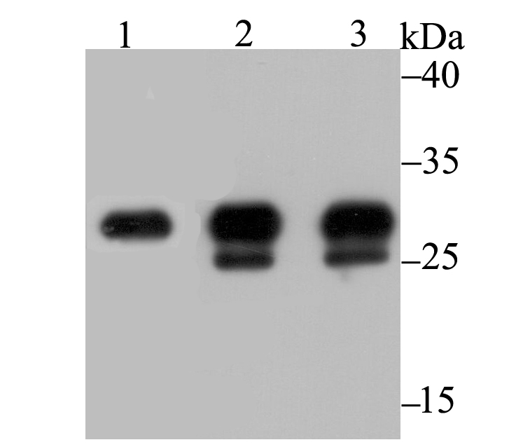
Fig1: Western blot analysis of Carbonic Anhydrase I on different lysates using anti-Carbonic Anhydrase I antibody at 1/1,000 dilution. Positive control: Lane 1: Human colon Lane 2: Mouse spleen Lane 3: Mouse colonApplication
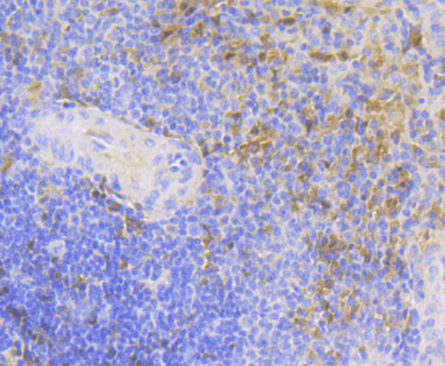
Fig2: Immunohistochemical analysis of paraffin-embedded human spleen tissue using anti-Carbonic Anhydrase I antibody. Counter stained with hematoxylin.Application
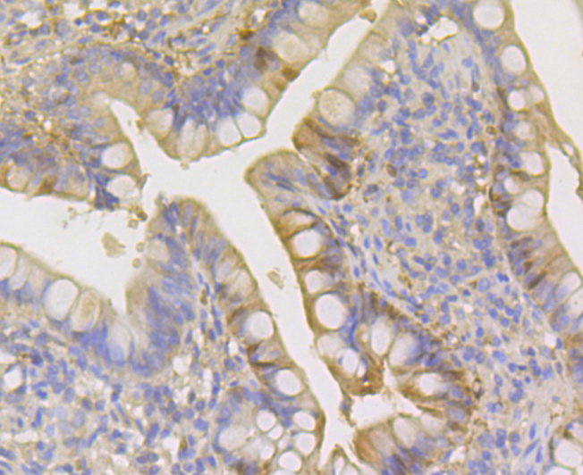
Fig3: Immunohistochemical analysis of paraffin-embedded human small intestine tissue using anti-Carbonic Anhydrase I antibody. Counter stained with hematoxylin.Application
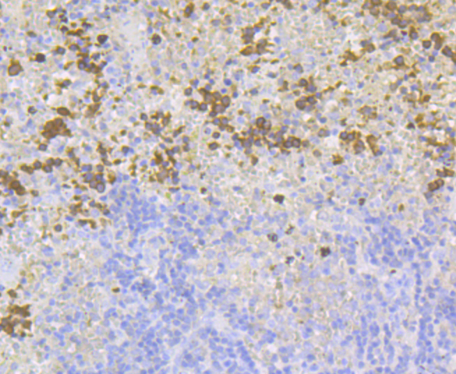
Fig4: Immunohistochemical analysis of paraffin-embedded mouse spleen tissue using anti-Carbonic Anhydrase I antibody. Counter stained with hematoxylin.| Product Name | Carbonic Anhydrase I [JG38-71] |
|---|---|
| Antibody Type | Primary Antibodies |
| Antigen Alias | CA 1 antibody CA I antibody CA-I antibody CA1 antibody CAB antibody CAH1_HUMAN antibody CAI antibody CAN antibody Car 1 antibody Car1 antibody Carbonate dehydratase I antibody Carbonic anhydrase 1 antibody Carbonic anhydrase A antibody Carbonic anhydrase B antibody Carbonic anhydrase B, formerly antibody Carbonic anhydrase I antibody Carbonic dehydratase antibody ECK0125 antibody JW0122 antibody yadF antibody |
| Product description | Carbonic anhydrases (CAs), also designated carbonate dehydratases or carbonate hydrolyases, form a large family of genes that encode zinc metalloenzymes of great physiologic importance. As catalysts of the reversible hydration of carbon dioxide, these enzymes participate in a variety of biologic processes, including respiration, acid-base balance, bone resorption and calcification as well as the formation of aqueous humor, cerebrospinal fluid, saliva and gastric acid. Genes in the α-carbonic anhydrase family encode either active carbonic anhydrase isozymes or "acatalytic" (devoid of CO2 hydration activity) carbonic anhydrase-related proteins. Human CA I (CA1) is encoded by the CA1 gene, which maps to a region on chromosome 8 that harbors a cluster of CA genes. CA I localizes to the cytoplasm and research indicates that a severe deficiency of CA I does not result in any obvious hematological or renal consequences. |
| Immunogen | Recombinant protein within human Carbonic Anhydrase I aa 1-200. |
| Clonality | Monoclonal |
|---|---|
| Isotype | IgG |
| Host Species | Recombinant rabbit |
| Tested Applications | WBIHC |
| WB:1:500-1:2,000 IHC:1:50-1:200 |
|
| Species Reactivity | HumanMouseRat |
| Concentration | 1mg/ml |
| Alternative Names | CA 1 antibody
CA I antibody
CA-I antibody
CA1 antibody
CAB antibody
CAH1_HUMAN antibody
CAI antibody
CAN antibody
Car 1 antibody
Car1 antibody
Carbonate dehydratase I antibody
Carbonic anhydrase 1 antibody
Carbonic anhydrase A antibody
Carbonic anhydrase B antibody
Carbonic anhydrase B formerly antibody Carbonic anhydrase I antibody Carbonic dehydratase antibody ECK0125 antibody JW0122 antibody yadF antibody |
|---|---|
| Molecular Weight(MW) | 29 kDa |
| Cellular Localization | Cytoplasm. |
Application
Fig1: Western blot analysis of Carbonic Anhydrase I on different lysates using anti-Carbonic Anhydrase I antibody at 1/1,000 dilution. Positive control: Lane 1: Human colon Lane 2: Mouse spleen Lane 3: Mouse colon
Application
Fig2: Immunohistochemical analysis of paraffin-embedded human spleen tissue using anti-Carbonic Anhydrase I antibody. Counter stained with hematoxylin.
Application
Fig3: Immunohistochemical analysis of paraffin-embedded human small intestine tissue using anti-Carbonic Anhydrase I antibody. Counter stained with hematoxylin.
Application
Fig4: Immunohistochemical analysis of paraffin-embedded mouse spleen tissue using anti-Carbonic Anhydrase I antibody. Counter stained with hematoxylin.| Positive Control | Human colon tissue lysate, mouse spleen tissue, mouse colon tissue lysate, human spleen tissue, human small intestine tissue. |
|---|---|
| Application Notes | WB:1:500-1:2,000 IHC:1:50-1:200 |
| Form | Liquid |
|---|---|
| Storage Instructions | Store at +4℃ after thawing. Aliquot store at -20℃. Avoid repeated freeze / thaw cycles. |
| Storage Buffer | 1*TBS (pH7.4), 1%BSA, 40%Glycerol. Preservative: 0.05% Sodium Azide. |


