Application
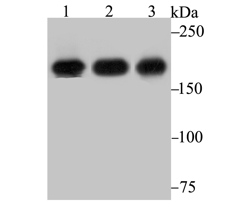
Fig1: Western blot analysis of IQGAP1 on different lysates using anti-IQGAP1 antibody at 1/1,000 dilution. Positive control: Lane 1: HUVEC Lane 2: A431 Lane 3: Mouse placentaApplication
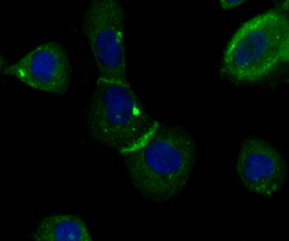
Fig2: ICC staining IQGAP1 in A431 cells (green). The nuclear counter stain is DAPI (blue). Cells were fixed in paraformaldehyde, permeabilised with 0.25% Triton X100/PBS.Application
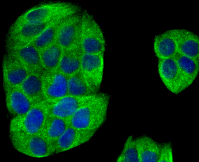
Fig3: ICC staining IQGAP1 in Hela cells (green). The nuclear counter stain is DAPI (blue). Cells were fixed in paraformaldehyde, permeabilised with 0.25% Triton X100/PBS.Application
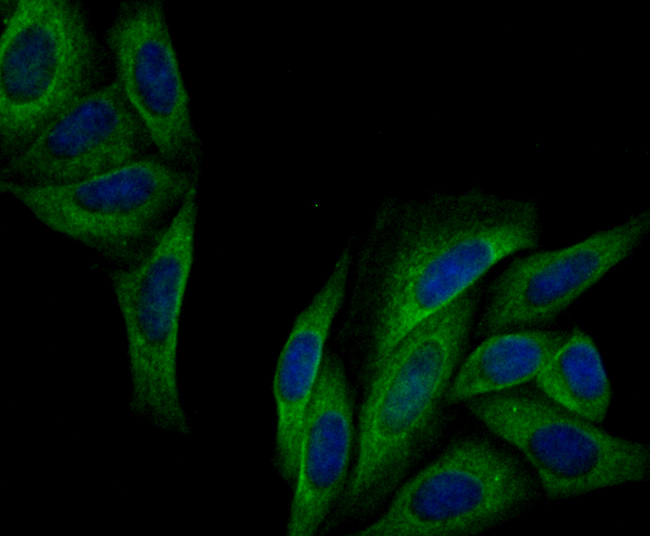
Fig4: ICC staining IQGAP1 in SiHa cells (green). The nuclear counter stain is DAPI (blue). Cells were fixed in paraformaldehyde, permeabilised with 0.25% Triton X100/PBS.Application
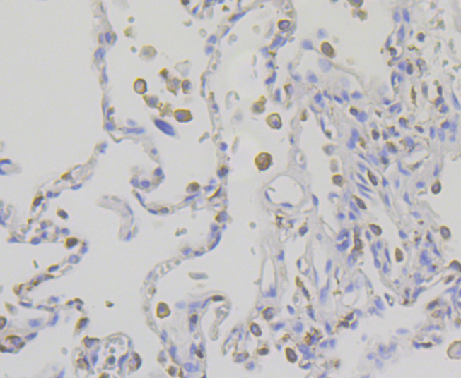
Fig5: Immunohistochemical analysis of paraffin-embedded human lung cancer tissue using anti-IQGAP1 antibody. Counter stained with hematoxylin.Application
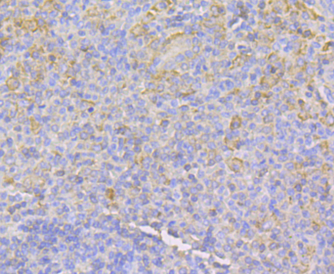
Fig6: Immunohistochemical analysis of paraffin-embedded human spleen tissue using anti-IQGAP1 antibody. Counter stained with hematoxylin.Application
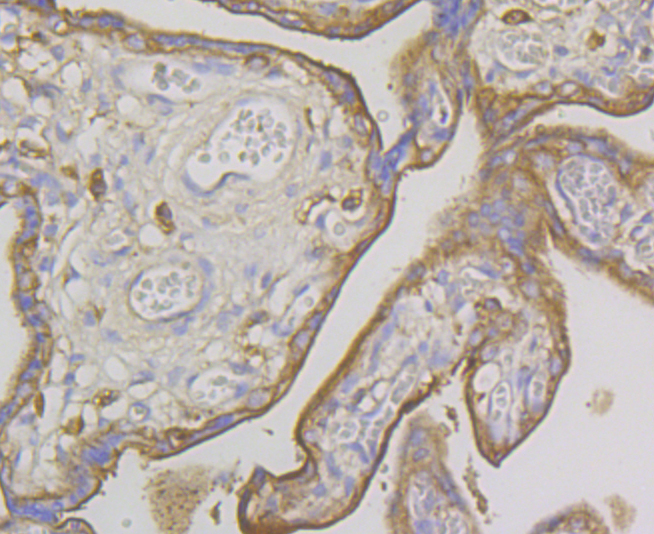
Fig7: Immunohistochemical analysis of paraffin-embedded human placenta tissue using anti-IQGAP1 antibody. Counter stained with hematoxylin.Application
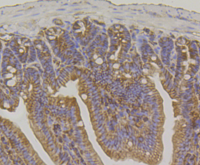
Fig8: Immunohistochemical analysis of paraffin-embedded mouse colon tissue using anti-IQGAP1 antibody. Counter stained with hematoxylin.Application
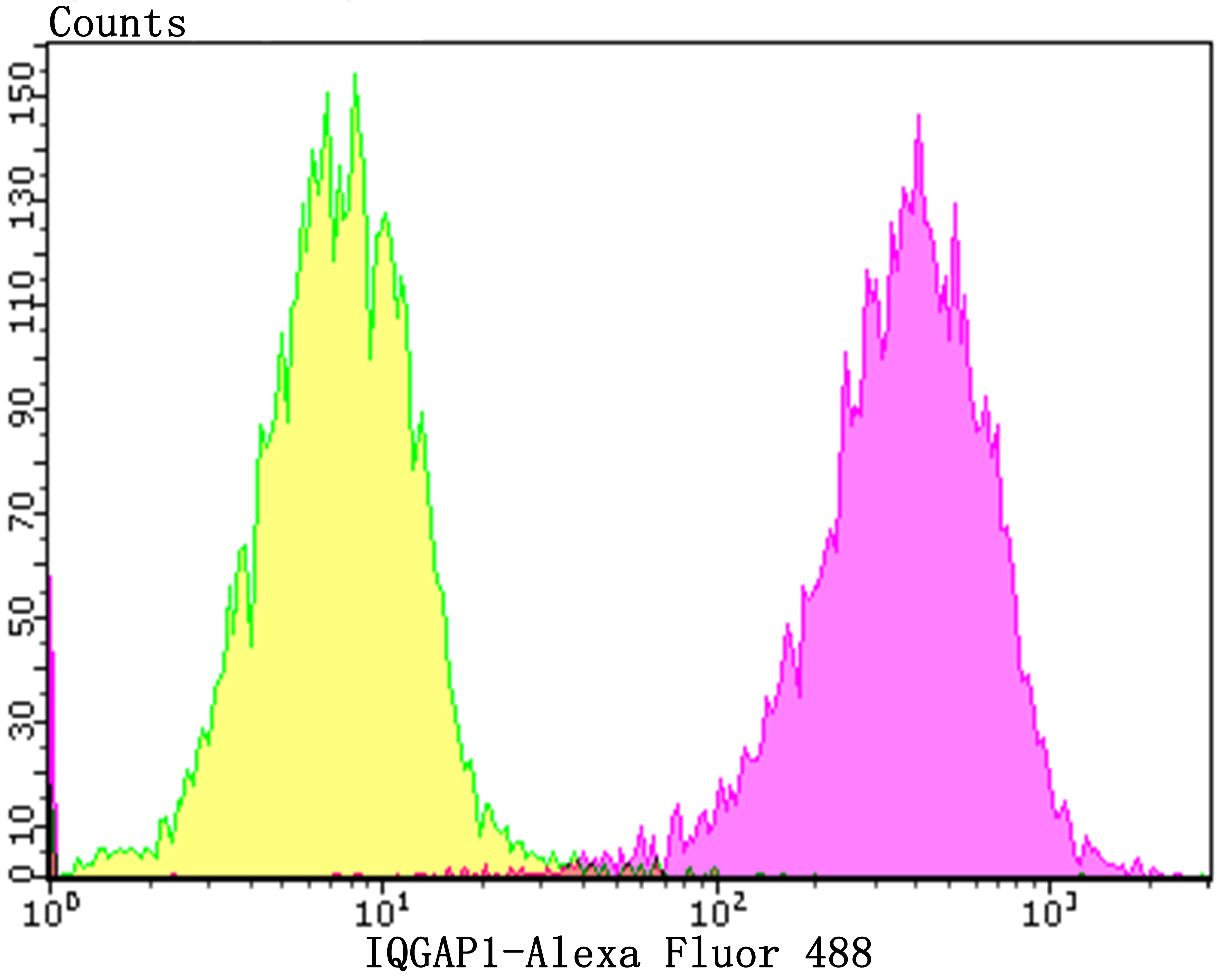
Fig9 Flow cytometric analysis of SiHa cells with IQGAP1 antibody at 1/100 dilution (purple) compared with an unlabelled control (cells without incubation with primary antibody; yellow). Alexa Fluor 488-conjugated goat anti-rabbit IgG was used as the secondary antibody.| Product Name | IQGAP1 [JG38-74] |
|---|---|
| Antibody Type | Primary Antibodies |
| Antigen Alias | HUMORFA01 antibody IQ motif containing GTPase activating protein 1 antibody IQGA1_HUMAN antibody Iqgap1 antibody KIAA0051 antibody p195 antibody Ras GTPase activating like protein 1 antibody Ras GTPase-activating-like protein IQGAP1 antibody RasGAP-like with IQ motifs antibody SAR1 antibody |
| Product description | IQGAP1, for IQ motif containing GTPase activating protein, is a Ras GAP-related, Actin-binding protein that interacts with the small GTPases Cdc42 and Rac1. The C-terminus of IQGAP1 is essential for interacting with Cdc42 and, in addition, IQGAP1 contains a WW domain and a predicted N-terminal coiled-coil region, which may be involved in IQGAP dimerization. Expression of IQGAP1 is highest in placenta, lung and kidney, where it co-localizes with Cdc42 to the cytoskeleton and assists with Cdc42 in mediating the regulation of cell proliferation, polarity and cell morphology. IQGAP1 regulates cadherin-mediated cell adhesion via binding to E-cadherin, b-catenin and a-catenin. This association induces the accumulation of these proteins at the site of cell-cell contact. IQGAP1 is negatively regulated by calmodulin, which binds to IQGAP1 in a calcium-dependent manner and disrupts IQGAP1 from associating with Cdc42. |
| Immunogen | Recombinant protein corresponding to N-terminal human IQGAP1. |
| Clonality | Monoclonal |
|---|---|
| Isotype | IgG |
| Host Species | Recombinant rabbit |
| Tested Applications | WBICCIFIHCFC |
| WB:1:500-1:2,000 ICC/IF:1:50-1:200 IHC:1:50-1:200 FC:1:50-1:100 |
|
| Species Reactivity | HumanMouse |
| Concentration | 1mg/ml |
| Alternative Names | HUMORFA01 antibody IQ motif containing GTPase activating protein 1 antibody IQGA1_HUMAN antibody Iqgap1 antibody KIAA0051 antibody p195 antibody Ras GTPase activating like protein 1 antibody Ras GTPase-activating-like protein IQGAP1 antibody RasGAP-like with IQ motifs antibody SAR1 antibody |
|---|---|
| Molecular Weight(MW) | 189 kDa |
| Cellular Localization | Cell membrane. Cytoplasm. Nucleus. |
Application
Fig1: Western blot analysis of IQGAP1 on different lysates using anti-IQGAP1 antibody at 1/1,000 dilution. Positive control: Lane 1: HUVEC Lane 2: A431 Lane 3: Mouse placenta
Application
Fig2: ICC staining IQGAP1 in A431 cells (green). The nuclear counter stain is DAPI (blue). Cells were fixed in paraformaldehyde, permeabilised with 0.25% Triton X100/PBS.
Application
Fig3: ICC staining IQGAP1 in Hela cells (green). The nuclear counter stain is DAPI (blue). Cells were fixed in paraformaldehyde, permeabilised with 0.25% Triton X100/PBS.
Application
Fig4: ICC staining IQGAP1 in SiHa cells (green). The nuclear counter stain is DAPI (blue). Cells were fixed in paraformaldehyde, permeabilised with 0.25% Triton X100/PBS.
Application
Fig5: Immunohistochemical analysis of paraffin-embedded human lung cancer tissue using anti-IQGAP1 antibody. Counter stained with hematoxylin.
Application
Fig6: Immunohistochemical analysis of paraffin-embedded human spleen tissue using anti-IQGAP1 antibody. Counter stained with hematoxylin.
Application
Fig7: Immunohistochemical analysis of paraffin-embedded human placenta tissue using anti-IQGAP1 antibody. Counter stained with hematoxylin.
Application
Fig8: Immunohistochemical analysis of paraffin-embedded mouse colon tissue using anti-IQGAP1 antibody. Counter stained with hematoxylin.
Application
Fig9 Flow cytometric analysis of SiHa cells with IQGAP1 antibody at 1/100 dilution (purple) compared with an unlabelled control (cells without incubation with primary antibody; yellow). Alexa Fluor 488-conjugated goat anti-rabbit IgG was used as the secondary antibody.| Positive Control | HUVEC, A431, mouse placenta tissue lysate, Hela, SiHa, human lung cancer tissue, human spleen tissue, human placenta tissue, mouse colon tissue. |
|---|---|
| Application Notes | WB:1:500-1:2,000 ICC/IF:1:50-1:200 IHC:1:50-1:200 FC:1:50-1:100 |
| Form | Liquid |
|---|---|
| Storage Instructions | Store at +4℃ after thawing. Aliquot store at -20℃. Avoid repeated freeze / thaw cycles. |
| Storage Buffer | 1*TBS (pH7.4), 1%BSA, 40%Glycerol. Preservative: 0.05% Sodium Azide. |


