Application
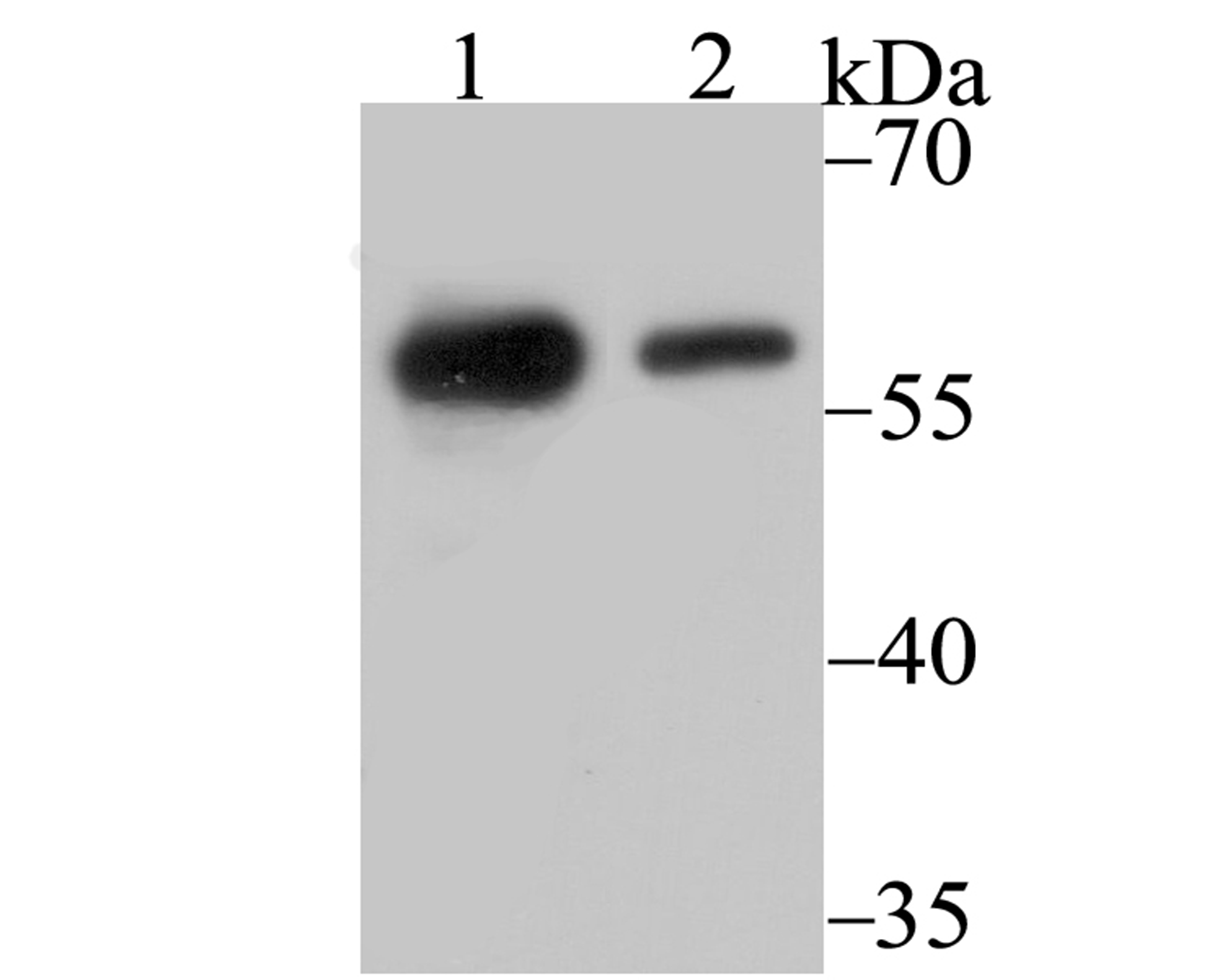
Fig1: Western blot analysis of p54nrb on 293 (1) and SH-SY-5Y (2) cell lysate using anti-p54nrb antibody at 1/500 dilution.Application
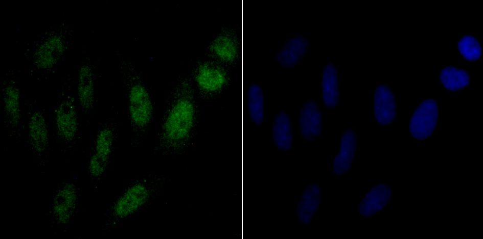
Fig2: ICC staining p54nrb in SiHa cells (green). The nuclear counter stain is DAPI (blue). Cells were fixed in paraformaldehyde, permeabilised with 0.25% Triton X100/PBS.Application
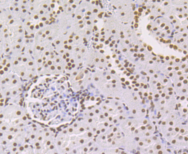
Fig3: Immunohistochemical analysis of paraffin-embedded rat kidney tissue using anti- p54nrb antibody. Counter stained with hematoxylin.Application
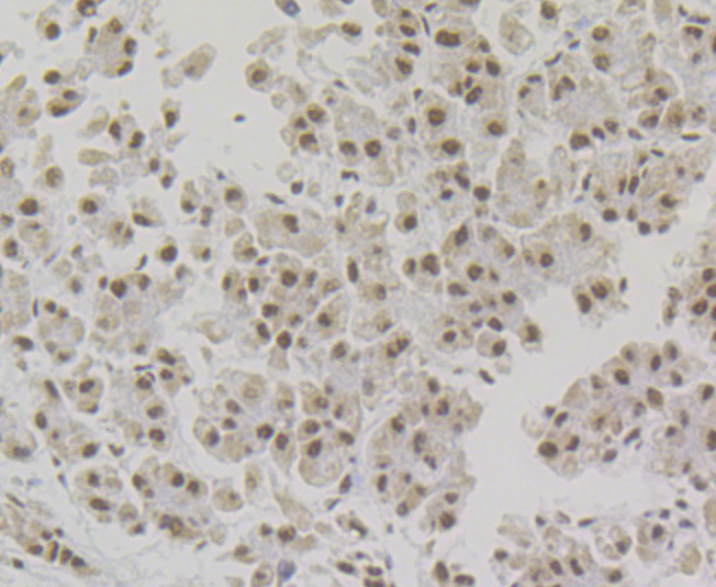
Fig4: Immunohistochemical analysis of paraffin-embedded human liver tissue using anti- p54nrb antibody. Counter stained with hematoxylin.Application
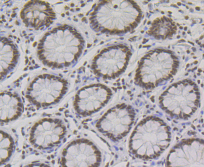
Fig5: Immunohistochemical analysis of paraffin-embedded human colon tissue using anti- p54nrb antibody. Counter stained with hematoxylin.Application
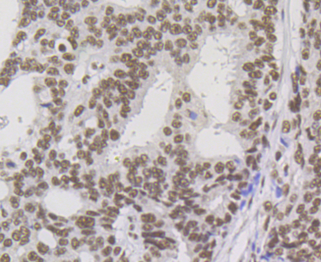
Fig6: Immunohistochemical analysis of paraffin-embedded human prostate cancer tissue using anti-p54nrb antibody. Counter stained with hematoxylin.Application
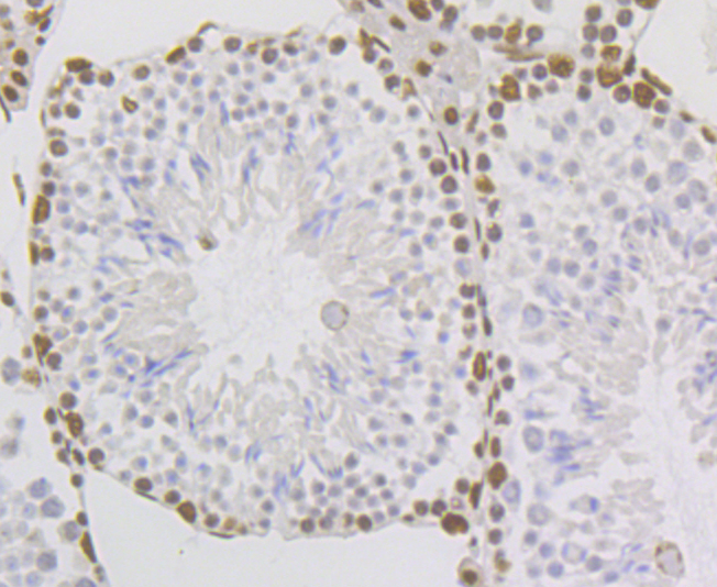
Fig7: Immunohistochemical analysis of paraffin-embedded mouse testis tissue using anti- p54nrb antibody. Counter stained with hematoxylin.| Product Name | p54nrb [JG87-38] |
|---|---|
| Antibody Type | Primary Antibodies |
| Antigen Alias | 52 kDa subunit antibody 54 kDa nuclear RNA and DNA binding protein antibody 54 kDa nuclear RNA- and DNA-binding protein antibody 55 kDa nuclear protein antibody DNA binding p52/p100 complex 52 kDa subunit antibody DNA-binding p52/p100 complex antibody NMT 55 antibody NMT55 antibody Non Pou domain containing octamer (ATGCAAAT) binding protein antibody Non POU domain containing octamer binding antibody Non POU domain containing octamer binding protein antibody Non-POU domain-containing octamer-binding protein antibody Nono antibody NonO protein antibody NONO_HUMAN antibody NRB 54 antibody NRB antibody NRB54 antibody Nuclear RNA binding protein 54kD antibody P54 antibody p54(nrb) antibody p54nrb antibody PPP1R114 antibody Protein phosphatase 1 regulatory subunit 114 antibody |
| Product description | Found in both primary and transformed human cells, paraspeckles are discrete bodies in the interchromatin nucleoplasmic space which contain p54/nrb (nuclear RNA-binding protein) and at least two other RNA-binding proteins, paraspeckle protein 1 (PSP1) and paraspeckle protein 2 (PSP2). Paraspeckles often co-localize with splicing speckles, which are the site of splicing factor accumulation. Paraspeckle proteins, including p54/nrb, move dynamically between the nucleolus and paraspeckles and translocate to distinct caps in the nucleolar periphery when transcription is inhibited. Originally purified from HeLa cells, the nuclear p54/nrb has two RNA recognition motifs and shares extensive homology with both the human splicing factor PSF and Drosophila NONA/BJ6, which is required for normal vision and courtship. The shared domain between these proteins is termed a DBHS (Drosophila behavior, human splicing) domain and may play a role in regulating various pathways at the level of pre-mRNA splicing. |
| Immunogen | Recombinant protein of C-terminal human p54nrb. |
| Clonality | Monoclonal |
|---|---|
| Isotype | IgG |
| Host Species | Recombinant rabbit |
| Tested Applications | WBICCIFIHC |
| WB:1:500-1:2,000 ICC:1:50 IHC:1:50-1:200 |
|
| Species Reactivity | HumanMouseRat |
| Concentration | 1mg/ml |
| Alternative Names | 52 kDa subunit antibody 54 kDa nuclear RNA and DNA binding protein antibody 54 kDa nuclear RNA- and DNA-binding protein antibody 55 kDa nuclear protein antibody DNA binding p52/p100 complex 52 kDa subunit antibody DNA-binding p52/p100 complex antibody NMT 55 antibody NMT55 antibody Non Pou domain containing octamer (ATGCAAAT) binding protein antibody Non POU domain containing octamer binding antibody Non POU domain containing octamer binding protein antibody Non-POU domain-containing octamer-binding protein antibody Nono antibody NonO protein antibody NONO_HUMAN antibody NRB 54 antibody NRB antibody NRB54 antibody Nuclear RNA binding protein 54kD antibody P54 antibody p54(nrb) antibody p54nrb antibody PPP1R114 antibody Protein phosphatase 1 regulatory subunit 114 antibody |
|---|---|
| Molecular Weight(MW) | 54 kDa |
| Cellular Localization | Nucleus. |
Application
Fig1: Western blot analysis of p54nrb on 293 (1) and SH-SY-5Y (2) cell lysate using anti-p54nrb antibody at 1/500 dilution.
Application
Fig2: ICC staining p54nrb in SiHa cells (green). The nuclear counter stain is DAPI (blue). Cells were fixed in paraformaldehyde, permeabilised with 0.25% Triton X100/PBS.
Application
Fig3: Immunohistochemical analysis of paraffin-embedded rat kidney tissue using anti- p54nrb antibody. Counter stained with hematoxylin.
Application
Fig4: Immunohistochemical analysis of paraffin-embedded human liver tissue using anti- p54nrb antibody. Counter stained with hematoxylin.
Application
Fig5: Immunohistochemical analysis of paraffin-embedded human colon tissue using anti- p54nrb antibody. Counter stained with hematoxylin.
Application
Fig6: Immunohistochemical analysis of paraffin-embedded human prostate cancer tissue using anti-p54nrb antibody. Counter stained with hematoxylin.
Application
Fig7: Immunohistochemical analysis of paraffin-embedded mouse testis tissue using anti- p54nrb antibody. Counter stained with hematoxylin.| Positive Control | 293, SH-SY-5Y, SiHa, rat kidney tissue, human liver tissue, human colon tissue, human prostate cancer tissue, mouse testis tissue. |
|---|---|
| Application Notes | WB:1:500-1:2,000 ICC:1:50 IHC:1:50-1:200 |
| Form | Liquid |
|---|---|
| Storage Instructions | Store at +4℃ after thawing. Aliquot store at -20℃. Avoid repeated freeze / thaw cycles. |
| Storage Buffer | 1*TBS (pH7.4), 1%BSA, 40%Glycerol. Preservative: 0.05% Sodium Azide. |


