Application
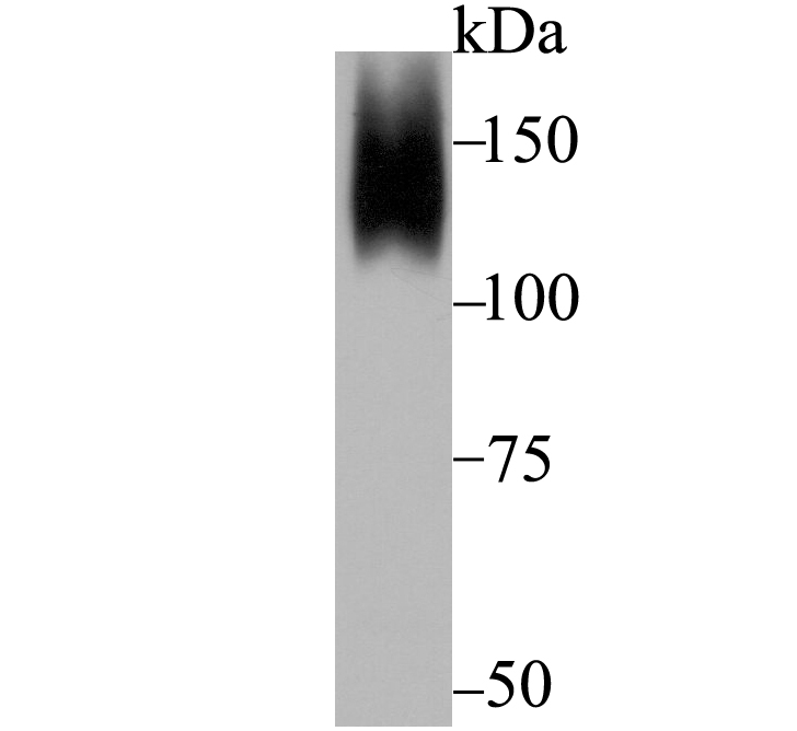
Fig1: Western blot analysis of ORP150 on mouse prostate tissue lysate using anti-ORP150 antibody at 1/5,000 dilution.Application
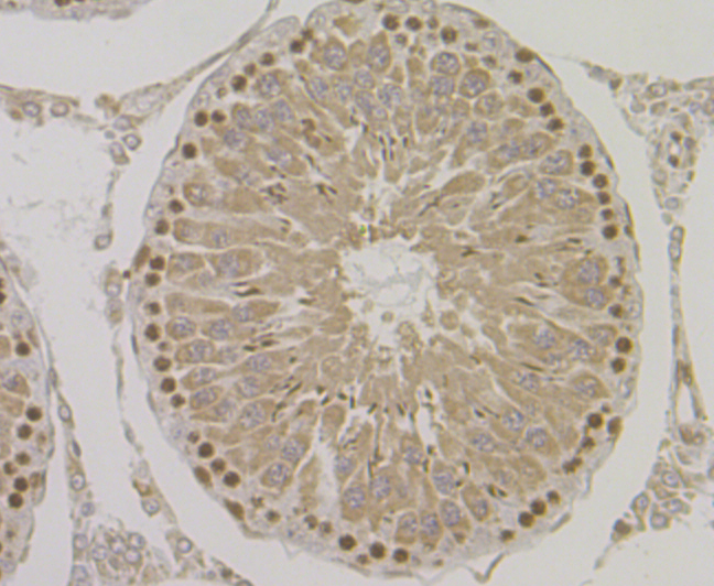
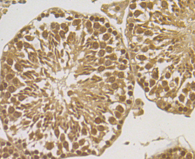
Fig2: Immunohistochemical analysis of paraffin-embedded rat testis tissue using anti-ORP150 antibody. Counter stained with hematoxylin.Application
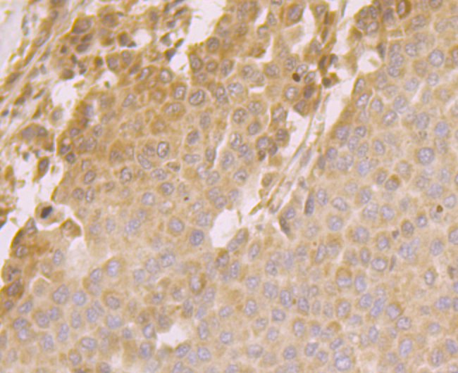
Fig3: Immunohistochemical analysis of paraffin-embedded human liver cancer tissue using anti-ORP150 antibody. Counter stained with hematoxylin.Application
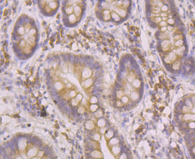
Fig4: Immunohistochemical analysis of paraffin-embedded human small intestine tissue using anti-ORP150 antibody. Counter stained with hematoxylin.Application
Fig5: Immunohistochemical analysis of paraffin-embedded mouse testis tissue using anti-ORP150 antibody. Counter stained with hematoxylin.Application
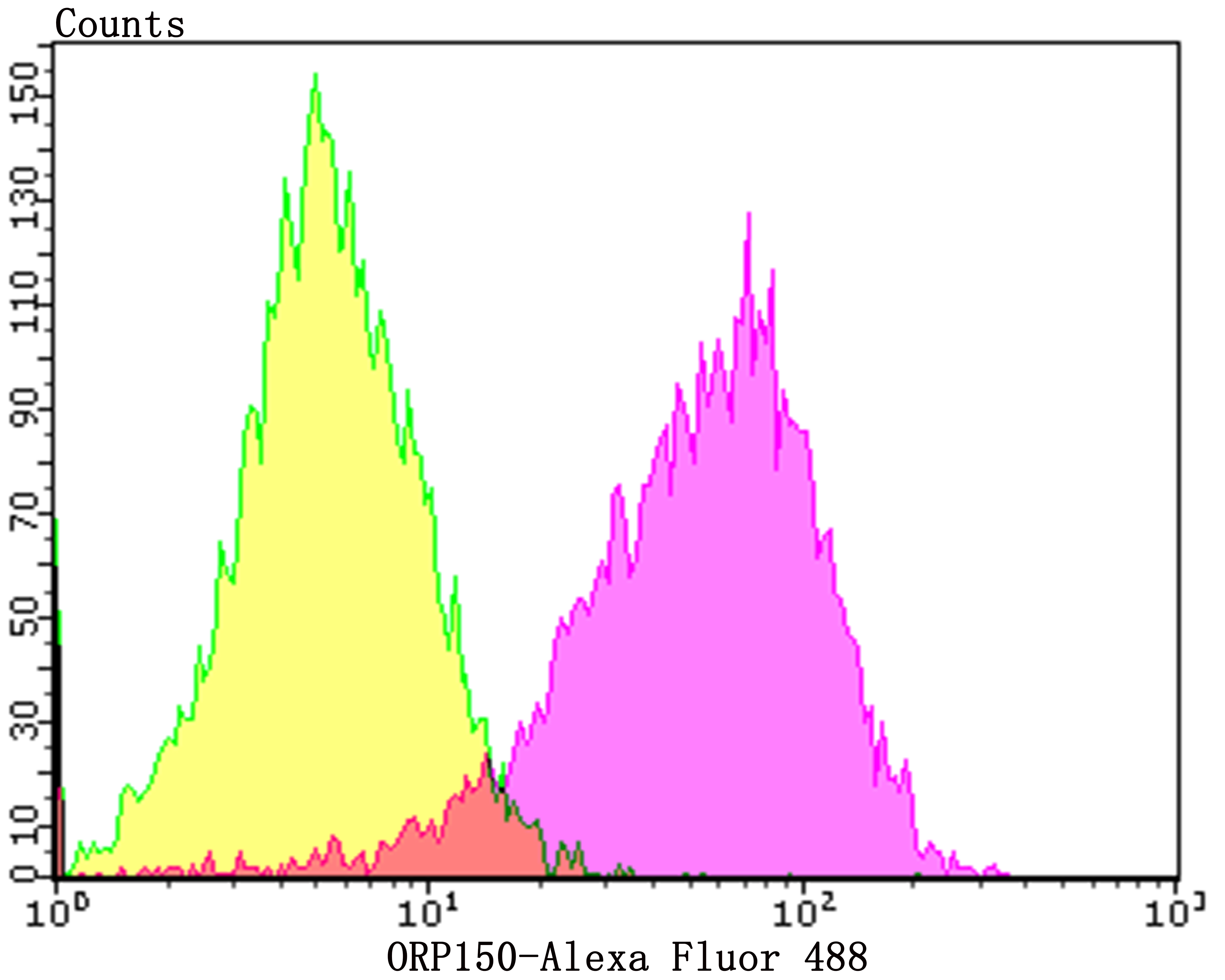
Fig6: Flow cytometric analysis of HepG2 cells with ORP150 antibody at 1/100 dilution (purple) compared with an unlabelled control (cells without incubation with primary antibody; yellow). Alexa Fluor 488-conjugated goat anti-rabbit IgG was used as the secondary antibody.| Product Name | ORP150 [JG39-11] |
|---|---|
| Antibody Type | Primary Antibodies |
| Antigen Alias | 150 kDa oxygen regulated protein antibody 150 kDa oxygen-regulated protein antibody 170 kDa glucose regulated protein antibody 170 kDa glucose-regulated protein antibody DKFZp686N08236 antibody FLJ94899 antibody FLJ97572 antibody glucose regulated protein 170 antibody Grp 170 antibody GRP-170 antibody Grp170 antibody HSP 12A antibody HSP12A antibody Hyou1 antibody HYOU1_HUMAN antibody Hypoxia up regulated 1 antibody hypoxia up-regulated 1 precursor antibody Hypoxia up-regulated protein 1 antibody Hypoxia upregulated 1 antibody Orp 150 antibody ORP-150 antibody Orp150 antibody oxygen regulated protein (150kD) antibody |
| Product description | ORP150, also known as HYOU1 (hypoxia upregulated 1), is a 999 amino acid protein that localizes to the lumen of the endoplasmic reticulum (ER) and is a member of the heat shock protein 70 (HSP 70) family. ORP150 is highly expressed in tissues that have well-developed ERs and a large number of secretory proteins (such as liver and pancreas). It is expressed at lower levels in kidney and brain, and plays an essential role in cytoprotective cellular responses to hypoxia (oxygen deprivation). Specifically, ORP150 participates in protein folding and secretion in the ER and functions to protect cells from hypoxia-induced apoptosis, thereby playing a crucial role in cell survival. ORP150 expression is upregulated in a variety of tumors, such as breast cancer, suggesting an important role in tumorigenesis. The gene encoding ORP150 has two translation initiation sites, resulting in a truncated transcript that lacks an ER signal peptide, but is thought to function as a housekeeping protein in the cytoplasm. |
| Immunogen | Recombinant protein within human ORP150 aa 800-950. |
| Clonality | Monoclonal |
|---|---|
| Isotype | IgG |
| Host Species | Recombinant rabbit |
| Tested Applications | WBIHCFC |
| WB:1:5,000-1:20,000 IHC:1:50-1:200 FC:1:50-1:100 |
|
| Species Reactivity | HumanMouseRat |
| Concentration | 1mg/ml |
| Alternative Names | 150 kDa oxygen regulated protein antibody 150 kDa oxygen-regulated protein antibody 170 kDa glucose regulated protein antibody 170 kDa glucose-regulated protein antibody DKFZp686N08236 antibody FLJ94899 antibody FLJ97572 antibody glucose regulated protein 170 antibody Grp 170 antibody GRP-170 antibody Grp170 antibody HSP 12A antibody HSP12A antibody Hyou1 antibody HYOU1_HUMAN antibody Hypoxia up regulated 1 antibody hypoxia up-regulated 1 precursor antibody Hypoxia up-regulated protein 1 antibody Hypoxia upregulated 1 antibody Orp 150 antibody ORP-150 antibody Orp150 antibody oxygen regulated protein (150kD) antibody |
|---|---|
| Molecular Weight(MW) | 111 kDa |
| Cellular Localization | Endoplasmic reticulum. |
Application
Fig1: Western blot analysis of ORP150 on mouse prostate tissue lysate using anti-ORP150 antibody at 1/5,000 dilution.
Application
Fig2: Immunohistochemical analysis of paraffin-embedded rat testis tissue using anti-ORP150 antibody. Counter stained with hematoxylin.
Application
Fig3: Immunohistochemical analysis of paraffin-embedded human liver cancer tissue using anti-ORP150 antibody. Counter stained with hematoxylin.
Application
Fig4: Immunohistochemical analysis of paraffin-embedded human small intestine tissue using anti-ORP150 antibody. Counter stained with hematoxylin.
Application
Fig5: Immunohistochemical analysis of paraffin-embedded mouse testis tissue using anti-ORP150 antibody. Counter stained with hematoxylin.
Application
Fig6: Flow cytometric analysis of HepG2 cells with ORP150 antibody at 1/100 dilution (purple) compared with an unlabelled control (cells without incubation with primary antibody; yellow). Alexa Fluor 488-conjugated goat anti-rabbit IgG was used as the secondary antibody.| Positive Control | HepG2, mouse prostate tissue lysate, rat testis tissue, human liver cancer tissue, human small intestine tissue, mouse testis tissue. |
|---|---|
| Application Notes | WB:1:5,000-1:20,000 IHC:1:50-1:200 FC:1:50-1:100 |
| Form | Liquid |
|---|---|
| Storage Instructions | Store at +4℃ after thawing. Aliquot store at -20℃. Avoid repeated freeze / thaw cycles. |
| Storage Buffer | 1*TBS (pH7.4), 1%BSA, 40%Glycerol. Preservative: 0.05% Sodium Azide. |


