Application
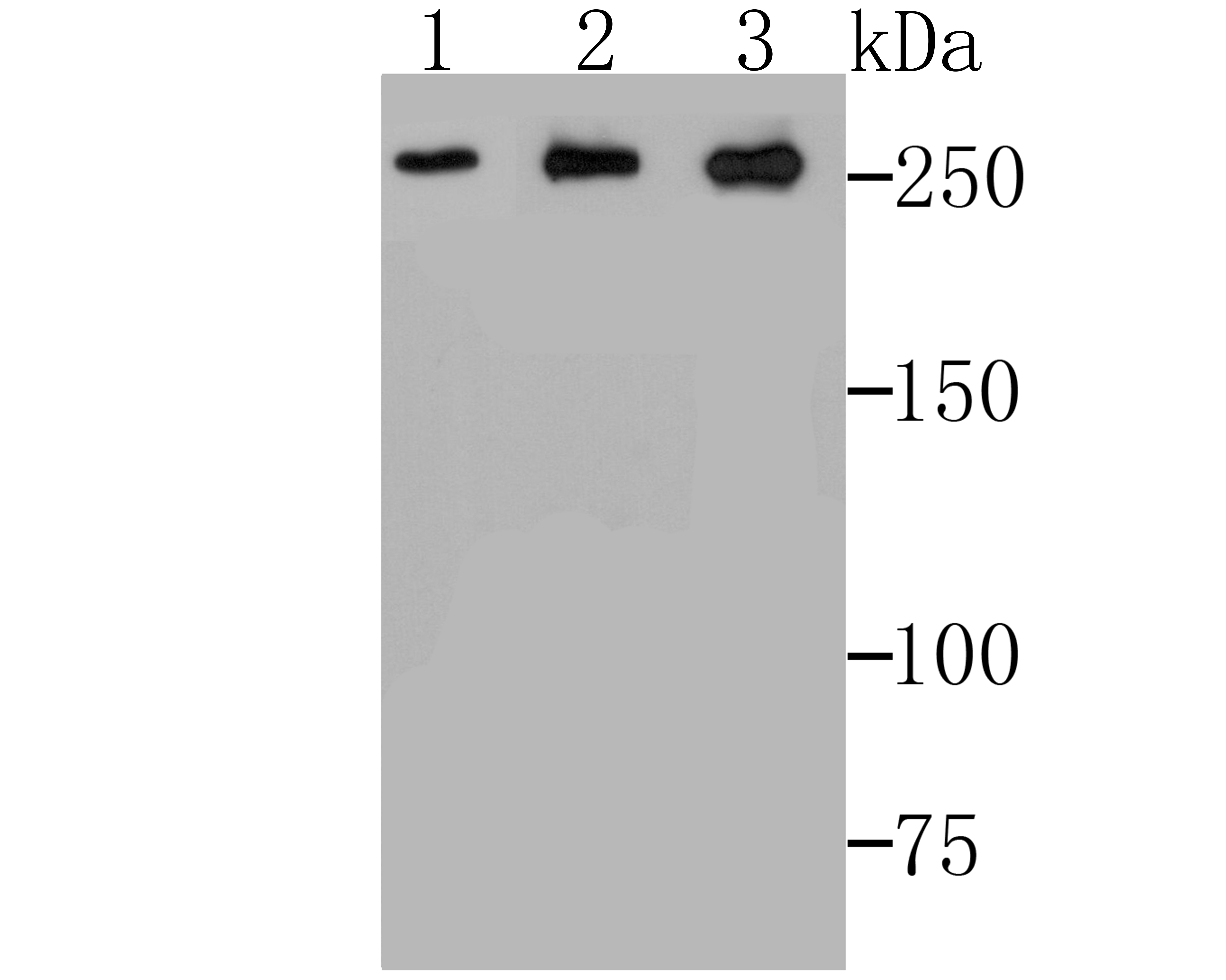
Fig1: Western blot analysis of ADAMTS13 on SiHa cell (1), PC-3M cell (2) and A549 cell lysates using anti-ADAMTS13 antibody at 1/500 dilution.Application
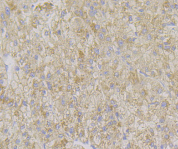
Fig2: Immunohistochemical analysis of paraffin-embedded human liver tissue using anti-ADAMTS13 antibody. Counter stained with hematoxylin.Application
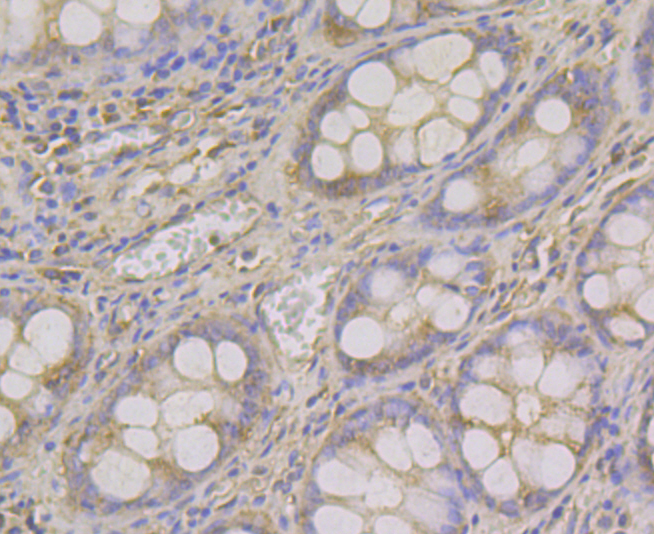
Fig3: Immunohistochemical analysis of paraffin-embedded human colon tissue using anti-ADAMTS13 antibody. Counter stained with hematoxylin.Application
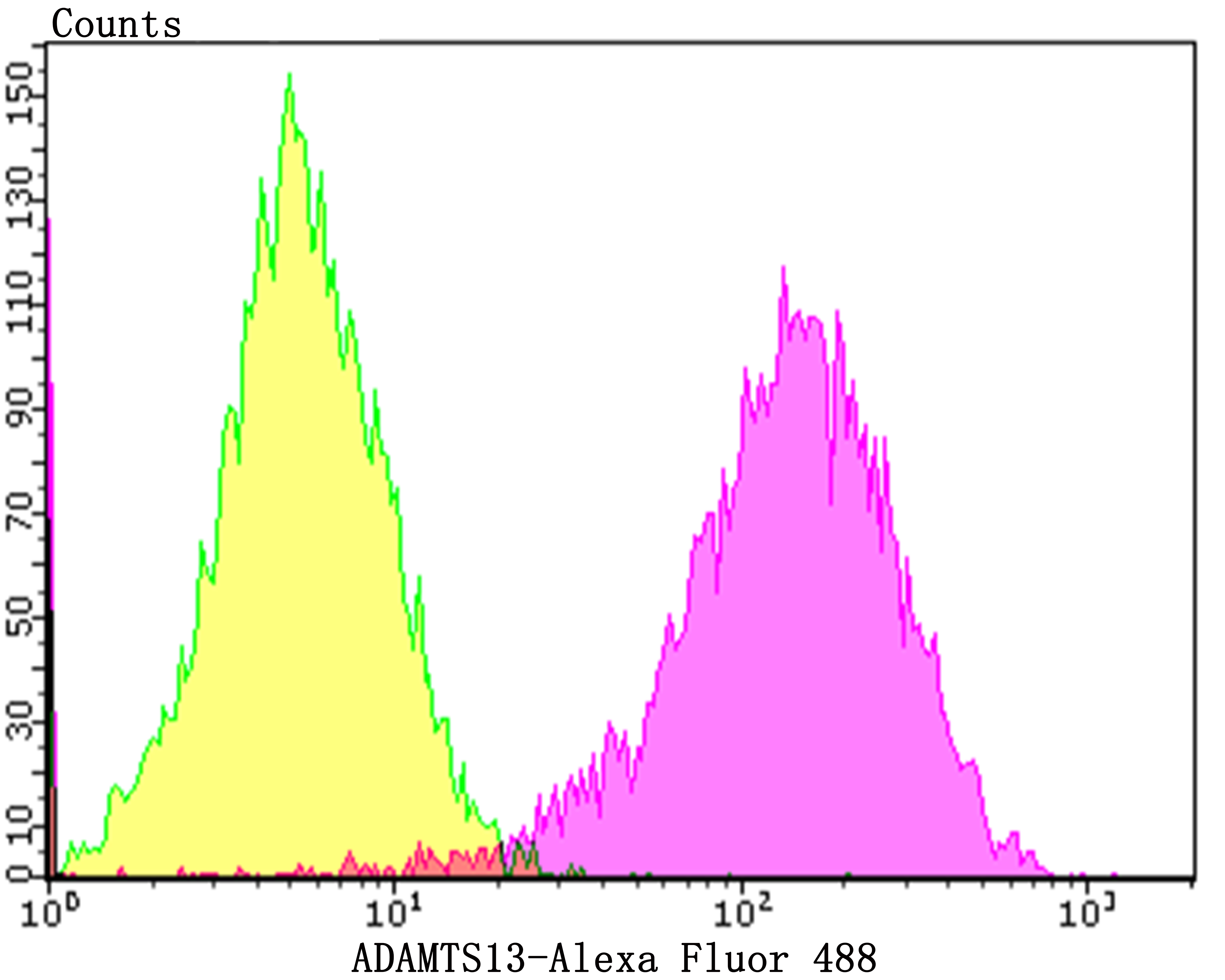
Fig4: Flow cytometric analysis of HepG2 cells with ADAMTS13 antibody at 1/100 dilution (purple) compared with an unlabelled control (cells without incubation with primary antibody; yellow). Alexa Fluor 488-conjugated goat anti-rabbit IgG was used as the secondary antibody.| Product Name | ADAMTS13 [JG39-21] |
|---|---|
| Antibody Type | Primary Antibodies |
| Antigen Alias | A disintegrin and metalloproteinase with thrombospondin motifs 13 antibody A disintegrin like and metalloprotease (reprolysin type) with thrombospondin type 1 motif 13 antibody A disintegrin like and metalloprotease with thrombospondin type 1 motif 13 antibody ADAM metallopeptidase with thrombospondin type 1 motif 13 antibody ADAM TS antibody ADAM-TS 13 antibody ADAM-TS13 antibody ADAMTS 13 antibody ADAMTS-13 antibody ADAMTS13 antibody ADAMTS13 protein antibody ATS13_HUMAN antibody C9orf8 antibody TTP antibody Von Willebrand factor cleaving protease antibody von Willebrand factor-cleaving protease antibody vWF cleaving protease antibody vWF CP antibody vWF-cleaving protease antibody vWF-CP antibody vWFCP antibody |
| Product description | ADAMTS13 (a disintegrin and metalloproteinase with a thrombospondin type 1 motif, member 13)—also known as von Willebrand factor-cleaving protease (VWFCP)—is a zinc-containing metalloprotease enzyme that cleaves von Willebrand factor (vWf), a large protein involved in blood clotting. It is secreted in blood and degrades large vWf multimers, decreasing their activity. Glycosylated. O-fucosylated by POFUT2 on a serine or a threonine residue found within the consensus sequence C1-X2-(S/T)-C2-G of the TSP type-1 repeat domains where C1 and C2 are the first and second cysteine residue of the repeat, respectively. Fucosylated repeats can then be further glycosylated by the addition of a beta-1,3-glucose residue by the glucosyltransferase, B3GALTL. |
| Immunogen | Recombinant protein within human ADAMTS13 aa 150-350. |
| Clonality | Monoclonal |
|---|---|
| Isotype | IgG |
| Host Species | Recombinant rabbit |
| Tested Applications | WBIHCFC |
| WB:1:500-1:2,000 IHC:1:50-1:200 FC:1:50-1:100 |
|
| Species Reactivity | Human |
| Concentration | 1mg/ml |
| Alternative Names | A disintegrin and metalloproteinase with thrombospondin motifs 13 antibody A disintegrin like and metalloprotease (reprolysin type) with thrombospondin type 1 motif 13 antibody A disintegrin like and metalloprotease with thrombospondin type 1 motif 13 antibody ADAM metallopeptidase with thrombospondin type 1 motif 13 antibody ADAM TS antibody ADAM-TS 13 antibody ADAM-TS13 antibody ADAMTS 13 antibody ADAMTS-13 antibody ADAMTS13 antibody ADAMTS13 protein antibody ATS13_HUMAN antibody C9orf8 antibody TTP antibody Von Willebrand factor cleaving protease antibody von Willebrand factor-cleaving protease antibody vWF cleaving protease antibody vWF CP antibody vWF-cleaving protease antibody vWF-CP antibody vWFCP antibody |
|---|---|
| Molecular Weight(MW) | Predicted band size 154 kDa |
| Cellular Localization | Secreted. |
| SwissProt ID | Q76LX8 |
|---|
Application
Fig1: Western blot analysis of ADAMTS13 on SiHa cell (1), PC-3M cell (2) and A549 cell lysates using anti-ADAMTS13 antibody at 1/500 dilution.
Application
Fig2: Immunohistochemical analysis of paraffin-embedded human liver tissue using anti-ADAMTS13 antibody. Counter stained with hematoxylin.
Application
Fig3: Immunohistochemical analysis of paraffin-embedded human colon tissue using anti-ADAMTS13 antibody. Counter stained with hematoxylin.
Application
Fig4: Flow cytometric analysis of HepG2 cells with ADAMTS13 antibody at 1/100 dilution (purple) compared with an unlabelled control (cells without incubation with primary antibody; yellow). Alexa Fluor 488-conjugated goat anti-rabbit IgG was used as the secondary antibody.| Positive Control | SiHa cell, PC-3M, A549, HepG2, human liver tissue, human colon tissue. |
|---|---|
| Application Notes | WB:1:500-1:2,000 IHC:1:50-1:200 FC:1:50-1:100 |
| Form | Liquid |
|---|---|
| Storage Instructions | Store at +4℃ after thawing. Aliquot store at -20℃. Avoid repeated freeze / thaw cycles. |
| Storage Buffer | 1*TBS (pH7.4), 1%BSA, 40%Glycerol. Preservative: 0.05% Sodium Azide. |


