WB
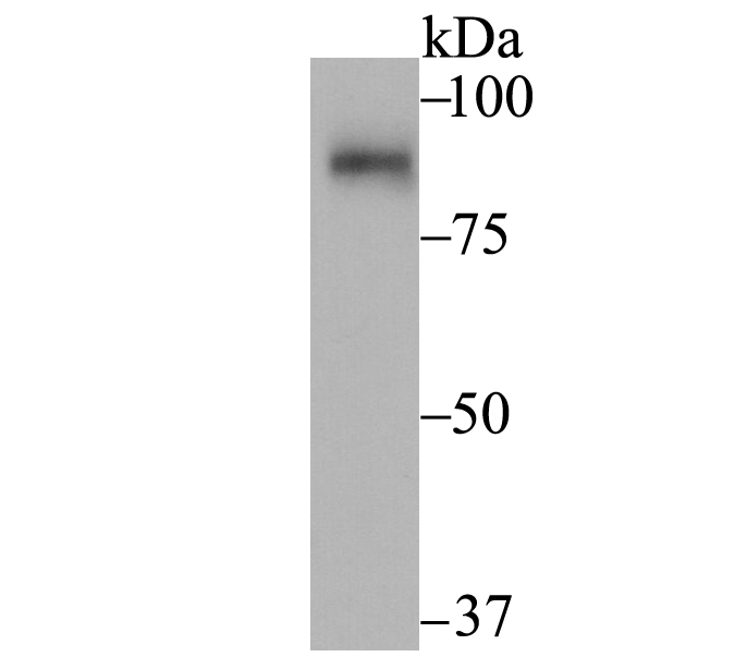
Western blot analysis of SUN1 on rat kidney tissue lysate using anti-SUN1 antibody at 1/2,000 dilution.WB
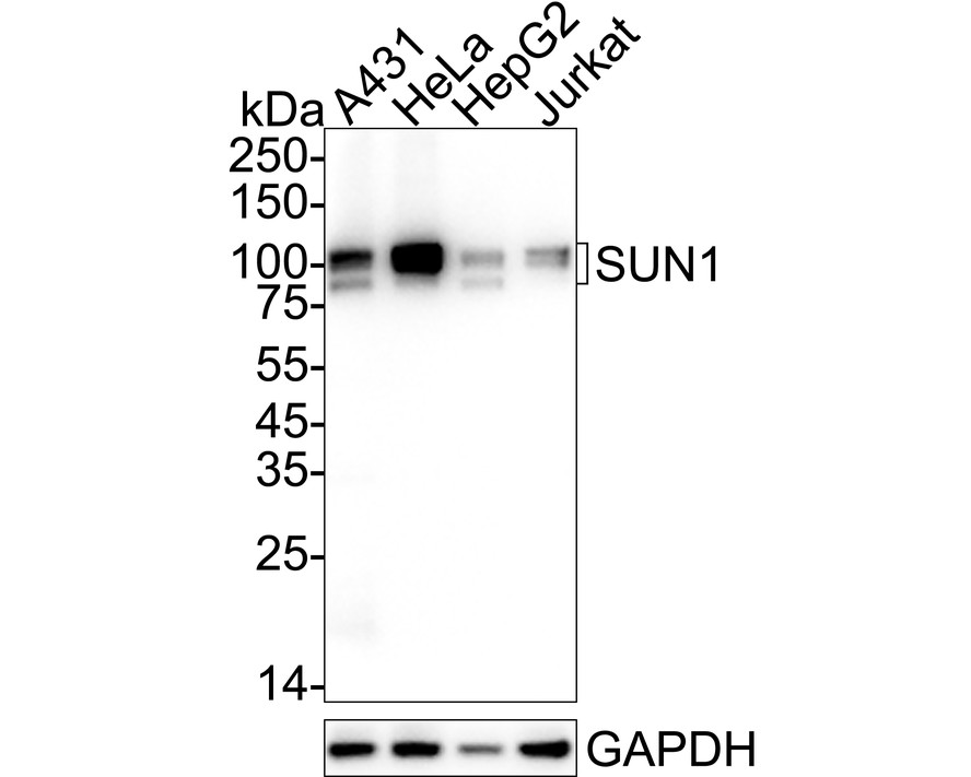
Western blot analysis of SUN1 on different lysates with Rabbit anti-SUN1 antibody at 1/2,000 dilution. Lane 1: A431 cell lysate, Lane 2: HeLa cell lysate, Lane 3: HepG2 cell lysate, Lane 4: Jurkat cell lysate, Lysates/proteins at 20 µg/Lane. Exposure time: 1 minute; 4-20% SDS-PAGE gel. Proteins were transferred to a PVDF membrane and blocked with 5% NFDM/TBST for 1 hour at room temperature. The primary antibody at 1/2,000 dilution was used in 5% NFDM/TBST at 4℃ overnight. Goat Anti-Rabbit IgG - HRP Secondary Antibody at 1/50,000 dilution was used for 1 hour at room temperature.IHC
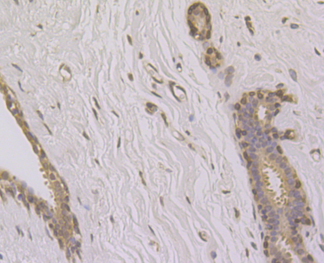
Immunohistochemical analysis of paraffin-embedded human breast tissue using anti-SUN1 antibody. Counter stained with hematoxylin.IHC
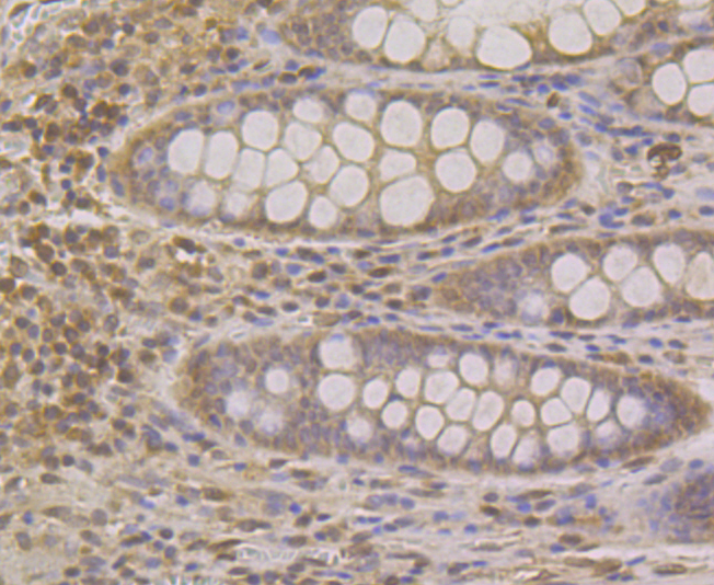
Immunohistochemical analysis of paraffin-embedded human colon tissue using anti-SUN1 antibody. Counter stained with hematoxylin.ICC/IF
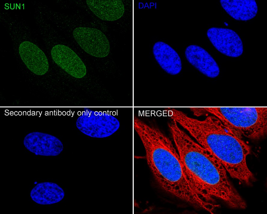
Immunocytochemistry analysis of HeLa cells labeling SUN1 with Rabbit anti-SUN1 antibody at 1/100 dilution. Cells were fixed in 4% paraformaldehyde for 15 minutes at room temperature, permeabilized with 0.1% Triton X-100 in PBS for 15 minutes at room temperature, then blocked with 1% BSA in 10% negative goat serum for 1 hour at room temperature. Cells were then incubated with Rabbit anti-SUN1 antibody at 1/100 dilution in 1% BSA in PBST overnight at 4 ℃. Goat Anti-Rabbit IgG H&L (iFluor™ 488) was used as the secondary antibody at 1/1,000 dilution. PBS instead of the primary antibody was used as the secondary antibody only control. Nuclear DNA was labelled in blue with DAPI. Beta tubulin (red) was stained at 1/100 dilution overnight at +4℃. Goat Anti-Mouse IgG H&L (iFluor™ 594) was used as the secondary antibody at 1/1,000 dilution.| Product Name | SUN1 Recombinant Rabbit Monoclonal Antibody |
|---|---|
| Antibody Type | Primary Antibodies |
| Product description | UNC84A (UNC84 homolog A), also known as SUN1, is a multi-pass nuclear membrane protein that is involved in nuclear anchoring and migration. Highly expressed in heart, brain and testis, UNC84A functions as an A-type Lamin-binding protein that forms a link between the inner and outer nuclear envelope membranes. This link acts as a structural bridge between the nuclear interior and the Actin cytoskeleton and is essential for proper localization of nuclear envelope proteins. Additionally, UNC84A may be involved in telomere attachment and in normal testis development. UNC84A contains one UNC84 (SUN) domain and exists as four isoforms due to alternative splicing events. |
| Immunogen | Recombinant protein within Human SUN1 aa 90-240 / 812. |
| Clonality | monoclonal |
|---|---|
| Isotype | IgG |
| Host Species | Recombinant rabbit |
| Tested Applications | ICC/IFIHCWB |
| WB:1:2000 IHC:1:50-1:200 ICC/IF:1:50-1:200 |
|
| Species Reactivity | HumanRat |
| Concentration | 1mg/ml |
| Purification | Protein A |
| Alternative Names | Protein unc-84 homolog A antibody Sad1 and UNC84 domain containing 1 antibody Sad1/unc 84 protein like 1 antibody Sad1/unc-84 protein-like 1 antibody Sun domain-containing protein 1 antibody Sun1 antibody SUN1_HUMAN antibody UNC 84 homolog A (C elegans) antibody UNC 84A antibody UNC84 A antibody UNC84 antibody UNC84 homolog A (C elegans) antibody UNC84A antibody |
|---|---|
| Molecular Weight(MW) | 87kDa(Observed band size: 90/100kDa) |
| Cellular Localization | Nucleus. |
| SwissProt ID | O94901 |
|---|
WB
Western blot analysis of SUN1 on rat kidney tissue lysate using anti-SUN1 antibody at 1/2,000 dilution.
WB
Western blot analysis of SUN1 on different lysates with Rabbit anti-SUN1 antibody at 1/2,000 dilution. Lane 1: A431 cell lysate, Lane 2: HeLa cell lysate, Lane 3: HepG2 cell lysate, Lane 4: Jurkat cell lysate, Lysates/proteins at 20 µg/Lane. Exposure time: 1 minute; 4-20% SDS-PAGE gel. Proteins were transferred to a PVDF membrane and blocked with 5% NFDM/TBST for 1 hour at room temperature. The primary antibody at 1/2,000 dilution was used in 5% NFDM/TBST at 4℃ overnight. Goat Anti-Rabbit IgG - HRP Secondary Antibody at 1/50,000 dilution was used for 1 hour at room temperature.
IHC
Immunohistochemical analysis of paraffin-embedded human breast tissue using anti-SUN1 antibody. Counter stained with hematoxylin.
IHC
Immunohistochemical analysis of paraffin-embedded human colon tissue using anti-SUN1 antibody. Counter stained with hematoxylin.
ICC/IF
Immunocytochemistry analysis of HeLa cells labeling SUN1 with Rabbit anti-SUN1 antibody at 1/100 dilution. Cells were fixed in 4% paraformaldehyde for 15 minutes at room temperature, permeabilized with 0.1% Triton X-100 in PBS for 15 minutes at room temperature, then blocked with 1% BSA in 10% negative goat serum for 1 hour at room temperature. Cells were then incubated with Rabbit anti-SUN1 antibody at 1/100 dilution in 1% BSA in PBST overnight at 4 ℃. Goat Anti-Rabbit IgG H&L (iFluor™ 488) was used as the secondary antibody at 1/1,000 dilution. PBS instead of the primary antibody was used as the secondary antibody only control. Nuclear DNA was labelled in blue with DAPI. Beta tubulin (red) was stained at 1/100 dilution overnight at +4℃. Goat Anti-Mouse IgG H&L (iFluor™ 594) was used as the secondary antibody at 1/1,000 dilution.| Positive Control | Rat kidney tissue lysate, human colon tissue, human breast tissue, SH-SY-5Y, A431. |
|---|---|
| Application Notes | WB:1:2000 IHC:1:50-1:200 ICC/IF:1:50-1:200 |
| Form | Liquid |
|---|---|
| Storage Instructions | Store at +4℃ after thawing. Aliquot store at -20℃. Avoid repeated freeze / thaw cycles. |
| Storage Buffer | 1*TBS (pH7.4), 1%BSA, 40%Glycerol. Preservative: 0.05% Sodium Azide. |


