Application
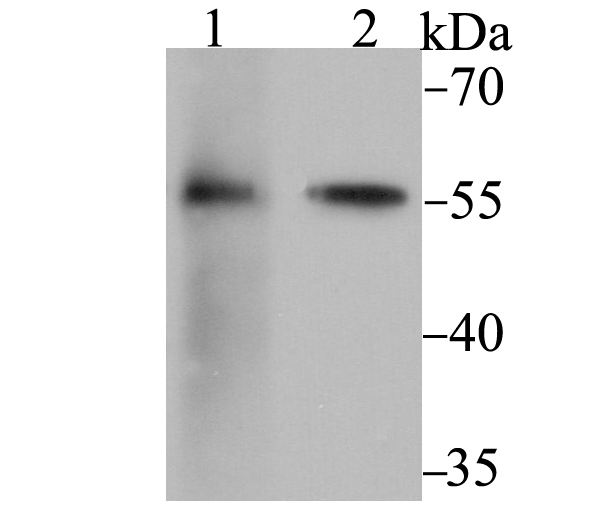
Fig1: Western blot analysis of FKBP52 on 293 (1) and SK-Br-3 (2) cell lysate using anti-FKBP52 antibody at 1/2,000 dilution.Application
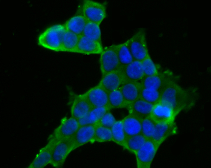
Fig2: ICC staining FKBP52 in 293T cells (green). The nuclear counter stain is DAPI (blue). Cells were fixed in paraformaldehyde, permeabilised with 0.25% Triton X100/PBS.Application
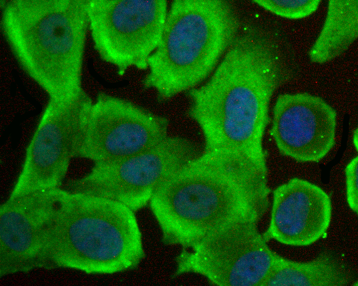
Fig3: ICC staining FKBP52 in MCF-7 cells (green). The nuclear counter stain is DAPI (blue). Cells were fixed in paraformaldehyde, permeabilised with 0.25% Triton X100/PBS.Application
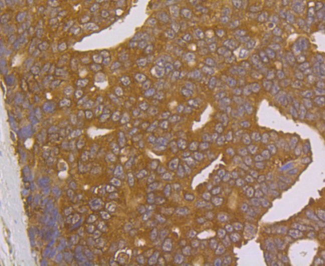
Fig4: Immunohistochemical analysis of paraffin-embedded human prostate cancer tissue using anti-FKBP52 antibody. Counter stained with hematoxylin.Application
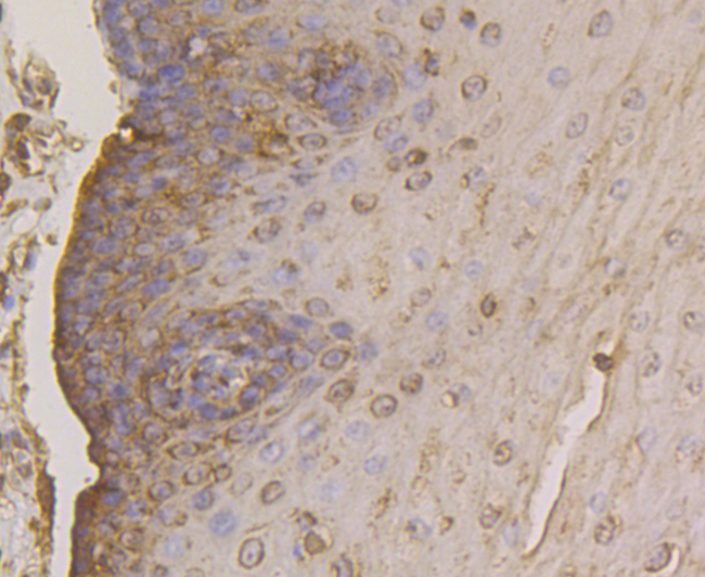
Fig5: Immunohistochemical analysis of paraffin-embedded human esophagus tissue using anti-FKBP52 antibody. Counter stained with hematoxylin.Application
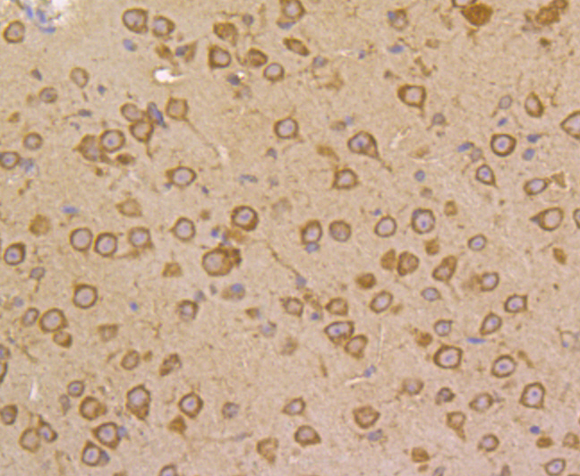
Fig6: Immunohistochemical analysis of paraffin-embedded mouse brain tissue using anti-FKBP52 antibody. Counter stained with hematoxylin.Application
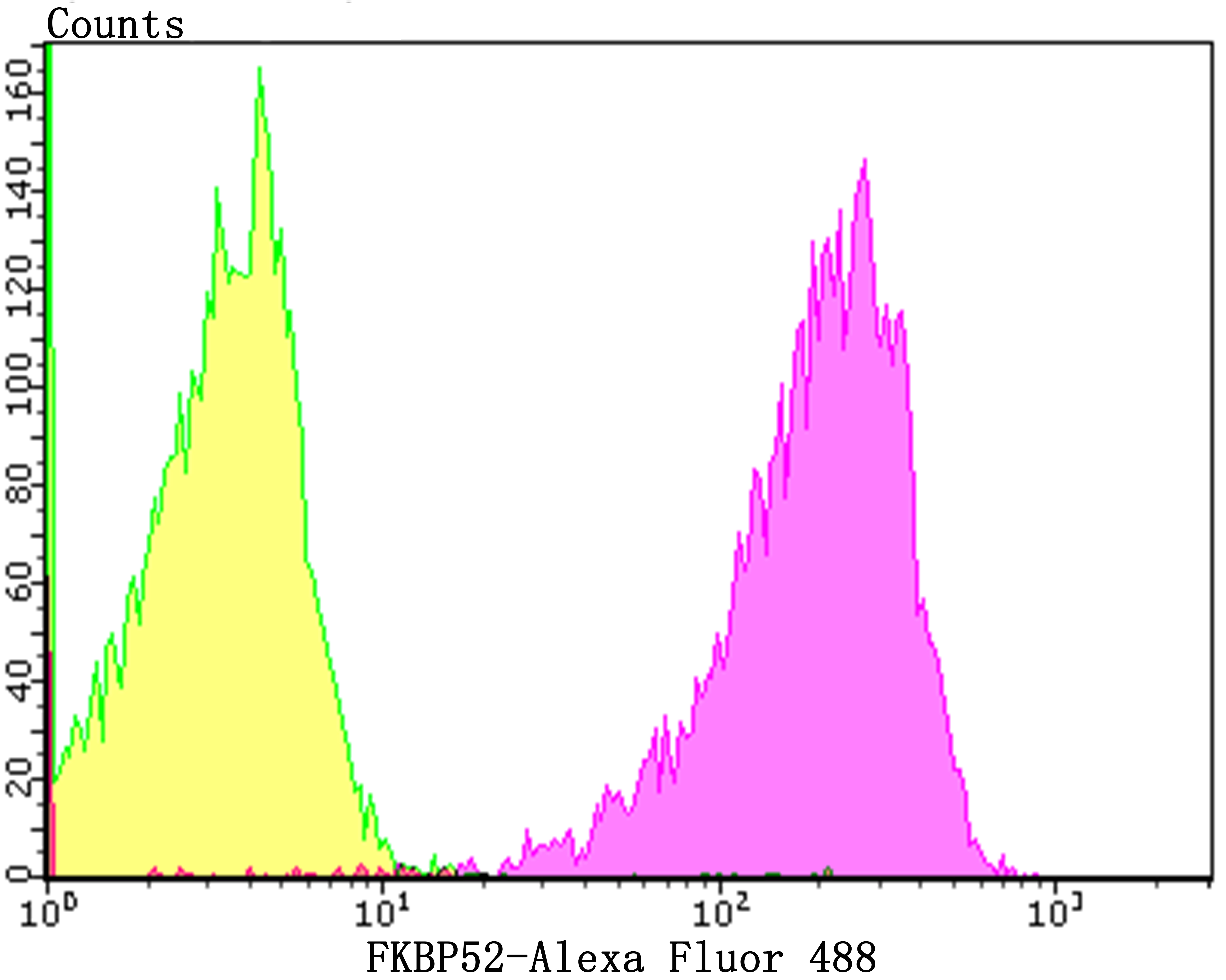
Fig7: Flow cytometric analysis of 293T cells with FKBP52 antibody at 1/100 dilution (purple) compared with an unlabelled control (cells without incubation with primary antibody; yellow). Alexa Fluor 488-conjugated goat anti-rabbit IgG was used as the secondary antibody.| Product Name | FKBP52 [JG96-30] |
|---|---|
| Antibody Type | Primary Antibodies |
| Antigen Alias | 51 kDa FK506-binding protein antibody 52 kDa FK506 binding protein antibody 52 kDa FK506-binding protein antibody 52 kDa FKBP antibody 59 kDa immunophilin antibody FK506 binding protein 4 antibody FK506-binding protein 4 antibody FKBP 4 antibody FKBP 52 antibody FKBP 59 antibody FKBP-4 antibody FKBP-52 antibody FKBP4 antibody FKBP4_HUMAN antibody FKBP51 antibody FKBP52 protein antibody FKBP59 antibody HBI antibody Hsp 56 antibody HSP binding immunophilin antibody HSP-binding immunophilin antibody Hsp56 antibody Immunophilin FKBP52 antibody N-terminally processed antibody p52 antibody p59 antibody p59 protein antibody Peptidyl prolyl cis trans isomerase antibody Peptidyl-prolyl cis-trans isomerase FKBP4 antibody Peptidylprolyl cis trans isomerase antibody PPIase antibody PPIase FKBP4 antibody Rotamase antibody T cell FK506 binding protein 59kD antibody |
| Product description | Immunophilin protein with PPIase and co-chaperone activities. Component of steroid receptors heterocomplexes through interaction with heat-shock protein 90 (HSP90). May play a role in the intracellular trafficking of heterooligomeric forms of steroid hormone receptors between cytoplasm and nuclear compartments. The isomerase activity controls neuronal growth cones via regulation of TRPC1 channel opening. Acts also as a regulator of microtubule dynamics by inhibiting MAPT/TAU ability to promote microtubule assembly. May have a protective role against oxidative stress in mitochondria. |
| Immunogen | Recombinant protein within human FKBP52 aa 1-200. |
| Clonality | Monoclonal |
|---|---|
| Isotype | IgG |
| Host Species | Recombinant rabbit |
| Tested Applications | WBICCIHCFC |
| WB:1:1,000-1:2,000 ICC:1:50-1:200 IHC:1:50-1:200 FC:1:50-1:100 |
|
| Species Reactivity | HumanMouse |
| Concentration | 1mg/ml |
| Alternative Names | 51 kDa FK506-binding protein antibody 52 kDa FK506 binding protein antibody 52 kDa FK506-binding protein antibody 52 kDa FKBP antibody 59 kDa immunophilin antibody FK506 binding protein 4 antibody FK506-binding protein 4 antibody FKBP 4 antibody FKBP 52 antibody FKBP 59 antibody FKBP-4 antibody FKBP-52 antibody FKBP4 antibody FKBP4_HUMAN antibody FKBP51 antibody FKBP52 protein antibody FKBP59 antibody HBI antibody Hsp 56 antibody HSP binding immunophilin antibody HSP-binding immunophilin antibody Hsp56 antibody Immunophilin FKBP52 antibody N-terminally processed antibody p52 antibody p59 antibody p59 protein antibody Peptidyl prolyl cis trans isomerase antibody Peptidyl-prolyl cis-trans isomerase FKBP4 antibody Peptidylprolyl cis trans isomerase antibody PPIase antibody PPIase FKBP4 antibody Rotamase antibody T cell FK506 binding protein 59kD antibody |
|---|---|
| Molecular Weight(MW) | 52 kDa |
| Cellular Localization | Cytoplasm. Cytoskeleton. Microtubule. Mitochondrion. Nucleus. |
| SwissProt ID | Q02790 |
|---|
Application
Fig1: Western blot analysis of FKBP52 on 293 (1) and SK-Br-3 (2) cell lysate using anti-FKBP52 antibody at 1/2,000 dilution.
Application
Fig2: ICC staining FKBP52 in 293T cells (green). The nuclear counter stain is DAPI (blue). Cells were fixed in paraformaldehyde, permeabilised with 0.25% Triton X100/PBS.
Application
Fig3: ICC staining FKBP52 in MCF-7 cells (green). The nuclear counter stain is DAPI (blue). Cells were fixed in paraformaldehyde, permeabilised with 0.25% Triton X100/PBS.
Application
Fig4: Immunohistochemical analysis of paraffin-embedded human prostate cancer tissue using anti-FKBP52 antibody. Counter stained with hematoxylin.
Application
Fig5: Immunohistochemical analysis of paraffin-embedded human esophagus tissue using anti-FKBP52 antibody. Counter stained with hematoxylin.
Application
Fig6: Immunohistochemical analysis of paraffin-embedded mouse brain tissue using anti-FKBP52 antibody. Counter stained with hematoxylin.
Application
Fig7: Flow cytometric analysis of 293T cells with FKBP52 antibody at 1/100 dilution (purple) compared with an unlabelled control (cells without incubation with primary antibody; yellow). Alexa Fluor 488-conjugated goat anti-rabbit IgG was used as the secondary antibody.| Positive Control | 293, SK-Br-3, 293T, MCF-7, human prostate cancer tissue, human esophagus tissue, mouse brain tissue. |
|---|---|
| Application Notes | WB:1:1,000-1:2,000 ICC:1:50-1:200 IHC:1:50-1:200 FC:1:50-1:100 |
| Form | Liquid |
|---|---|
| Storage Instructions | Store at +4℃ after thawing. Aliquot store at -20℃. Avoid repeated freeze / thaw cycles. |
| Storage Buffer | 1*TBS (pH7.4), 1%BSA, 40%Glycerol. Preservative: 0.05% Sodium Azide. |


