Application
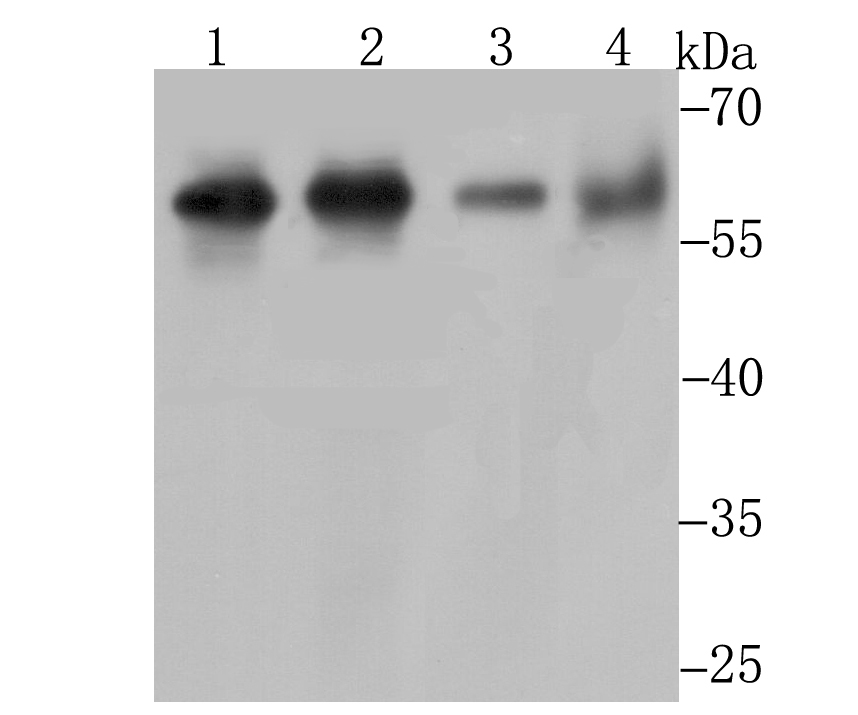
Fig1: Western blot analysis of Methionine Aminopeptidase 2 on Hela cell using anti-Methionine Aminopeptidase 2 antibody at 1/1,000 dilution. Positive control: Lane 1: Daudi Lane 2: K562 Lane 3: Mouse thymus Lane 4: Mouse kidneyApplication
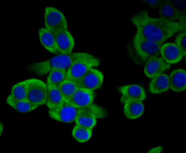
Fig2: ICC staining Methionine Aminopeptidase 2 in LOVO cells (green). The nuclear counter stain is DAPI (blue). Cells were fixed in paraformaldehyde, permeabilised with 0.25% Triton X100/PBS.Application
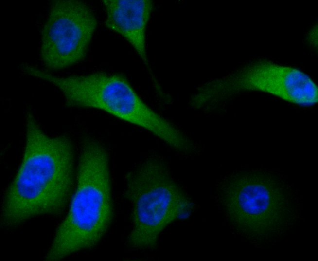
Fig3: ICC staining Methionine Aminopeptidase 2 in PC-3M cells (green). The nuclear counter stain is DAPI (blue). Cells were fixed in paraformaldehyde, permeabilised with 0.25% Triton X100/PBS.Application
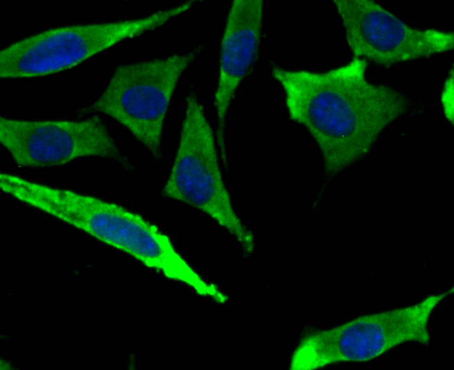
Fig4: ICC staining Methionine Aminopeptidase 2 in SH-SY-5Y cells (green). The nuclear counter stain is DAPI (blue). Cells were fixed in paraformaldehyde, permeabilised with 0.25% Triton X100/PBS.Application
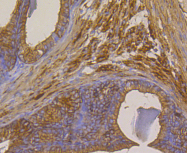
Fig5: Immunohistochemical analysis of paraffin-embedded rat seminal vesicle tissue using anti-Methionine Aminopeptidase 2 antibody. Counter stained with hematoxylin.Application
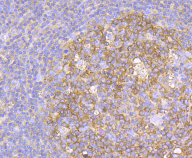
Fig6: Immunohistochemical analysis of paraffin-embedded human tonsil tissue using anti-Methionine Aminopeptidase 2 antibody. Counter stained with hematoxylin.Application
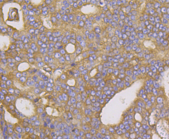
Fig7: Immunohistochemical analysis of paraffin-embedded human prostate cancer tissue using anti-Methionine Aminopeptidase 2 antibody. Counter stained with hematoxylin.Application
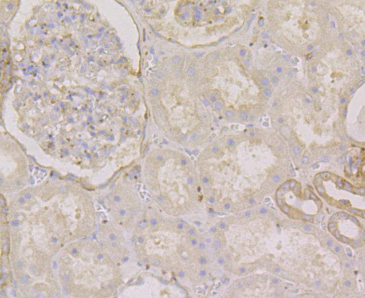
Fig8: Immunohistochemical analysis of paraffin-embedded human kidney tissue using anti-Methionine Aminopeptidase 2 antibody. Counter stained with hematoxylin.Application
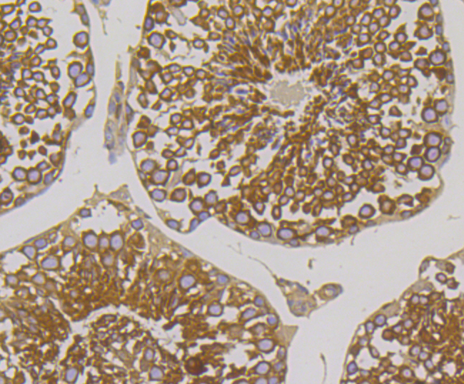
Fig9: Immunohistochemical analysis of paraffin-embedded mouse testis tissue using anti-Methionine Aminopeptidase 2 antibody. Counter stained with hematoxylin.Application
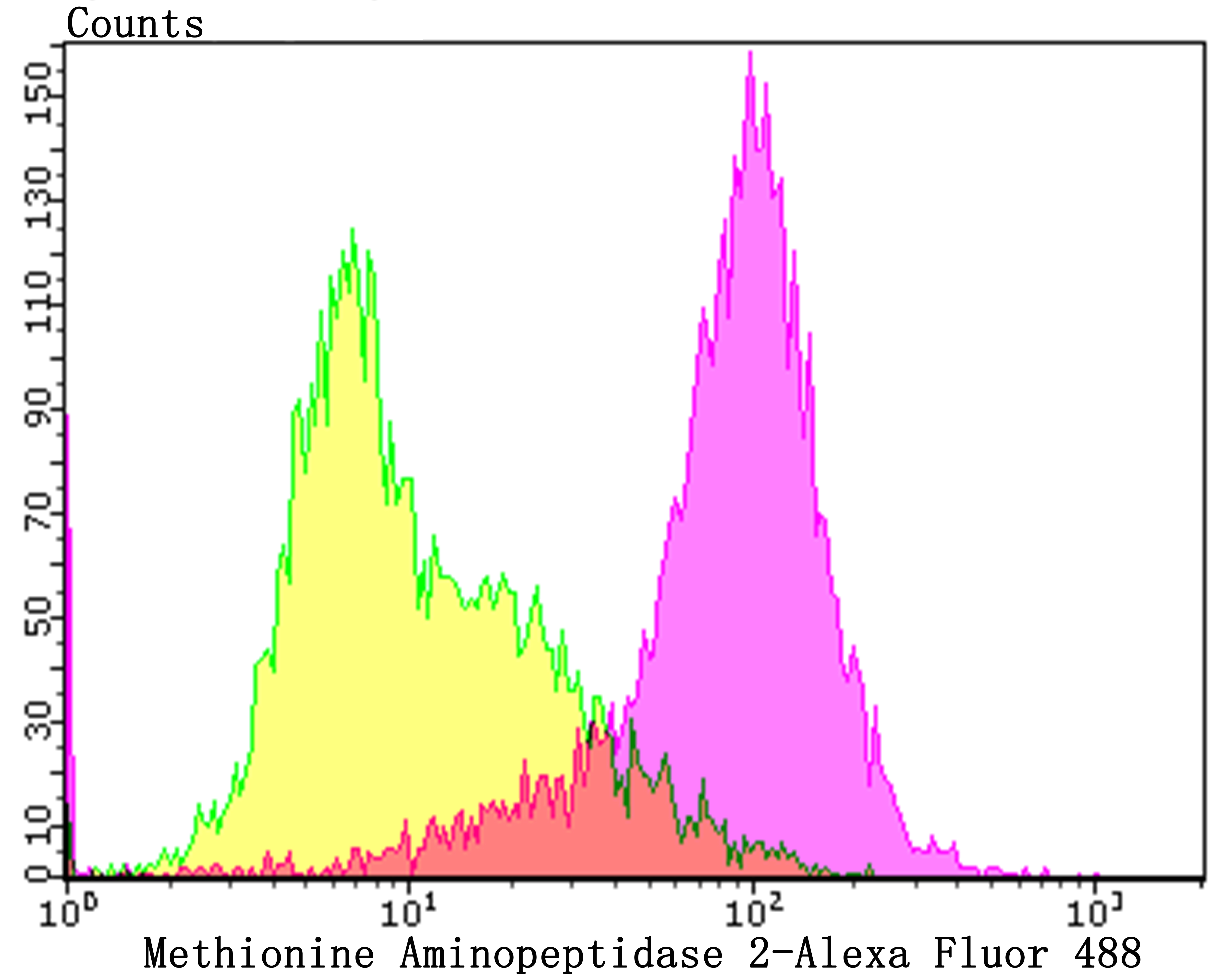
Fig10: Flow cytometric analysis of Daudi cells with Methionine Aminopeptidase 2 antibody at 1/100 dilution (purple) compared with an unlabelled control (cells without incubation with primary antibody; yellow). Alexa Fluor 488-conjugated goat anti-rabbit IgG was used as the secondary antibody.| Product Name | Methionine Aminopeptidase 2 [JG39-79] |
|---|---|
| Antibody Type | Primary Antibodies |
| Antigen Alias | A930035J23Rik antibody AI047573 antibody AL024412 antibody Amp2 antibody AU014659, antibody EIF 2 associated p67 homolog antibody EIF-2-associated p67 antibody Initiation factor 2 associated 67 kDa glycoprotein antibody Initiation factor 2-associated 67 kDa glycoprotein antibody MAP 2 antibody MAP2 antibody MAP2_HUMAN antibody MetAP 2 antibody Metap2 antibody Methionine aminopeptidase 2 antibody Methionyl aminopeptidase 2 antibody MGC102452 antibody MGC127390 antibody MGC53792 antibody MNPEP antibody p67 antibody p67eIF2 antibody Peptidase M antibody Peptidase M2 antibody |
| Product description | Methionine aminopeptidases (MetAP), also designated peptidase M proteins, are members of the M24 family of proteins. Both MetAP-1 and MetAP-2 release N-terminal amino acids, usually methionine, from nascent peptides and arylamines. Eukaryotes contain both MetAP-1 and MetAP-2, whereas prokaryotes possess only the MetAP-1 enzyme. MetAP-1 and MetAP-2 control cell proliferation in mammalian cells. MetAP-2 is highly conserved between human and Saccharomyces cerevisiae. Neurofibromin (NF1) regulates MetAp-2 and increased expression of MetAP-2 correlates with several forms of cancer. Inhibitors of MetAP-2 are potential targets in cancer therapeutics, particularly in NF1-associated tumor proliferation. Chemotherapeutic drugs such as ovalicin and fumagillin bind to the active site of and inhibit MetAp-2. |
| Immunogen | Recombinant protein within C-terminal human Methionine Aminopeptidase 2. |
| Clonality | Monoclonal |
|---|---|
| Isotype | IgG |
| Host Species | Recombinant rabbit |
| Tested Applications | WBICCIFIHCFC |
| WB:1:500-1:2,000 ICC/IF:1:50-1:200 IHC:1:50-1:200 FC:1:50-1:100 |
|
| Species Reactivity | HumanMouseRat |
| Concentration | 1mg/ml |
| Alternative Names | A930035J23Rik antibody
AI047573 antibody
AL024412 antibody
Amp2 antibody
AU014659 antibody EIF 2 associated p67 homolog antibody EIF-2-associated p67 antibody Initiation factor 2 associated 67 kDa glycoprotein antibody Initiation factor 2-associated 67 kDa glycoprotein antibody MAP 2 antibody MAP2 antibody MAP2_HUMAN antibody MetAP 2 antibody Metap2 antibody Methionine aminopeptidase 2 antibody Methionyl aminopeptidase 2 antibody MGC102452 antibody MGC127390 antibody MGC53792 antibody MNPEP antibody p67 antibody p67eIF2 antibody Peptidase M antibody Peptidase M2 antibody |
|---|---|
| Molecular Weight(MW) | Predicted band size 53 kDa |
| Cellular Localization | Cytoplasm. |
Application
Fig1: Western blot analysis of Methionine Aminopeptidase 2 on Hela cell using anti-Methionine Aminopeptidase 2 antibody at 1/1,000 dilution. Positive control: Lane 1: Daudi Lane 2: K562 Lane 3: Mouse thymus Lane 4: Mouse kidney
Application
Fig2: ICC staining Methionine Aminopeptidase 2 in LOVO cells (green). The nuclear counter stain is DAPI (blue). Cells were fixed in paraformaldehyde, permeabilised with 0.25% Triton X100/PBS.
Application
Fig3: ICC staining Methionine Aminopeptidase 2 in PC-3M cells (green). The nuclear counter stain is DAPI (blue). Cells were fixed in paraformaldehyde, permeabilised with 0.25% Triton X100/PBS.
Application
Fig4: ICC staining Methionine Aminopeptidase 2 in SH-SY-5Y cells (green). The nuclear counter stain is DAPI (blue). Cells were fixed in paraformaldehyde, permeabilised with 0.25% Triton X100/PBS.
Application
Fig5: Immunohistochemical analysis of paraffin-embedded rat seminal vesicle tissue using anti-Methionine Aminopeptidase 2 antibody. Counter stained with hematoxylin.
Application
Fig6: Immunohistochemical analysis of paraffin-embedded human tonsil tissue using anti-Methionine Aminopeptidase 2 antibody. Counter stained with hematoxylin.
Application
Fig7: Immunohistochemical analysis of paraffin-embedded human prostate cancer tissue using anti-Methionine Aminopeptidase 2 antibody. Counter stained with hematoxylin.
Application
Fig8: Immunohistochemical analysis of paraffin-embedded human kidney tissue using anti-Methionine Aminopeptidase 2 antibody. Counter stained with hematoxylin.
Application
Fig9: Immunohistochemical analysis of paraffin-embedded mouse testis tissue using anti-Methionine Aminopeptidase 2 antibody. Counter stained with hematoxylin.
Application
Fig10: Flow cytometric analysis of Daudi cells with Methionine Aminopeptidase 2 antibody at 1/100 dilution (purple) compared with an unlabelled control (cells without incubation with primary antibody; yellow). Alexa Fluor 488-conjugated goat anti-rabbit IgG was used as the secondary antibody.| Positive Control | Daudi, K562, mouse thymus tissue lysate, mouse kidney tissue, LOVO, PC-3M, SH-SY-5Y, rat seminal vesicle tissue, human tonsil tissue, human prostate cancer tissue, human kidney tissue, mouse testis tissue. |
|---|---|
| Application Notes | WB:1:500-1:2,000 ICC/IF:1:50-1:200 IHC:1:50-1:200 FC:1:50-1:100 |
| Form | Liquid |
|---|---|
| Storage Instructions | Store at +4℃ after thawing. Aliquot store at -20℃. Avoid repeated freeze / thaw cycles. |
| Storage Buffer | 1*TBS (pH7.4), 1%BSA, 40%Glycerol. Preservative: 0.05% Sodium Azide. |


