Application
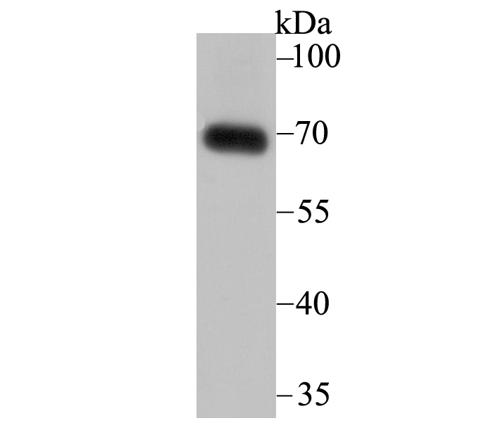
Fig1: Western blot analysis of SNX1 on human skin tissue lysate using anti-SNX1 antibody at 1/2,000 dilution.Application
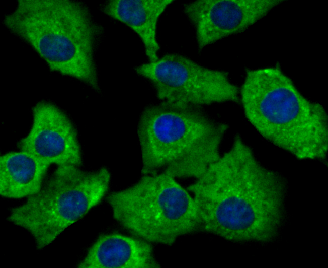
Fig2: ICC staining SNX1 in A549 cells (green). The nuclear counter stain is DAPI (blue). Cells were fixed in paraformaldehyde, permeabilised with 0.25% Triton X100/PBS.Application
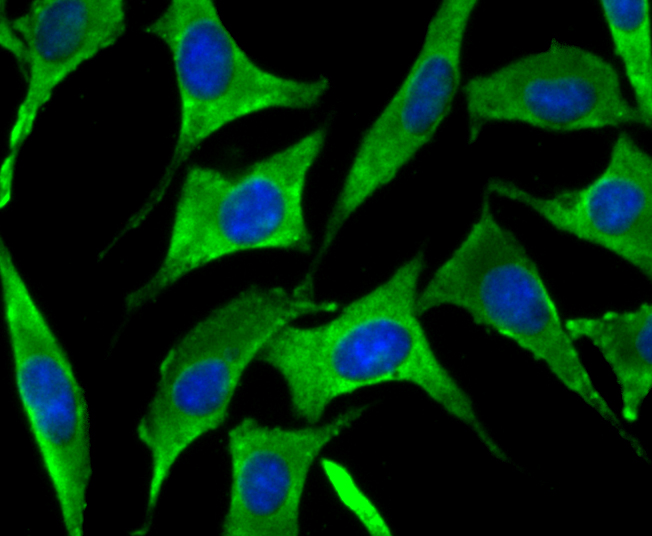
Fig3: ICC staining SNX1 in SH-SY-5Y cells (green). The nuclear counter stain is DAPI (blue). Cells were fixed in paraformaldehyde, permeabilised with 0.25% Triton X100/PBS.Application
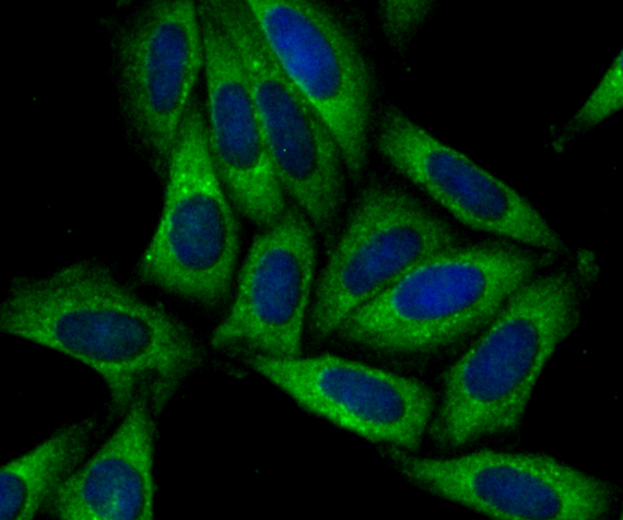
Fig4: ICC staining SNX1 in SiHa cells (green). The nuclear counter stain is DAPI (blue). Cells were fixed in paraformaldehyde, permeabilised with 0.25% Triton X100/PBS.Application
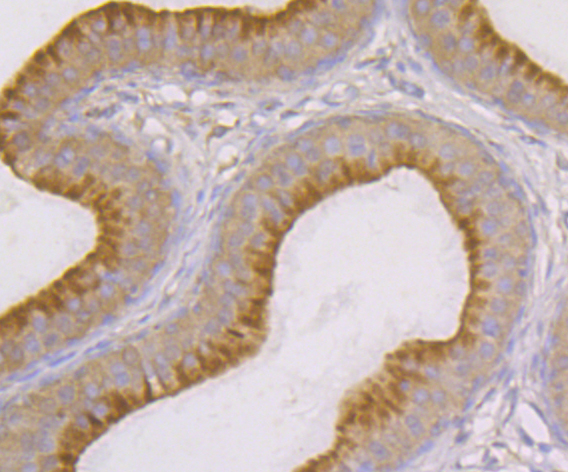
Fig5: Immunohistochemical analysis of paraffin-embedded rat epididymis tissue using anti-SNX1 antibody. Counter stained with hematoxylin.Application
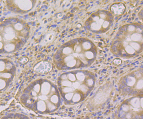
Fig6: Immunohistochemical analysis of paraffin-embedded human colon tissue using anti-SNX1 antibody. Counter stained with hematoxylin.Application
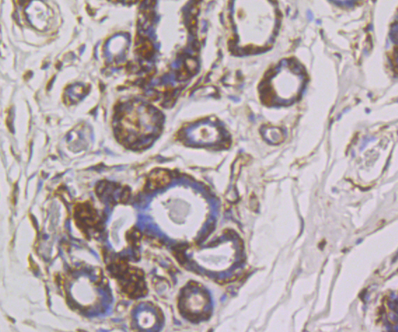
Fig7: Immunohistochemical analysis of paraffin-embedded human breast cancer tissue using anti-SNX1 antibody. Counter stained with hematoxylin.Application
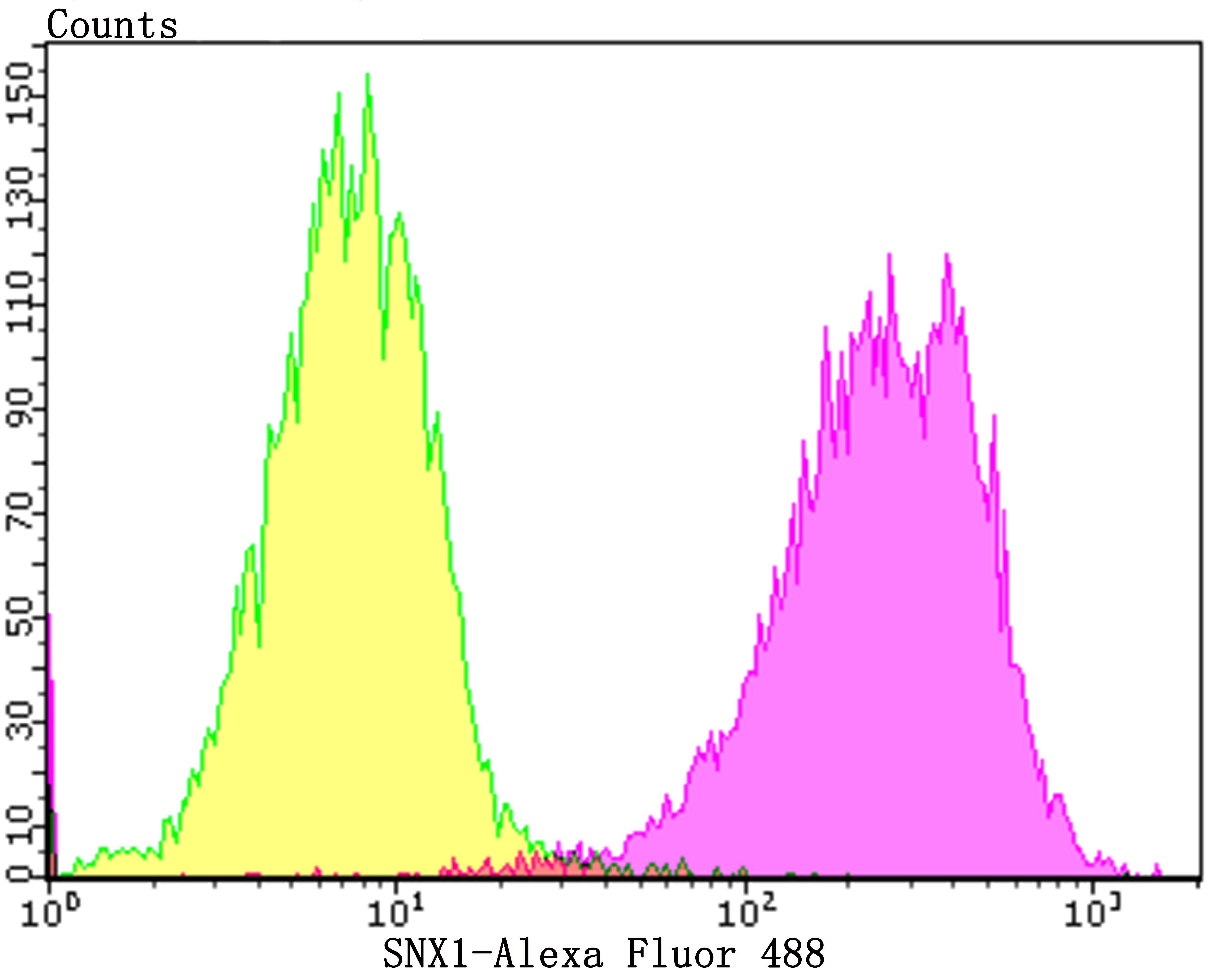
Fig8: Flow cytometric analysis of SiHa cells with SNX1 antibody at 1/100 dilution (purple) compared with an unlabelled control (cells without incubation with primary antibody; yellow). Alexa Fluor 488-conjugated goat anti-rabbit IgG was used as the secondary antibody.| Product Name | SNX1 [JG40-06] |
|---|---|
| Antibody Type | Primary Antibodies |
| Antigen Alias | HsT17379 antibody MGC8664 antibody SNX 1 antibody SNX 1a antibody Snx1 antibody SNX1_HUMAN antibody SNX1A antibody Sorting nexin 1 antibody Sorting nexin 1A antibody Sorting nexin-1 antibody Vps5 antibody |
| Product description | Sorting nexin 1 (SNX1) is a member of a large family of hydrophilic proteins that interact with a variety of receptor types and are involved in intracellular trafficking. SNX1 and the related splice variant, SNX1A, bind the epidermal growth factor (EGF) receptor, facilitate its transport to lysosome, and thereby contribute to the degradation of the receptor. SNX2 and SNX4 share a high degree of amino acid similarity with SNX1, as they all contain a characteristic phox homology (PX) domain. These proteins are all partially associated with cellular membranes, and they, likewise, associate with EGF, PDGF and insulin receptor tyrosine kinases. These nexins are widely expressed and yet have various tissue distribution patterns. Additionally, the sorting nexins can associate with each other and with a variety of other cellular proteins, suggesting that they exist as part of multisubunit complexes. The related protein, SNX3, comprises a distinct subgroup of nexins that share less sequence similarity outside of the PX domain and have dramatically different binding affinities for the tyrosine kinase receptors. |
| Immunogen | Recombinant protein within human SNX1 aa 50-250. |
| Clonality | Monoclonal |
|---|---|
| Isotype | IgG |
| Host Species | Recombinant rabbit |
| Tested Applications | WBICCIFIHCFC |
| WB:1:1,000-1:2,000 ICC/IF:1:50-1:200 IHC:1:50-1:200 FC:1:50-1:100 |
|
| Species Reactivity | HumanMouseRat |
| Concentration | 1mg/ml |
| Alternative Names | HsT17379 antibody MGC8664 antibody SNX 1 antibody SNX 1a antibody Snx1 antibody SNX1_HUMAN antibody SNX1A antibody Sorting nexin 1 antibody Sorting nexin 1A antibody Sorting nexin-1 antibody Vps5 antibody |
|---|---|
| Molecular Weight(MW) | 63 kDa |
| Cellular Localization | Endosome. Golgi apparatus. |
Application
Fig1: Western blot analysis of SNX1 on human skin tissue lysate using anti-SNX1 antibody at 1/2,000 dilution.
Application
Fig2: ICC staining SNX1 in A549 cells (green). The nuclear counter stain is DAPI (blue). Cells were fixed in paraformaldehyde, permeabilised with 0.25% Triton X100/PBS.
Application
Fig3: ICC staining SNX1 in SH-SY-5Y cells (green). The nuclear counter stain is DAPI (blue). Cells were fixed in paraformaldehyde, permeabilised with 0.25% Triton X100/PBS.
Application
Fig4: ICC staining SNX1 in SiHa cells (green). The nuclear counter stain is DAPI (blue). Cells were fixed in paraformaldehyde, permeabilised with 0.25% Triton X100/PBS.
Application
Fig5: Immunohistochemical analysis of paraffin-embedded rat epididymis tissue using anti-SNX1 antibody. Counter stained with hematoxylin.
Application
Fig6: Immunohistochemical analysis of paraffin-embedded human colon tissue using anti-SNX1 antibody. Counter stained with hematoxylin.
Application
Fig7: Immunohistochemical analysis of paraffin-embedded human breast cancer tissue using anti-SNX1 antibody. Counter stained with hematoxylin.
Application
Fig8: Flow cytometric analysis of SiHa cells with SNX1 antibody at 1/100 dilution (purple) compared with an unlabelled control (cells without incubation with primary antibody; yellow). Alexa Fluor 488-conjugated goat anti-rabbit IgG was used as the secondary antibody.| Positive Control | Human skin tissue lysate, A549, SH-SY-5Y, SiHa, rat epididymis tissue, human colon tissue, human breast cancer tissue. |
|---|---|
| Application Notes | WB:1:1,000-1:2,000 ICC/IF:1:50-1:200 IHC:1:50-1:200 FC:1:50-1:100 |
| Form | Liquid |
|---|---|
| Storage Instructions | Store at +4℃ after thawing. Aliquot store at -20℃. Avoid repeated freeze / thaw cycles. |
| Storage Buffer | 1*TBS (pH7.4), 1%BSA, 40%Glycerol. Preservative: 0.05% Sodium Azide. |


