Application
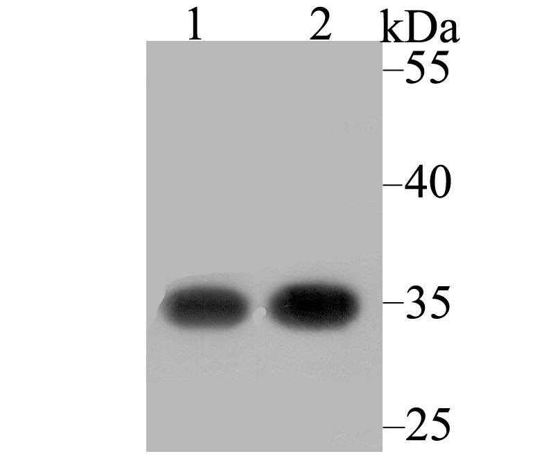
Fig1: Western blot analysis of RACK1 on different lysates using anti-RACK1 antibody at 1/2,000 dilution. Positive control: Lane 1: SH-SY-5Y Lane 2: Rat small intestineApplication
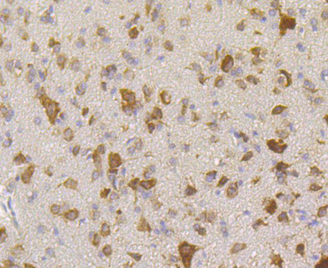
Fig2: Immunohistochemical analysis of paraffin-embedded rat cerebellum tissue using anti-RACK1 antibody. Counter stained with hematoxylin.Application
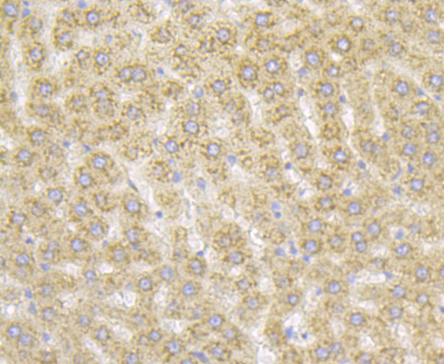
Fig3: Immunohistochemical analysis of paraffin-embedded rat liver tissue using anti-RACK1 antibody. Counter stained with hematoxylin.Application
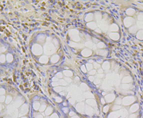
Fig4: Immunohistochemical analysis of paraffin-embedded human colon tissue using anti-RACK1 antibody. Counter stained with hematoxylin.Application
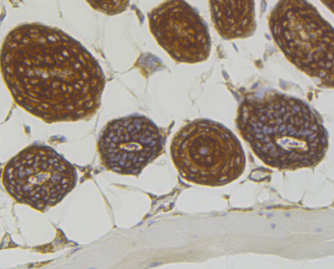
Fig5: Immunohistochemical analysis of paraffin-embedded mouse skin tissue using anti-RACK1 antibody. Counter stained with hematoxylin.Application
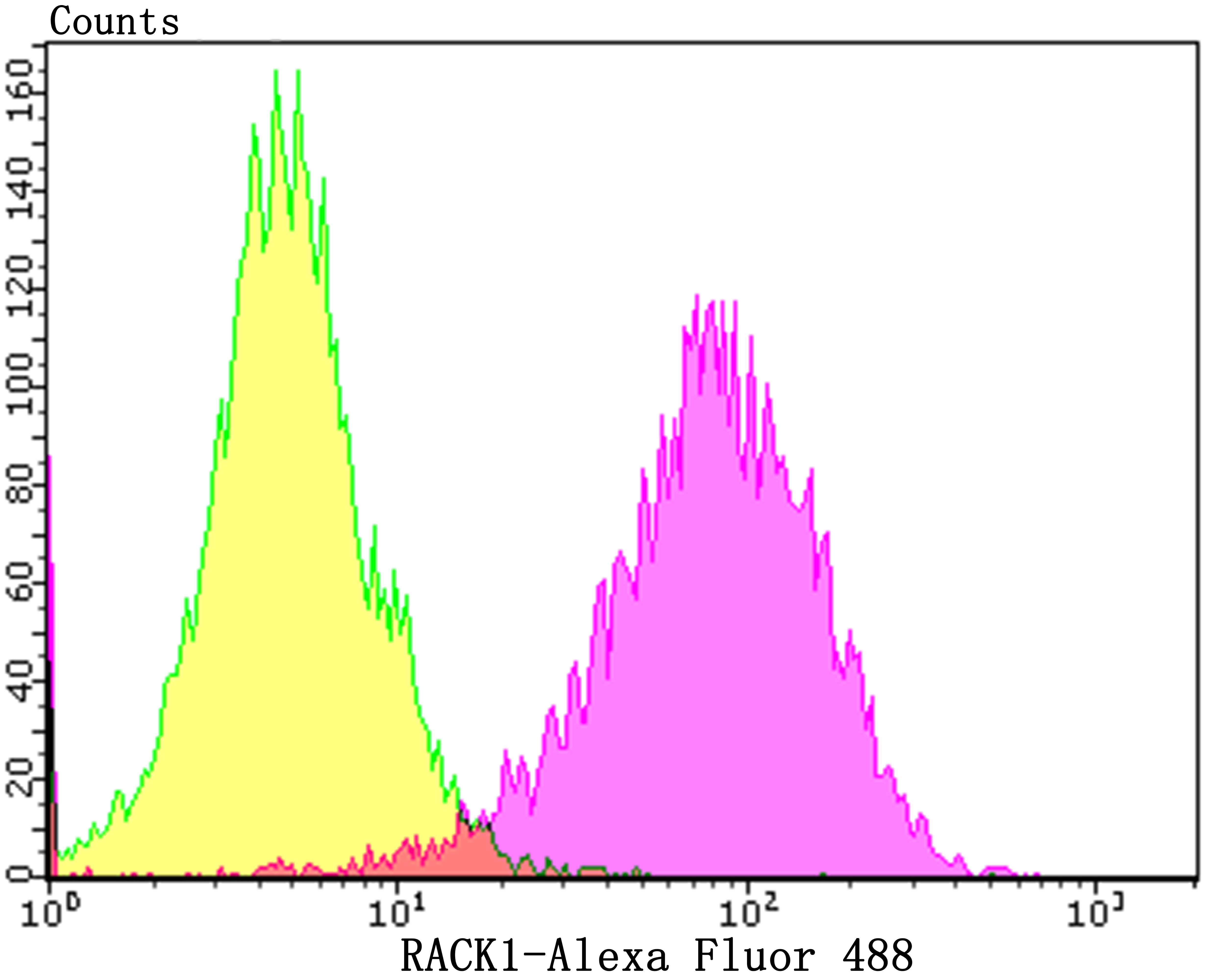
Fig6: Flow cytometric analysis of SH-SY-5Y cells with RACK1 antibody at 1/100 dilution (purple) compared with an unlabelled control (cells without incubation with primary antibody; yellow). Alexa Fluor 488-conjugated goat anti-rabbit IgG was used as the secondary antibody.| Product Name | RACK1 [JG40-26] |
|---|---|
| Antibody Type | Primary Antibodies |
| Antigen Alias | Cell proliferation-inducing gene 21 protein antibody GBLP_HUMAN antibody Gnb2-rs1 antibody Gnb2l1 antibody Guanine nucleotide binding protein (G protein) beta polypeptide 2 like 1 antibody Guanine nucleotide binding protein beta polypeptide 2 like 1 antibody Guanine nucleotide binding protein beta subunit 2 like 1 antibody Guanine nucleotide binding protein beta subunit like protein 12.3 antibody Guanine nucleotide binding protein subunit beta 2 like 1 antibody Guanine nucleotide binding protein subunit beta like protein 12.3 antibody Guanine nucleotide-binding protein subunit beta-2-like 1 antibody Guanine nucleotide-binding protein subunit beta-like protein 12.3 antibody H12.3 antibody HLC-7 antibody Human lung cancer oncogene 7 protein antibody lung cancer oncogene 7 antibody OTTHUMP00000223704 antibody OTTHUMP00000223870 antibody OTTHUMP00000223891 antibody OTTHUMP00000223893 antibody OTTHUMP00000223900 antibody OTTHUMP00000223902 antibody OTTHUMP00000223930 antibody OTTHUMP00000223931 antibody PIG21 antibody Proliferation inducing gene 21 antibody Protein homologous to chicken B complex protein guanine nucleotide binding antibody RACK1 antibody Receptor for activated C kinase 1 antibody Receptor for activated C kinase antibody Receptor of activated protein kinase C 1 antibody |
| Product description | Members of the protein kinase C (PKC) family play a key regulatory role in a variety of cellular functions, including cell growth and differentiation, gene expression, hormone secretion and membrane function. Receptor for activated C kinases, termed RACKs, are intracellular receptors for activated PKC that may be involved in the activation-induced translocation of PKC. RACK1 (receptor for activated C kinase 1) is a 317 amino acid G protein b subunit-like protein that functions as a RACK and inhibits the activity of Src tyrosine kinases. In response to PKC activation, the intracellular localization of RACK1 and PKC bII changes, and RACK1 and PKC bII co-localize to the same sites. RACK1 is therefore thought to be a shuttling protein for PKC bII. A deficit in RACK1 may be associated with impaired PKC activation in the aging brain. The RACK1 gene is conserved in chimpanzee, dog, cow, mouse, rat, chicken, zebrafish, fruit fly, mosquito, C.elegans, S.pombe, S.cerevisiae, K.lactis, E.gossypii, M.grisea, N.crassa, A.thaliana, rice and P.falciparum, and maps to human chromosome 5q35.3. |
| Immunogen | Recombinant protein corresponding to C-terminal human RACK1. |
| Clonality | Monoclonal |
|---|---|
| Isotype | IgG |
| Host Species | Recombinant rabbit |
| Tested Applications | WBIHCFC |
| WB:1:1,000-1:2,000 IHC:1:50-1:200 FC:1:50-1:100 |
|
| Species Reactivity | HumanMouseRat |
| Concentration | 1mg/ml |
| Alternative Names | Cell proliferation-inducing gene 21 protein antibody GBLP_HUMAN antibody Gnb2-rs1 antibody Gnb2l1 antibody Guanine nucleotide binding protein (G protein) beta polypeptide 2 like 1 antibody Guanine nucleotide binding protein beta polypeptide 2 like 1 antibody Guanine nucleotide binding protein beta subunit 2 like 1 antibody Guanine nucleotide binding protein beta subunit like protein 12.3 antibody Guanine nucleotide binding protein subunit beta 2 like 1 antibody Guanine nucleotide binding protein subunit beta like protein 12.3 antibody Guanine nucleotide-binding protein subunit beta-2-like 1 antibody Guanine nucleotide-binding protein subunit beta-like protein 12.3 antibody H12.3 antibody HLC-7 antibody Human lung cancer oncogene 7 protein antibody lung cancer oncogene 7 antibody OTTHUMP00000223704 antibody OTTHUMP00000223870 antibody OTTHUMP00000223891 antibody OTTHUMP00000223893 antibody OTTHUMP00000223900 antibody OTTHUMP00000223902 antibody OTTHUMP00000223930 antibody OTTHUMP00000223931 antibody PIG21 antibody Proliferation inducing gene 21 antibody Protein homologous to chicken B complex protein guanine nucleotide binding antibody RACK1 antibody Receptor for activated C kinase 1 antibody Receptor for activated C kinase antibody Receptor of activated protein kinase C 1 antibody |
|---|---|
| Molecular Weight(MW) | 35 kDa |
| Cellular Localization | Cytoplasm. Cell membrane. Nucleus. |
Application
Fig1: Western blot analysis of RACK1 on different lysates using anti-RACK1 antibody at 1/2,000 dilution. Positive control: Lane 1: SH-SY-5Y Lane 2: Rat small intestine
Application
Fig2: Immunohistochemical analysis of paraffin-embedded rat cerebellum tissue using anti-RACK1 antibody. Counter stained with hematoxylin.
Application
Fig3: Immunohistochemical analysis of paraffin-embedded rat liver tissue using anti-RACK1 antibody. Counter stained with hematoxylin.
Application
Fig4: Immunohistochemical analysis of paraffin-embedded human colon tissue using anti-RACK1 antibody. Counter stained with hematoxylin.
Application
Fig5: Immunohistochemical analysis of paraffin-embedded mouse skin tissue using anti-RACK1 antibody. Counter stained with hematoxylin.
Application
Fig6: Flow cytometric analysis of SH-SY-5Y cells with RACK1 antibody at 1/100 dilution (purple) compared with an unlabelled control (cells without incubation with primary antibody; yellow). Alexa Fluor 488-conjugated goat anti-rabbit IgG was used as the secondary antibody.| Positive Control | SH-SY-5Y, rat small intestine tissue lysate, rat cerebellum tissue, rat liver tissue, human colon tissue, mouse skin tissue. |
|---|---|
| Application Notes | WB:1:1,000-1:2,000 IHC:1:50-1:200 FC:1:50-1:100 |
| Form | Liquid |
|---|---|
| Storage Instructions | Store at +4℃ after thawing. Aliquot store at -20℃. Avoid repeated freeze / thaw cycles. |
| Storage Buffer | 1*TBS (pH7.4), 1%BSA, 40%Glycerol. Preservative: 0.05% Sodium Azide. |


