Application
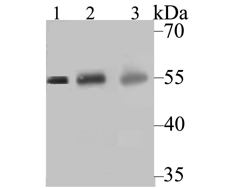
Fig1: Western blot analysis of CYP27A1 on different tissue lysates using anti-CYP27A1 antibody at 1/500 dilution. Positive control: Lane 1: Rat heart Lane 2: Human liver Lane 2: Human kidneyApplication
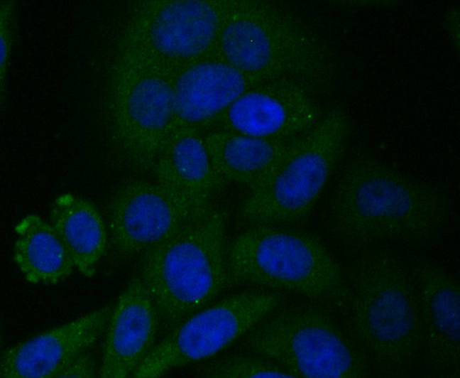
Fig2: ICC staining CYP27A1 in HepG2 cells (green). The nuclear counter stain is DAPI (blue). Cells were fixed in paraformaldehyde, permeabilised with 0.25% Triton X100/PBS.Application
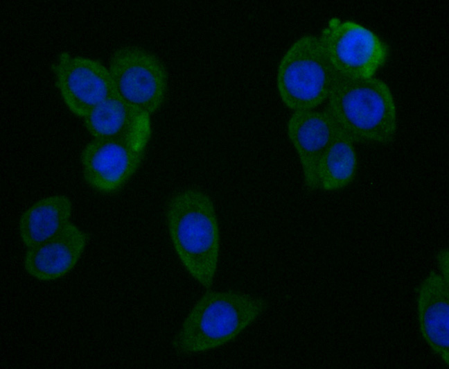
Fig3: ICC staining CYP27A1 in LOVO cells (green). The nuclear counter stain is DAPI (blue). Cells were fixed in paraformaldehyde, permeabilised with 0.25% Triton X100/PBS.Application
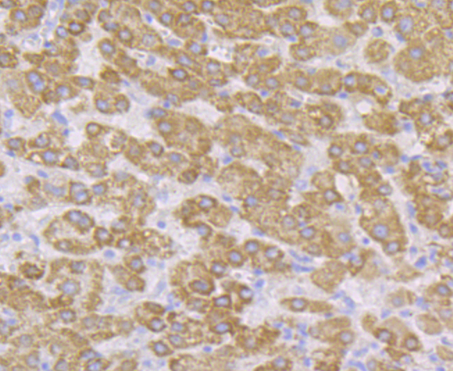
Fig4: Immunohistochemical analysis of paraffin-embedded human liver cancer tissue using anti-CYP27A1 antibody. Counter stained with hematoxylin.Application
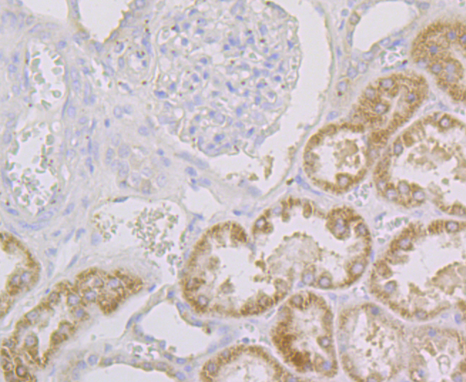
Fig5: Immunohistochemical analysis of paraffin-embedded human kidney tissue using anti-CYP27A1 antibody. Counter stained with hematoxylin.Application
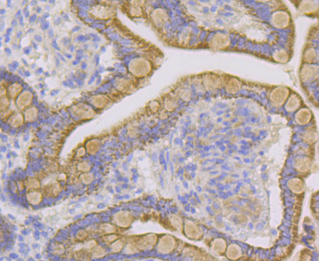
Fig6: Immunohistochemical analysis of paraffin-embedded human small intestine tissue using anti-CYP27A1 antibody. Counter stained with hematoxylin.Application
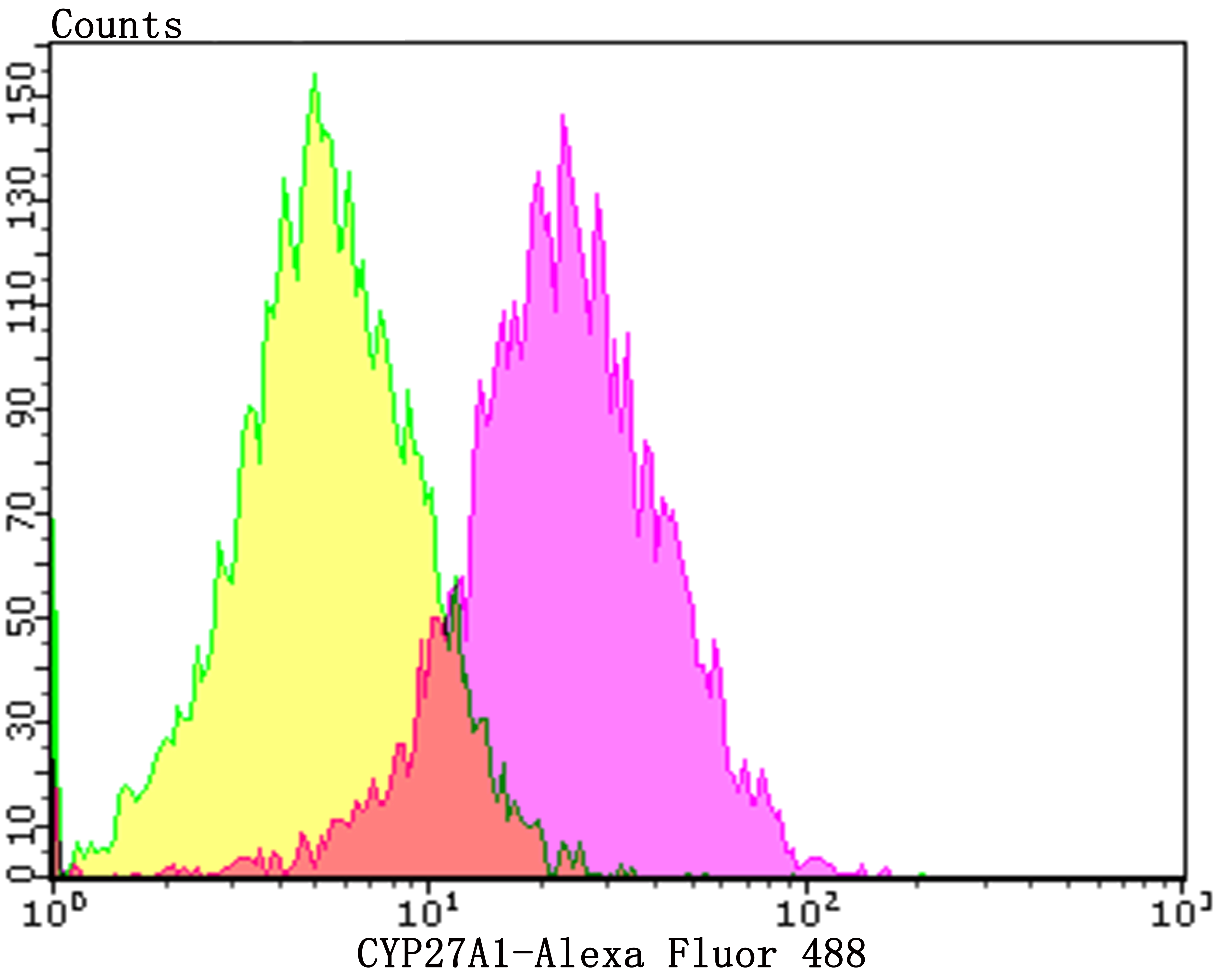
Fig7: Flow cytometric analysis of HepG2 cells with CYP27A1 antibody at 1/100 dilution (purple) compared with an unlabelled control (cells without incubation with primary antibody; yellow). Alexa Fluor 488-conjugated goat anti-rabbit IgG was used as the secondary antibody.| Product Name | CYP27A1 [JG40-36] |
|---|---|
| Antibody Type | Primary Antibodies |
| Antigen Alias | 12-alpha-triol 27-hydroxylase antibody 5-beta-cholestane-3-alpha antibody 5-beta-cholestane-3-alpha, 7-alpha, 12-alpha-triol 26-hydroxylase antibody 5-beta-cholestane-3-alpha, 7-alpha, 12-alpha-triol 27-hydroxylase antibody 5-beta-cholestane-3-alpha,7-alpha,12-alpha-triol 27-hydroxylase antibody 7-alpha antibody Cholestanetriol 26 monooxygenase antibody CP27 antibody CP27A_HUMAN antibody CTX antibody CYP antibody CYP27 antibody CYP27A1 antibody Cytochrome P 450C27/25 antibody Cytochrome P-450C27/25 antibody Cytochrome P450 27 antibody Cytochrome P450 27 mitochondrial antibody Cytochrome P450 family 27 subfamily A member 1 antibody Cytochrome P450 family 27 subfamily A polypeptide 1 antibody Cytochrome P450 subfamily XXVIIA (steroid 27-hydroxylase cerebrotendinous xanthomatosis) polypeptide 1 antibody mitochondrial antibody Sterol 26 hydroxylase antibody Sterol 26 hydroxylase mitochondrial antibody Sterol 26-hydroxylase antibody Sterol 27 hydroxylase antibody Sterol 27-hydroxylase antibody Vitamin D(3) 25 hydroxylase antibody Vitamin D(3) 25-hydroxylase antibody |
| Product description | P450 enzymes constitute a family of monooxygenase enzymes that are involved in the metabolism of a wide array of endo-genous and xenobiotic compounds. P450 enzymes can be classified, based on their sequence similarities, into distinct subfamilies, which include CYP1A and CYP2A. Other P450 family members include CYP19, also designated aromatase (P450arom), which catalyzes the conversion of C19 steroids to estrogens in various tissues, including placenta, gonads, adipose tissue, skin and brain. CYP19 expression is controlled by hormonally regulated promoters in different tissues and increased aromatase activity is associated with familial gynecomastia. Also, a polymorphic allele of CYP19 (repeat (TTTA)12) is present in a majority of breast cancer patients. P450 cholesterol 7a-hydroxylase, CYP7A1, is the rate limiting enzyme of bile acid synthesis in the liver, and its expression is mediated by the bile acid receptor FXR. CYP27A1 catalyzes vitamin D3 25-hydroxylation and is localized to the mitochondria in kidney and liver. |
| Immunogen | Recombinant protein corresponding to human CYP27A1 300-500aa. |
| Clonality | Monoclonal |
|---|---|
| Isotype | IgG |
| Host Species | Recombinant rabbit |
| Tested Applications | WBICCIHCFC |
| WB:1:500-1:2,000 ICC:1:50-1:200 IHC:1:50-1:200 FC:1:50-1:100 |
|
| Species Reactivity | HumanMouseRat |
| Concentration | 1mg/ml |
| Alternative Names | 12-alpha-triol 27-hydroxylase antibody
5-beta-cholestane-3-alpha antibody
5-beta-cholestane-3-alpha 7-alpha 12-alpha-triol 26-hydroxylase antibody 5-beta-cholestane-3-alpha 7-alpha 12-alpha-triol 27-hydroxylase antibody 5-beta-cholestane-3-alpha 7-alpha 12-alpha-triol 27-hydroxylase antibody 7-alpha antibody Cholestanetriol 26 monooxygenase antibody CP27 antibody CP27A_HUMAN antibody CTX antibody CYP antibody CYP27 antibody CYP27A1 antibody Cytochrome P 450C27/25 antibody Cytochrome P-450C27/25 antibody Cytochrome P450 27 antibody Cytochrome P450 27 mitochondrial antibody Cytochrome P450 family 27 subfamily A member 1 antibody Cytochrome P450 family 27 subfamily A polypeptide 1 antibody Cytochrome P450 subfamily XXVIIA (steroid 27-hydroxylase cerebrotendinous xanthomatosis) polypeptide 1 antibody mitochondrial antibody Sterol 26 hydroxylase antibody Sterol 26 hydroxylase mitochondrial antibody Sterol 26-hydroxylase antibody Sterol 27 hydroxylase antibody Sterol 27-hydroxylase antibody Vitamin D(3) 25 hydroxylase antibody Vitamin D(3) 25-hydroxylase antibody |
|---|---|
| Molecular Weight(MW) | 60 kDa |
| Cellular Localization | Mitochondrion membrane. |
Application
Fig1: Western blot analysis of CYP27A1 on different tissue lysates using anti-CYP27A1 antibody at 1/500 dilution. Positive control: Lane 1: Rat heart Lane 2: Human liver Lane 2: Human kidney
Application
Fig2: ICC staining CYP27A1 in HepG2 cells (green). The nuclear counter stain is DAPI (blue). Cells were fixed in paraformaldehyde, permeabilised with 0.25% Triton X100/PBS.
Application
Fig3: ICC staining CYP27A1 in LOVO cells (green). The nuclear counter stain is DAPI (blue). Cells were fixed in paraformaldehyde, permeabilised with 0.25% Triton X100/PBS.
Application
Fig4: Immunohistochemical analysis of paraffin-embedded human liver cancer tissue using anti-CYP27A1 antibody. Counter stained with hematoxylin.
Application
Fig5: Immunohistochemical analysis of paraffin-embedded human kidney tissue using anti-CYP27A1 antibody. Counter stained with hematoxylin.
Application
Fig6: Immunohistochemical analysis of paraffin-embedded human small intestine tissue using anti-CYP27A1 antibody. Counter stained with hematoxylin.
Application
Fig7: Flow cytometric analysis of HepG2 cells with CYP27A1 antibody at 1/100 dilution (purple) compared with an unlabelled control (cells without incubation with primary antibody; yellow). Alexa Fluor 488-conjugated goat anti-rabbit IgG was used as the secondary antibody.| Positive Control | Rat heart tissue, human liver tissue, human kidney tissue, human small intestine tissue, HepG2, LOVO. |
|---|---|
| Application Notes | WB:1:500-1:2,000 ICC:1:50-1:200 IHC:1:50-1:200 FC:1:50-1:100 |
| Form | Liquid |
|---|---|
| Storage Instructions | Store at +4℃ after thawing. Aliquot store at -20℃. Avoid repeated freeze / thaw cycles. |
| Storage Buffer | 1*TBS (pH7.4), 1%BSA, 40%Glycerol. Preservative: 0.05% Sodium Azide. |


