Application
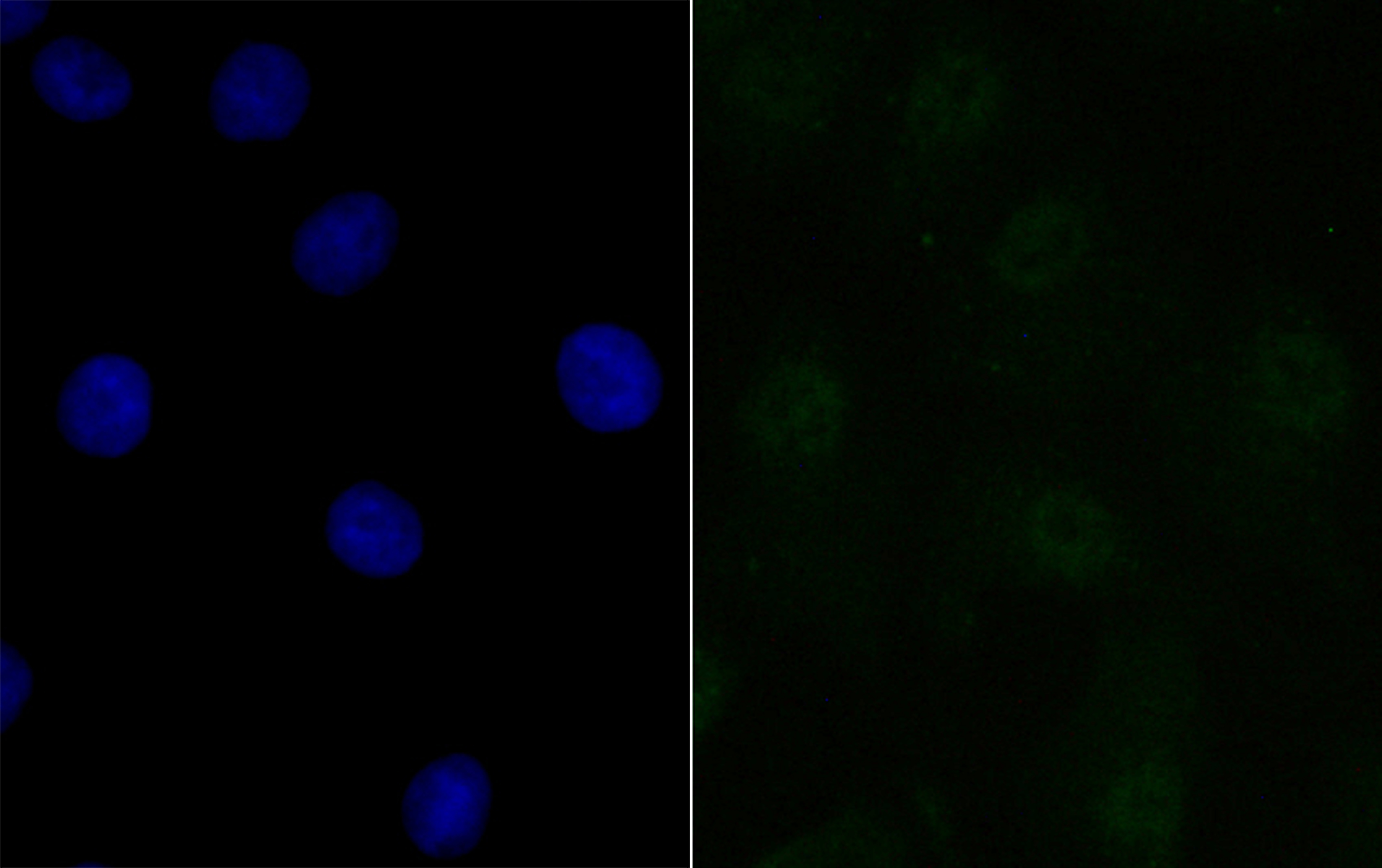
Fig1: ICC staining BANF1 in A549 cells (green). The nuclear counter stain is DAPI (blue). Cells were fixed in paraformaldehyde, permeabilised with 0.25% Triton X100/PBS.Application
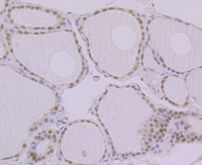
Fig2: Immunohistochemical analysis of paraffin-embedded human thyroid gland tissue using anti-BANF1 antibody. Counter stained with hematoxylin.Application
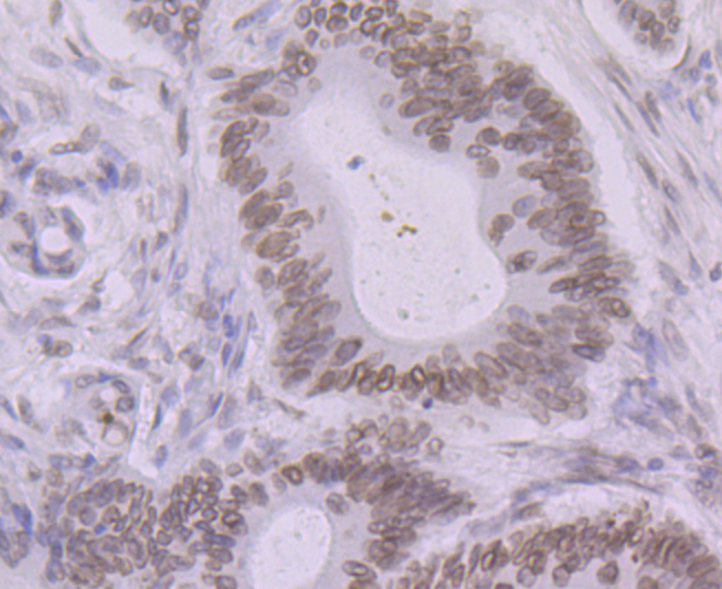
Fig3: Immunohistochemical analysis of paraffin-embedded human colon cancer tissue using anti-BANF1 antibody. Counter stained with hematoxylin.Application
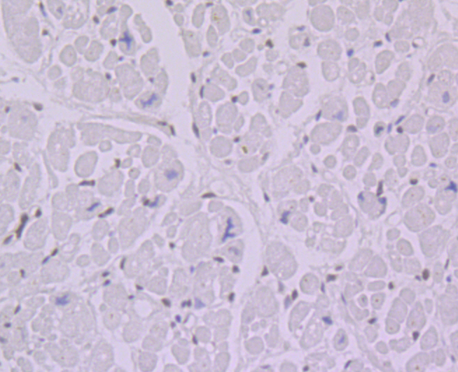
Fig4: Immunohistochemical analysis of paraffin-embedded human heart tissue using anti-BANF1 antibody. Counter stained with hematoxylin.Application
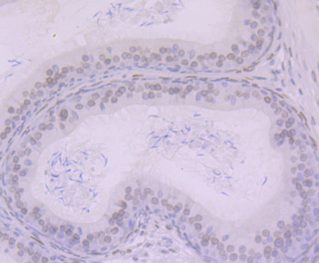
Fig5: Immunohistochemical analysis of paraffin-embedded rat epididymis tissue using anti-BANF1 antibody. Counter stained with hematoxylin.Application
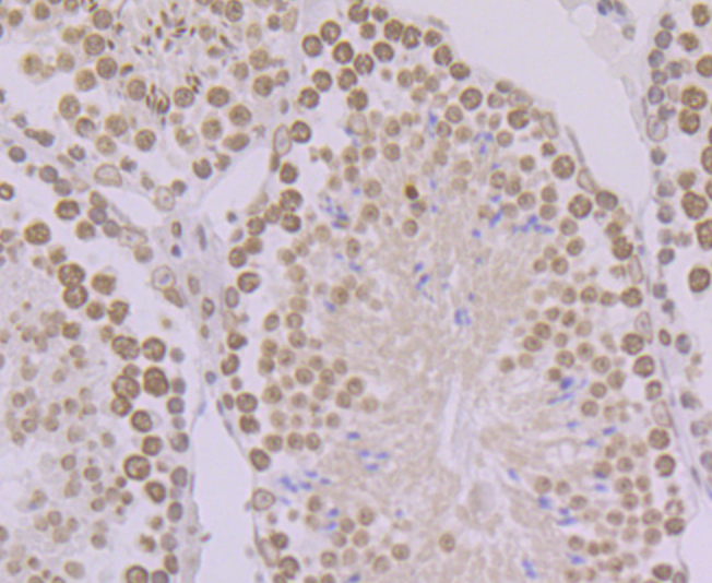
Fig6: Immunohistochemical analysis of paraffin-embedded mouse testis tissue using anti-BANF1 antibody. Counter stained with hematoxylin.Application
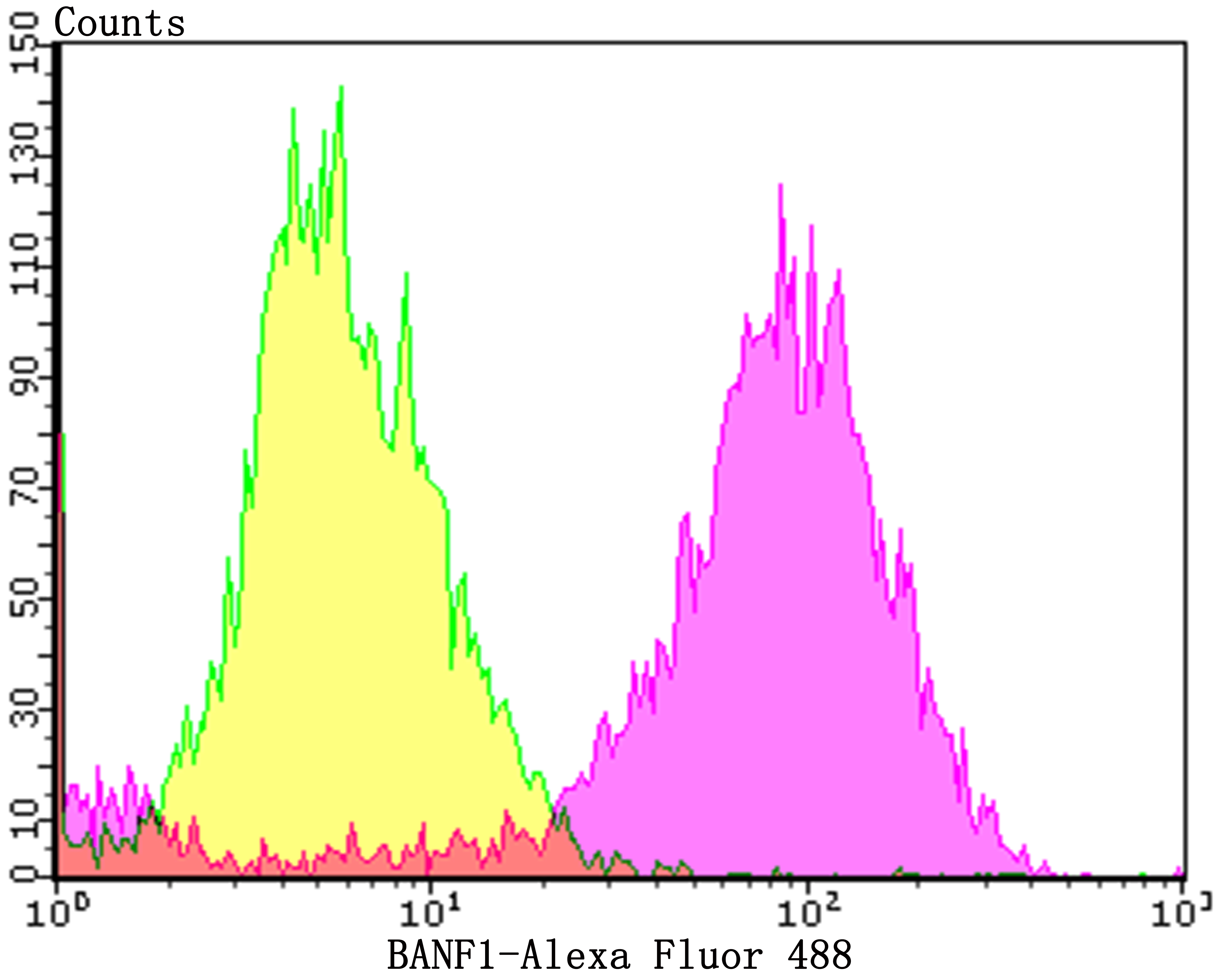
Fig7: Flow cytometric analysis of MCF-7 cells with BANF1 antibody at 1/100 dilution (purple) compared with an unlabelled control (cells without incubation with primary antibody; yellow). Alexa Fluor 488-conjugated goat anti-rabbit IgG was used as the secondary antibody.| Product Name | BANF1 [JG40-43] |
|---|---|
| Antibody Type | Primary Antibodies |
| Antigen Alias | BAF antibody BAF_HUMAN antibody BANF 1 antibody BANF1 antibody Barrier to autointegration factor 1 antibody Barrier to autointegration factor antibody Barrier-to-autointegration factor antibody BCRG 1 antibody BCRG1 antibody BCRP 1 antibody BCRP1 antibody Breakpoint cluster region protein 1 antibody D14S1460 antibody MGC111161 antibody NGPS antibody |
| Product description | Barrier-to-autointegration factor (BAF) binds non-specifically to double stranded DNA, possibly to play a role in tissue- or cell type-specific gene expression by interacting with different homeodomain transcription factors. BAF compresses chromatin structure and interacts with the LEM domain of nuclear proteins to play a crucial role in membrane recruitment and chromatin decondensation during nuclear assembly. Additionally, retroviruses like HIV-1 incorporate BAF from host cells into preintegration complexes (PICs) to prevent autointegration of retroviral DNA and thereby promote integration of retroviral DNA into the host chromosome. |
| Immunogen | Full length recombinant protein of human BANF1. |
| Clonality | Monoclonal |
|---|---|
| Isotype | IgG |
| Host Species | Recombinant rabbit |
| Tested Applications | WBICCIHCFC |
| ICC:1:50-1:100 IHC:1:50-1:200 FC:1:50-1:100 WB:1:500 |
|
| Species Reactivity | HumanMouseRat |
| Concentration | 1mg/ml |
| Alternative Names | BAF antibody BAF_HUMAN antibody BANF 1 antibody BANF1 antibody Barrier to autointegration factor 1 antibody Barrier to autointegration factor antibody Barrier-to-autointegration factor antibody BCRG 1 antibody BCRG1 antibody BCRP 1 antibody BCRP1 antibody Breakpoint cluster region protein 1 antibody D14S1460 antibody MGC111161 antibody NGPS antibody |
|---|---|
| Molecular Weight(MW) | 10 kDa |
| Cellular Localization | Nucleus. Cytoplasm. |
Application
Fig1: ICC staining BANF1 in A549 cells (green). The nuclear counter stain is DAPI (blue). Cells were fixed in paraformaldehyde, permeabilised with 0.25% Triton X100/PBS.
Application
Fig2: Immunohistochemical analysis of paraffin-embedded human thyroid gland tissue using anti-BANF1 antibody. Counter stained with hematoxylin.
Application
Fig3: Immunohistochemical analysis of paraffin-embedded human colon cancer tissue using anti-BANF1 antibody. Counter stained with hematoxylin.
Application
Fig4: Immunohistochemical analysis of paraffin-embedded human heart tissue using anti-BANF1 antibody. Counter stained with hematoxylin.
Application
Fig5: Immunohistochemical analysis of paraffin-embedded rat epididymis tissue using anti-BANF1 antibody. Counter stained with hematoxylin.
Application
Fig6: Immunohistochemical analysis of paraffin-embedded mouse testis tissue using anti-BANF1 antibody. Counter stained with hematoxylin.
Application
Fig7: Flow cytometric analysis of MCF-7 cells with BANF1 antibody at 1/100 dilution (purple) compared with an unlabelled control (cells without incubation with primary antibody; yellow). Alexa Fluor 488-conjugated goat anti-rabbit IgG was used as the secondary antibody.| Positive Control | A549, MCF-7, Rat epididymis tissue, human thyroid gland tissue, human colon cancer tissue, human heart tissue, mouse testis tissue. |
|---|---|
| Application Notes | ICC:1:50-1:100 IHC:1:50-1:200 FC:1:50-1:100 WB:1:500 |
| Form | Liquid |
|---|---|
| Storage Instructions | Store at +4℃ after thawing. Aliquot store at -20℃. Avoid repeated freeze / thaw cycles. |
| Storage Buffer | 1*TBS (pH7.4), 1%BSA, 40%Glycerol. Preservative: 0.05% Sodium Azide. |


