Application
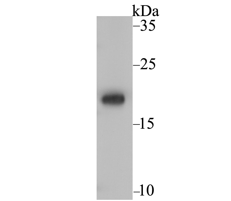
Fig1: Western blot analysis of Ndufs4 on rat heart tissue lysate using anti-Ndufs4 antibody at 1/1,000 dilution.Application
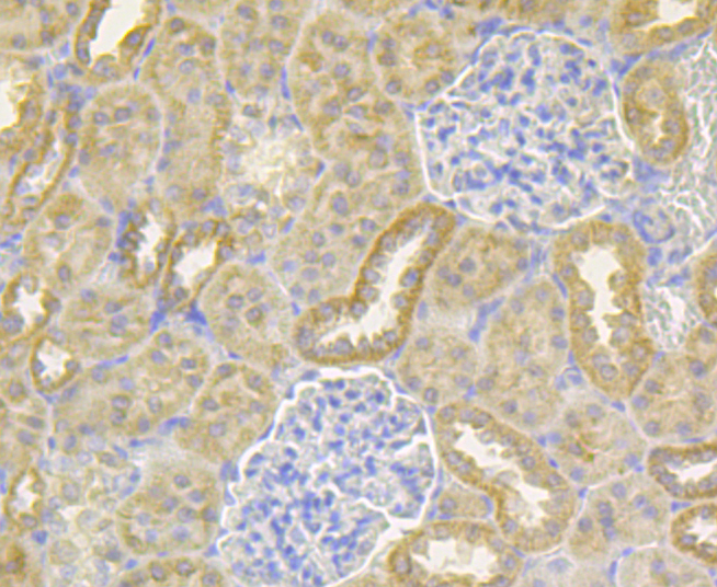
Fig2: Immunohistochemical analysis of paraffin-embedded rat kidney tissue using anti-Ndufs4 antibody. Counter stained with hematoxylin.Application
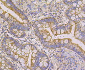
Fig3: Immunohistochemical analysis of paraffin-embedded human small intestine tissue using anti-Ndufs4 antibody. Counter stained with hematoxylin.Application
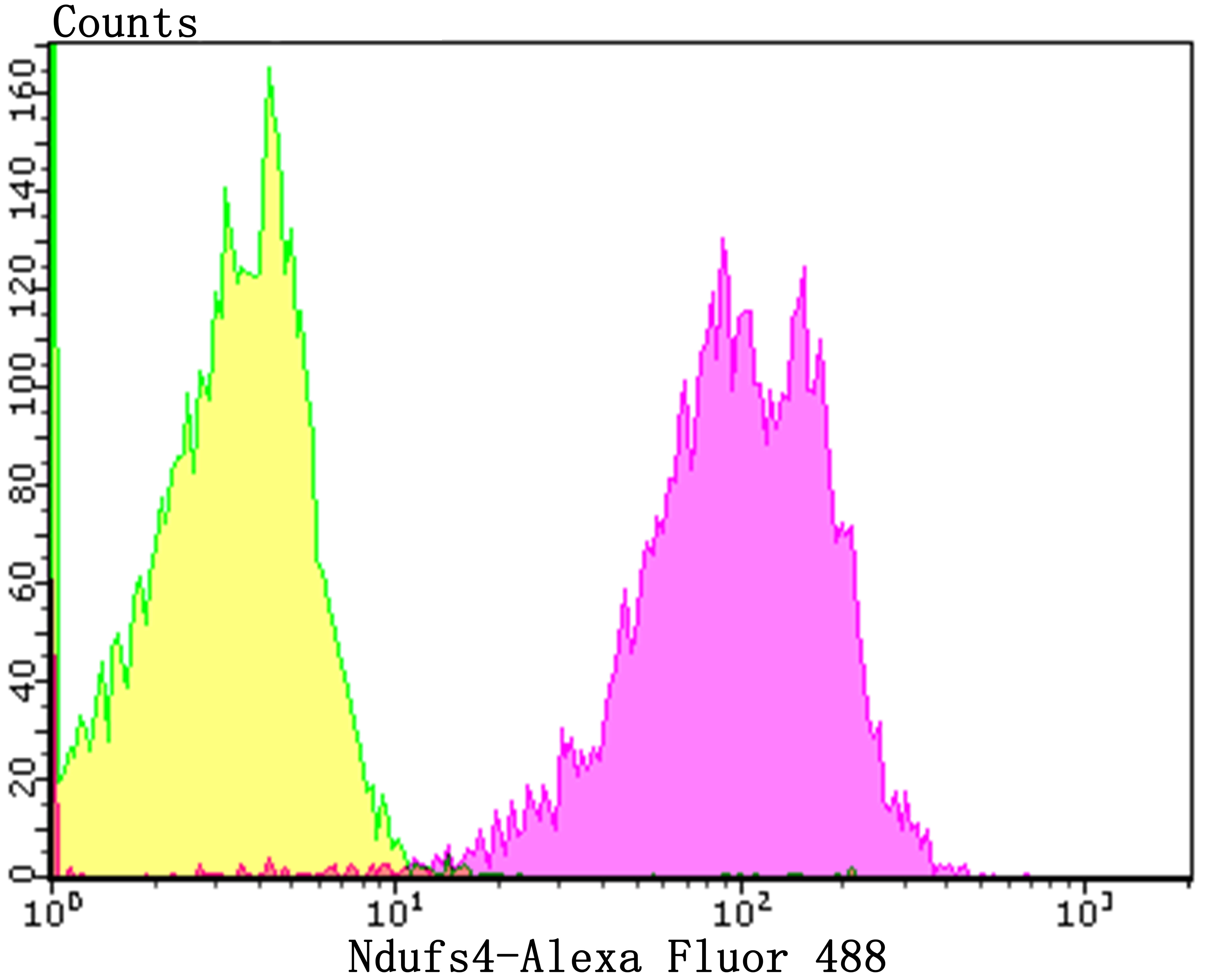
Fig4: Flow cytometric analysis of SH-SY-5Y cells with Ndufs4 antibody at 1/100 dilution (purple) compared with an unlabelled control (cells without incubation with primary antibody; yellow). Alexa Fluor 488-conjugated goat anti-rabbit IgG was used as the secondary antibody.| Product Name | Ndufs4 [JE40-47] |
|---|---|
| Antibody Type | Primary Antibodies |
| Antigen Alias | AQDQ antibody CI 18 antibody CI 18 kDa antibody CI AQDQ antibody CI-18 kDa antibody CI-AQDQ antibody Complex I 18 kDa antibody Complex I AQDQ antibody Complex I-18 kDa antibody Complex I-AQDQ antibody mitochondrial antibody mitochondrial respiratory chain complex I (18 KD subunit) antibody NADH coenzyme Q reductase antibody NADH dehydrogenase (ubiquinone) Fe S protein 4 18kDa antibody NADH dehydrogenase [ubiquinone] iron-sulfur protein 4 antibody NADH dehydrogenase antibody NADH ubiquinone oxidoreductase 18 kDa subunit antibody NADH-ubiquinone oxidoreductase 18 kDa subunit antibody NDUFS4 antibody NDUS4_HUMAN antibody |
| Product description | Complex 1 (also known as NADH dehydrogenase) of the electron transport chain (respiratory chain) is an enzymatic complex that catalyzes the transfer of electrons from NADH to ubiquinone. Free energy from the reaction is conserved in the transfer of protons into the intermembrane space to create an electrochemical proton gradient, a driving force for ATP synthesis. Complex 1 is a complicated, multi-protein, L-shaped complex composed of at least 45 different subunits and located in the mitochondrial inner membrane. NDUFS4 (NADH dehydrogenase (ubiquinone) Fe-S protein 4), also known as AQDQ or CI-18 (Complex I-18kDa protein), belongs to the Complex I NDUFS4 subunit family. NDUFS4 localizes to the matrix side of the inner membrane of the mitochondrion and functions as an accessory subunit of Complex I. Mutations in the gene encoding NDUFS4 can result in Complex I mitochondrial respiratory chain deficiency. Patients with this deficiency may exhibit cardiomyopathy, myopathy, liver failure and neurological disorders. |
| Immunogen | Recombinant protein within human Ndufs4 aa 43-175. |
| Clonality | Monoclonal |
|---|---|
| Isotype | IgG |
| Host Species | Recombinant rabbit |
| Tested Applications | WBIHCFCIP |
| WB:1:500-1:2,000 IHC:1:50-1:200 FC:1:50-1:100 IP:1:10-1:50 |
|
| Species Reactivity | HumanMouseRat |
| Concentration | 1mg/ml |
| Alternative Names | AQDQ antibody CI 18 antibody CI 18 kDa antibody CI AQDQ antibody CI-18 kDa antibody CI-AQDQ antibody Complex I 18 kDa antibody Complex I AQDQ antibody Complex I-18 kDa antibody Complex I-AQDQ antibody mitochondrial antibody mitochondrial respiratory chain complex I (18 KD subunit) antibody NADH coenzyme Q reductase antibody NADH dehydrogenase (ubiquinone) Fe S protein 4 18kDa antibody NADH dehydrogenase [ubiquinone] iron-sulfur protein 4 antibody NADH dehydrogenase antibody NADH ubiquinone oxidoreductase 18 kDa subunit antibody NADH-ubiquinone oxidoreductase 18 kDa subunit antibody NDUFS4 antibody NDUS4_HUMAN antibody |
|---|---|
| Molecular Weight(MW) | 20 kDa |
| Cellular Localization | Mitochondrion. |
Application
Fig1: Western blot analysis of Ndufs4 on rat heart tissue lysate using anti-Ndufs4 antibody at 1/1,000 dilution.
Application
Fig2: Immunohistochemical analysis of paraffin-embedded rat kidney tissue using anti-Ndufs4 antibody. Counter stained with hematoxylin.
Application
Fig3: Immunohistochemical analysis of paraffin-embedded human small intestine tissue using anti-Ndufs4 antibody. Counter stained with hematoxylin.
Application
Fig4: Flow cytometric analysis of SH-SY-5Y cells with Ndufs4 antibody at 1/100 dilution (purple) compared with an unlabelled control (cells without incubation with primary antibody; yellow). Alexa Fluor 488-conjugated goat anti-rabbit IgG was used as the secondary antibody.| Positive Control | Rat heart tissue lysate, rat kidney tissue, human small intestine tissue, SH-SY-5Y. |
|---|---|
| Application Notes | WB:1:500-1:2,000 IHC:1:50-1:200 FC:1:50-1:100 IP:1:10-1:50 |
| Form | Liquid |
|---|---|
| Storage Instructions | Store at +4℃ after thawing. Aliquot store at -20℃. Avoid repeated freeze / thaw cycles. |
| Storage Buffer | 1*TBS (pH7.4), 1%BSA, 40%Glycerol. Preservative: 0.05% Sodium Azide. |


