Application
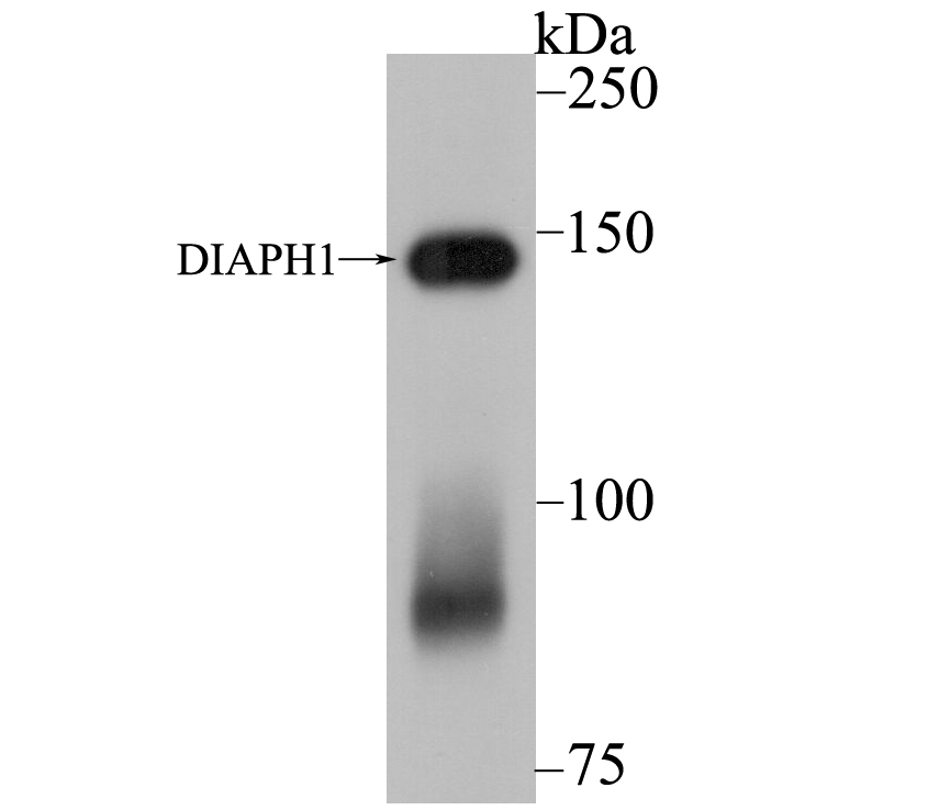
Fig1: Western blot analysis of DIAPH1 on SiHa cell lysate using anti-DIAPH1 antibody at 1/5,000 dilution.Application
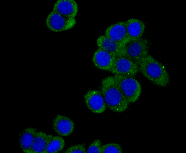
Fig2: ICC staining DIAPH1 in LOVO cells (green). The nuclear counter stain is DAPI (blue). Cells were fixed in paraformaldehyde, permeabilised with 0.25% Triton X100/PBS.Application
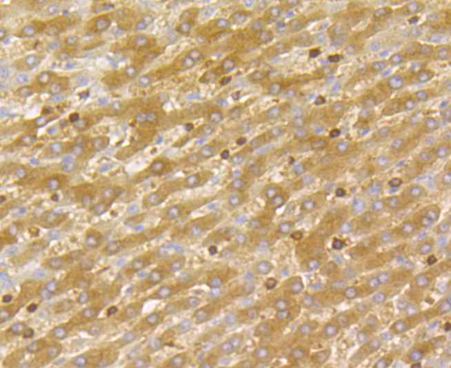
Fig3: Immunohistochemical analysis of paraffin-embedded human liver cancer tissue using anti-DIAPH1 antibody. Counter stained with hematoxylin.Application
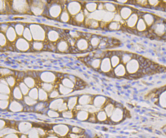
Fig4: Immunohistochemical analysis of paraffin-embedded human colon tissue using anti-DIAPH1 antibody. Counter stained with hematoxylin.| Product Name | DIAPH1 [JE40-59] |
|---|---|
| Antibody Type | Primary Antibodies |
| Antigen Alias | DIAPH1 antibody deafness, autosomal dominant 1 antibody DFNA1 antibody DIA1 antibody DIAP1 antibody DIAP1_HUMAN antibody DIAPH1 antibody Diaphanous homolog 1 (Drosophila) antibody diaphanous homolog 1 antibody Diaphanous related formin 1 antibody Diaphanous-related formin-1 antibody DRF1 antibody FLJ25265 antibody hDIA1 antibody LFHL1 antibody low frequency hearing loss 1 antibody p140DIA antibody Protein diaphanous homolog 1 antibody |
| Product description | Dia 1, also known as DIAPH1 (diaphanous homolog 1) or DRF1, a mammalian homolog of the Drosophila diaphanous gene, belongs to a family of formin homology (FH) proteins which are characterized by having tandemly aligned FH1 (formin homology 1) and FH2 (formin homology 2) domains in their carboxy terminal regions. Dia 1 contains a DAD (diaphanous autoregulatory) domain, which is involved in the elongation of actin filaments, and a GBD/FH3 (Rho GTPase-binding/formin homology 3) domain, which interacts with the DAD domain via autoinhibitory interactions to regulate the activation of Dia 1. Dia 1 is required for the assembly of F-actin structures, and regulates the polymerization and depolymerization of actin filaments. Localizing to the cell membrane, Dia 1 is expressed in a wide range of tissues, including brain, heart, lung and kidney. Defects to the gene encoding Dia 1 have been linked to deafness autosomal dominant type 1 (DFNA1), a disorder characterized by sensorineural hearing loss.. |
| Immunogen | Recombinant protein corresponding to N-terminal human DIAPH1. |
| Clonality | Monoclonal |
|---|---|
| Isotype | IgG |
| Host Species | Recombinant rabbit |
| Tested Applications | WBICCIFIHC |
| WB:1:5,000-1:10,000 ICC:1:50 IHC:1:50-1:100 |
|
| Species Reactivity | Human |
| Concentration | 1mg/ml |
| Alternative Names | DIAPH1 antibody
deafness autosomal dominant 1 antibody DFNA1 antibody DIA1 antibody DIAP1 antibody DIAP1_HUMAN antibody DIAPH1 antibody Diaphanous homolog 1 (Drosophila) antibody diaphanous homolog 1 antibody Diaphanous related formin 1 antibody Diaphanous-related formin-1 antibody DRF1 antibody FLJ25265 antibody hDIA1 antibody LFHL1 antibody low frequency hearing loss 1 antibody p140DIA antibody Protein diaphanous homolog 1 antibody |
|---|---|
| Molecular Weight(MW) | 141 kDa |
| Cellular Localization | Cytoskeleton. Plasma membrane. |
| SwissProt ID | O60610 |
|---|
Application
Fig1: Western blot analysis of DIAPH1 on SiHa cell lysate using anti-DIAPH1 antibody at 1/5,000 dilution.
Application
Fig2: ICC staining DIAPH1 in LOVO cells (green). The nuclear counter stain is DAPI (blue). Cells were fixed in paraformaldehyde, permeabilised with 0.25% Triton X100/PBS.
Application
Fig3: Immunohistochemical analysis of paraffin-embedded human liver cancer tissue using anti-DIAPH1 antibody. Counter stained with hematoxylin.
Application
Fig4: Immunohistochemical analysis of paraffin-embedded human colon tissue using anti-DIAPH1 antibody. Counter stained with hematoxylin.| Positive Control | SiHa, LOVO, human liver cancer tissue, human colon tissue. |
|---|---|
| Application Notes | WB:1:5,000-1:10,000 ICC:1:50 IHC:1:50-1:100 |
| Form | Liquid |
|---|---|
| Storage Instructions | Store at +4℃ after thawing. Aliquot store at -20℃. Avoid repeated freeze / thaw cycles. |
| Storage Buffer | 1*TBS (pH7.4), 1%BSA, 40%Glycerol. Preservative: 0.05% Sodium Azide. |


