Application
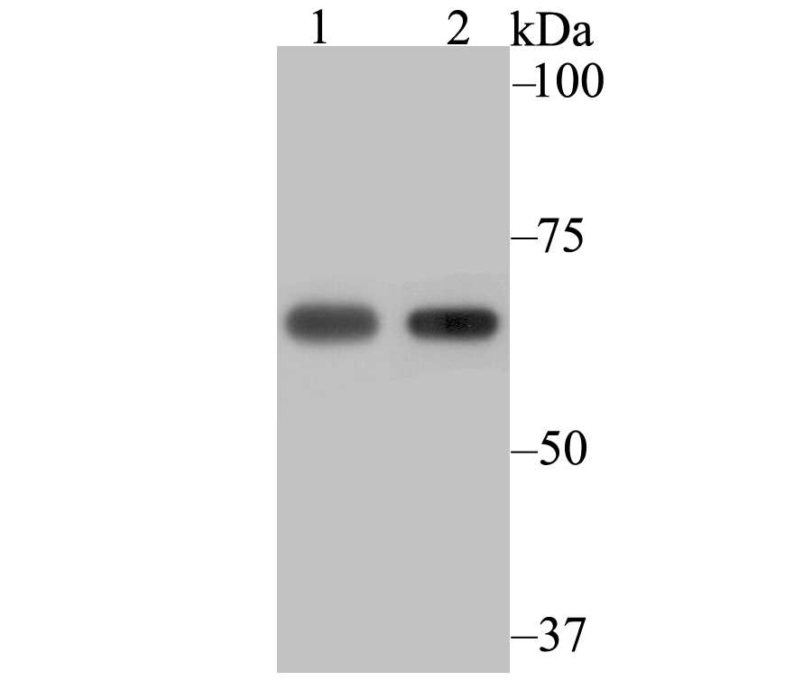
Fig1: Western blot analysis of NXF1 on 293 (1) and SiHa (2) cell lysate using anti-NXF1 antibody at 1/2,000 dilution.Application
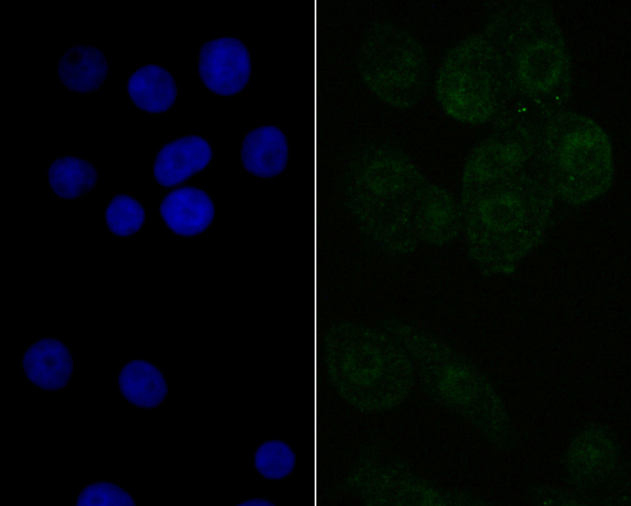
Fig2: ICC staining NXF1 in LOVO cells (green). The nuclear counter stain is DAPI (blue). Cells were fixed in paraformaldehyde, permeabilised with 0.25% Triton X100/PBS.| Product Name | NXF1 [JE40-63] |
|---|---|
| Antibody Type | Primary Antibodies |
| Antigen Alias | DKFZp667O0311 antibody DmNXF1 antibody MEX67 antibody MEX67, yeast, homolog of antibody Mex67p antibody mRNA export factor TAP antibody Mvb1 antibody Nuclear RNA export factor 1 antibody Nuclear RNA export factor 1 homolog (S. cerevisiae) antibody nxf 1 antibody NXF1 antibody NXF1_HUMAN antibody Protein small bristles antibody Sbr antibody TAP antibody Tip associating protein antibody Tip-associated protein antibody Tip-associating protein antibody |
| Product description | The constitutive transport element (CTE) of type D retroviruses serves as a signal of nuclear export for unspliced viral RNAs. TAP (tip-associating protein, also known as NXF1) mediates the export of CTE-containing simian type D retroviral RNAs through binding directly to the CTE. TAP is associated with a recognized mRNA export pathway and is a member of the multigene family of NXF proteins. NXF proteins belong to an evolutionarily conserved family of proteins, which are characterized by a leucine-rich-repeat domain (LRR) followed by a region known as the nuclear transport factor 2 (NTF2)-like domain. |
| Immunogen | Recombinant protein within c-terminal human NXF1. |
| Clonality | Monoclonal |
|---|---|
| Isotype | IgG |
| Host Species | Recombinant rabbit |
| Tested Applications | WBICCIP |
| WB:1:500-1:2,000 ICC:1:50-1:100 IP:1:10-1:50 |
|
| Species Reactivity | Human |
| Concentration | 1mg/ml |
| Alternative Names | DKFZp667O0311 antibody
DmNXF1 antibody
MEX67 antibody
MEX67 yeast homolog of antibody Mex67p antibody mRNA export factor TAP antibody Mvb1 antibody Nuclear RNA export factor 1 antibody Nuclear RNA export factor 1 homolog (S. cerevisiae) antibody nxf 1 antibody NXF1 antibody NXF1_HUMAN antibody Protein small bristles antibody Sbr antibody TAP antibody Tip associating protein antibody Tip-associated protein antibody Tip-associating protein antibody |
|---|---|
| Molecular Weight(MW) | 70 kDa |
| Cellular Localization | Nucleus. Cytoplasm. |
| SwissProt ID | Q9UBU9 |
|---|
Application
Fig1: Western blot analysis of NXF1 on 293 (1) and SiHa (2) cell lysate using anti-NXF1 antibody at 1/2,000 dilution.
Application
Fig2: ICC staining NXF1 in LOVO cells (green). The nuclear counter stain is DAPI (blue). Cells were fixed in paraformaldehyde, permeabilised with 0.25% Triton X100/PBS.| Positive Control | 293, SiHa. |
|---|---|
| Application Notes | WB:1:500-1:2,000 ICC:1:50-1:100 IP:1:10-1:50 |
| Form | Liquid |
|---|---|
| Storage Instructions | Store at +4℃ after thawing. Aliquot store at -20℃. Avoid repeated freeze / thaw cycles. |
| Storage Buffer | 1*TBS (pH7.4), 1%BSA, 40%Glycerol. Preservative: 0.05% Sodium Azide. |


