Application
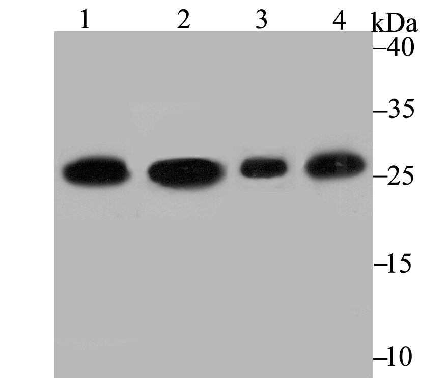
Fig1: Western blot analysis of PGAM1 on different lysates using anti-PGAM1 antibody at 1/2,000 dilution. Positive control: Lane 1: A431 Lane 2: A549 Lane 3: Rat brain Lane 4: Mouse brainApplication
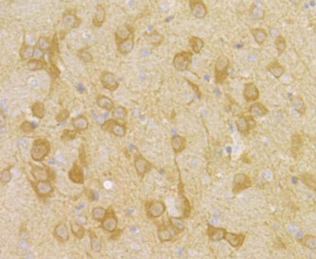
Fig2: Immunohistochemical analysis of paraffin-embedded rat brain tissue using anti-PGAM1 antibody. Counter stained with hematoxylin.Application
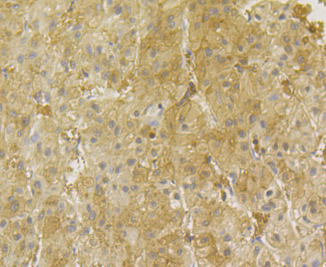
Fig3: Immunohistochemical analysis of paraffin-embedded human liver tissue using anti-PGAM1 antibody. Counter stained with hematoxylin.Application
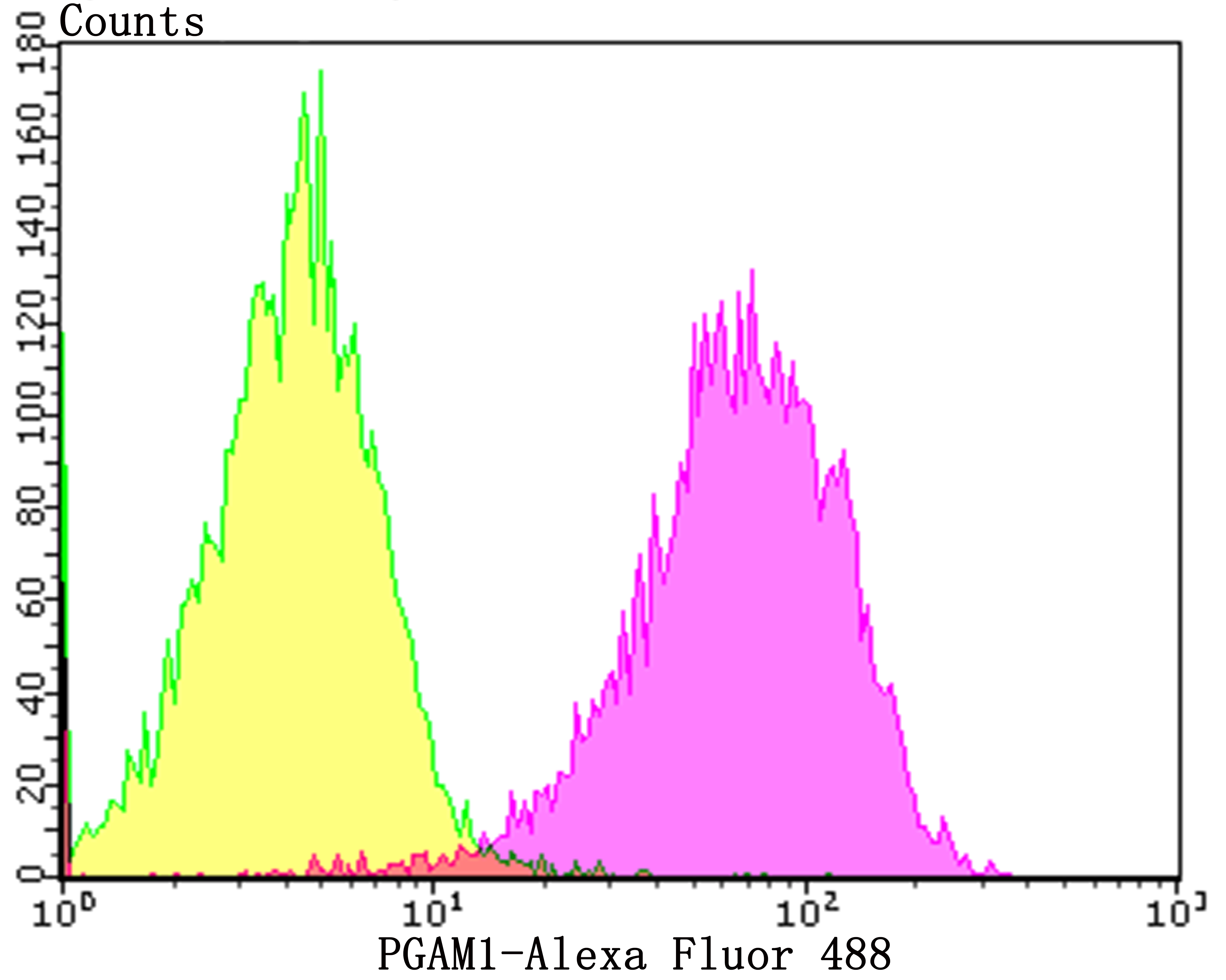
Fig4: Flow cytometric analysis of Jurkat cells with PGAM1 antibody at 1/100 dilution (purple) compared with an unlabelled control (cells without incubation with primary antibody; yellow). Alexa Fluor 488-conjugated goat anti-rabbit IgG was used as the secondary antibody.| Product Name | PGAM1 [JE40-67] |
|---|---|
| Antibody Type | Primary Antibodies |
| Antigen Alias | BPG dependent PGAM 1 antibody BPG dependent PGAM1 antibody BPG-dependent PGAM 1 antibody OTTHUMP00000059414 antibody PGAM 1 antibody PGAM A antibody PGAM B antibody PGAM-B antibody PGAM1 antibody PGAM1_HUMAN antibody PGAMA antibody PGAMB antibody Phosphoglycerate mutase 1 antibody Phosphoglycerate mutase 1 brain antibody Phosphoglycerate mutase A antibody Phosphoglycerate mutase A nonmuscle form antibody Phosphoglycerate mutase isozyme B antibody |
| Product description | Members of the PGAM (phosphoglycerate mutase) family of proteins are important components of glucose and 2,3-BPGA (2,3-bisphosphoglycerate) metabolism. They are responsible for catalyzing the transfer of phospho groups between the carbon atoms of phosphoglycerates. In mammals there are two types of PGAM isozymes: PGAM1 (also known as PGAMB) and PGAM2 (also known as PGAMA). In the cell, PGAM1 and PGAM2 exist as either homodimers or heterodimers and are responsible for the interconversion of 3-phosphoglycerate and 2-phosphoglycerate. PGAM2 homodimers are expressed in skeletal muscle, mature sperm cells and heart; PGAM1 homodimers are found in most other tissues; and PGAM1/PGAM2 heterodimers are found exclusively in the heart. PGAM4, also known as PGAM3, is a protein formerly considered to be specific to humans. Initially the PGAM4 gene was described as a pseudogene but it is now known to encode a functional protein at least 25 million years old. The gene encoding PGAM4 is believed to have originated by retrotransposition, with the original copy being the PGAM1 gene. |
| Immunogen | Recombinant protein within N-terminal human PGAM1. |
| Clonality | Monoclonal |
|---|---|
| Isotype | IgG |
| Host Species | Recombinant rabbit |
| Tested Applications | WBIHCFC |
| WB:1:500-1:2,000 IHC:1:50-1:100 FC:1:50-1:100 |
|
| Species Reactivity | HumanMouseRat |
| Concentration | 1mg/ml |
| Alternative Names | BPG dependent PGAM 1 antibody BPG dependent PGAM1 antibody BPG-dependent PGAM 1 antibody OTTHUMP00000059414 antibody PGAM 1 antibody PGAM A antibody PGAM B antibody PGAM-B antibody PGAM1 antibody PGAM1_HUMAN antibody PGAMA antibody PGAMB antibody Phosphoglycerate mutase 1 antibody Phosphoglycerate mutase 1 brain antibody Phosphoglycerate mutase A antibody Phosphoglycerate mutase A nonmuscle form antibody Phosphoglycerate mutase isozyme B antibody |
|---|---|
| Molecular Weight(MW) | 29 kDa |
| Cellular Localization | Cytosol. Extracellular region or secreted. Nucleus. |
Application
Fig1: Western blot analysis of PGAM1 on different lysates using anti-PGAM1 antibody at 1/2,000 dilution. Positive control: Lane 1: A431 Lane 2: A549 Lane 3: Rat brain Lane 4: Mouse brain
Application
Fig2: Immunohistochemical analysis of paraffin-embedded rat brain tissue using anti-PGAM1 antibody. Counter stained with hematoxylin.
Application
Fig3: Immunohistochemical analysis of paraffin-embedded human liver tissue using anti-PGAM1 antibody. Counter stained with hematoxylin.
Application
Fig4: Flow cytometric analysis of Jurkat cells with PGAM1 antibody at 1/100 dilution (purple) compared with an unlabelled control (cells without incubation with primary antibody; yellow). Alexa Fluor 488-conjugated goat anti-rabbit IgG was used as the secondary antibody.| Positive Control | A431, A549, rat brain tissue, mouse brain tissue, human liver tissue, Jurkat. |
|---|---|
| Application Notes | WB:1:500-1:2,000 IHC:1:50-1:100 FC:1:50-1:100 |
| Form | Liquid |
|---|---|
| Storage Instructions | Store at +4℃ after thawing. Aliquot store at -20℃. Avoid repeated freeze / thaw cycles. |
| Storage Buffer | 1*TBS (pH7.4), 1%BSA, 40%Glycerol. Preservative: 0.05% Sodium Azide. |


