Application
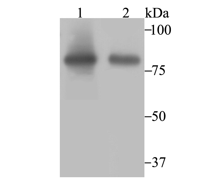
Fig1: Western blot analysis of Aconitase 2 on SiHa (1) and HL-60 (2) cell lysate using anti-Aconitase 2 antibody at 1/2,000 dilution.Application
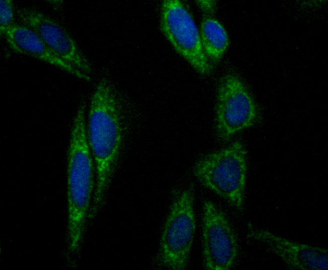
Fig2: ICC staining Aconitase 2 in SiHa cells (green). The nuclear counter stain is DAPI (blue). Cells were fixed in paraformaldehyde, permeabilised with 0.25% Triton X100/PBS.Application
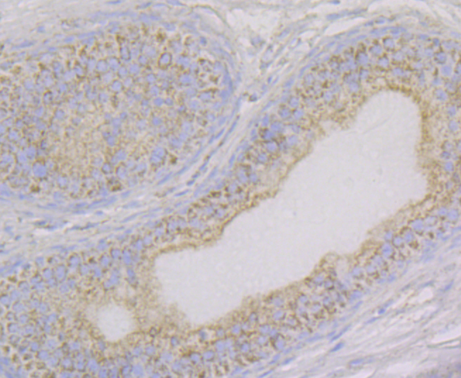
Fig3: Immunohistochemical analysis of paraffin-embedded rat epididymis tissue using anti-Aconitase 2 antibody. Counter stained with hematoxylin.Application
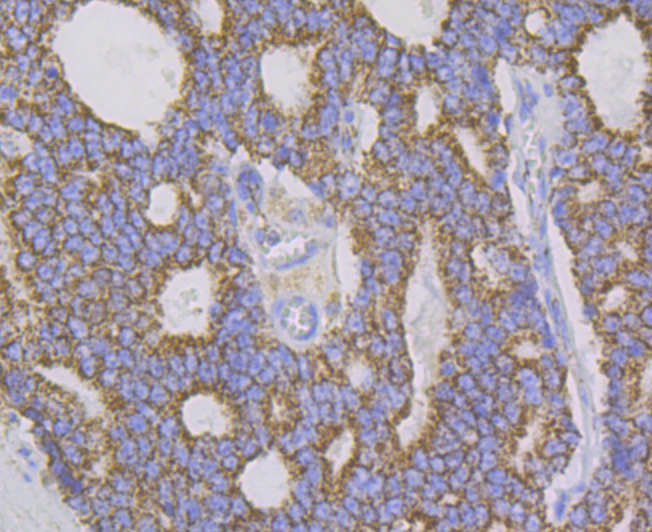
Fig4: Immunohistochemical analysis of paraffin-embedded human prostate cancer tissue using anti-Aconitase 2 antibody. Counter stained with hematoxylin.Application
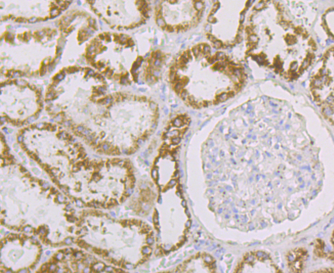
Fig5: Immunohistochemical analysis of paraffin-embedded human kidney tissue using anti-Aconitase 2 antibody. Counter stained with hematoxylin.Application
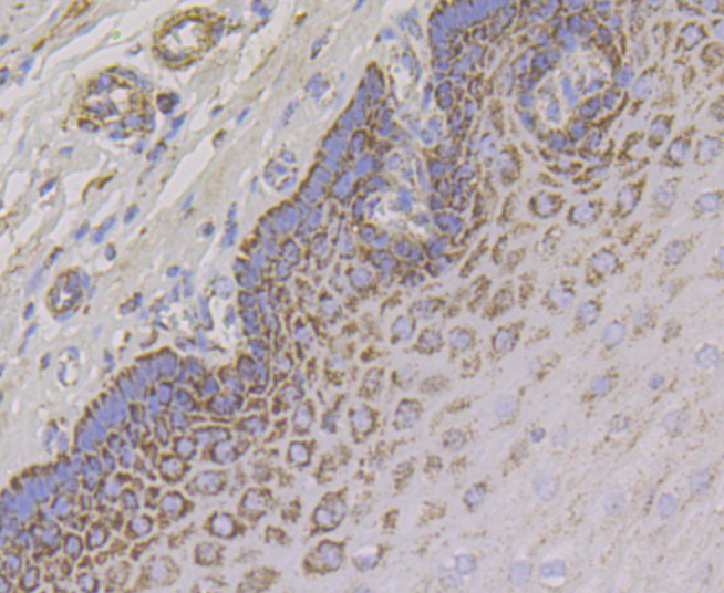
Fig6: Immunohistochemical analysis of paraffin-embedded human esophagus tissue using anti-Aconitase 2 antibody. Counter stained with hematoxylin.Application
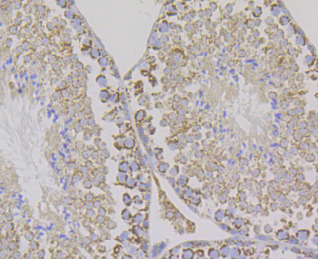
Fig7: Immunohistochemical analysis of paraffin-embedded mouse testis tissue using anti-Aconitase 2 antibody. Counter stained with hematoxylin.| Product Name | Aconitase 2 [JE40-83] |
|---|---|
| Antibody Type | Primary Antibodies |
| Antigen Alias | ACO 2 antibody ACO2 antibody ACON_HUMAN antibody aconitase 2 antibody Aconitase 2 mitochondrial antibody Aconitase antibody Aconitase2 antibody Aconitate hydratase antibody Aconitate hydratase mitochondrial antibody ACONM antibody Citrate hydro lyase antibody Citrate hydro-lyase antibody ICRD antibody mitochondrial antibody |
| Product description | Aconitase 2, mitochondrial is a protein that in humans is encoded by the ACO2 gene. The protein encoded by this gene belongs to the aconitase/IPM isomerase family. It is an enzyme that catalyzes the interconversion of citrate to isocitrate via cis-aconitate in the second step of the TCA cycle. This protein is encoded in the nucleus and functions in the mitochondrion. It was found to be one of the mitochondrial matrix proteins that are preferentially degraded by the serine protease 15 (PRSS15), also known as Lon protease, after oxidative modification. |
| Immunogen | Recombinant protein within human Aconitase 2 aa 1-150. |
| Clonality | Monoclonal |
|---|---|
| Isotype | IgG |
| Host Species | Recombinant rabbit |
| Tested Applications | WBICCIFIHCIP |
| WB:1:500-1:2,000 ICC:1:50-1:200 IHC:1:50-1:200 IP:1:10-1:50 |
|
| Species Reactivity | HumanMouseRat |
| Concentration | 1mg/ml |
| Alternative Names | ACO 2 antibody ACO2 antibody ACON_HUMAN antibody aconitase 2 antibody Aconitase 2 mitochondrial antibody Aconitase antibody Aconitase2 antibody Aconitate hydratase antibody Aconitate hydratase mitochondrial antibody ACONM antibody Citrate hydro lyase antibody Citrate hydro-lyase antibody ICRD antibody mitochondrial antibody |
|---|---|
| Molecular Weight(MW) | 85 kDa |
| Cellular Localization | Mitochondrion. |
Application
Fig1: Western blot analysis of Aconitase 2 on SiHa (1) and HL-60 (2) cell lysate using anti-Aconitase 2 antibody at 1/2,000 dilution.
Application
Fig2: ICC staining Aconitase 2 in SiHa cells (green). The nuclear counter stain is DAPI (blue). Cells were fixed in paraformaldehyde, permeabilised with 0.25% Triton X100/PBS.
Application
Fig3: Immunohistochemical analysis of paraffin-embedded rat epididymis tissue using anti-Aconitase 2 antibody. Counter stained with hematoxylin.
Application
Fig4: Immunohistochemical analysis of paraffin-embedded human prostate cancer tissue using anti-Aconitase 2 antibody. Counter stained with hematoxylin.
Application
Fig5: Immunohistochemical analysis of paraffin-embedded human kidney tissue using anti-Aconitase 2 antibody. Counter stained with hematoxylin.
Application
Fig6: Immunohistochemical analysis of paraffin-embedded human esophagus tissue using anti-Aconitase 2 antibody. Counter stained with hematoxylin.
Application
Fig7: Immunohistochemical analysis of paraffin-embedded mouse testis tissue using anti-Aconitase 2 antibody. Counter stained with hematoxylin.| Positive Control | SiHa, HL-60, rat epididymis tissue, human prostate cancer tissue, human kidney tissue, human esophagus tissue, mouse testis tissue. |
|---|---|
| Application Notes | WB:1:500-1:2,000 ICC:1:50-1:200 IHC:1:50-1:200 IP:1:10-1:50 |
| Form | Liquid |
|---|---|
| Storage Instructions | Store at +4℃ after thawing. Aliquot store at -20℃. Avoid repeated freeze / thaw cycles. |
| Storage Buffer | 1*TBS (pH7.4), 1%BSA, 40%Glycerol. Preservative: 0.05% Sodium Azide. |


