WB
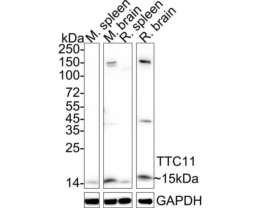
Western blot analysis of TTC11/FIS1 on different lysates with Rabbit anti-TTC11/FIS1 antibody at 1/1,000 dilution. Lane 1: Mouse spleen tissue lysate, Lane 2: Mouse brain tissue lysate, Lane 3: Rat spleen tissue lysate, Lane 4: Rat brain tissue lysate, Lysates/proteins at 40 µg/Lane. Exposure time: 25 seconds; 4-20% SDS-PAGE gel. Proteins were transferred to a PVDF membrane and blocked with 5% NFDM/TBST for 1 hour at room temperature. The primary antibody at 1/1,000 dilution was used in 5% NFDM/TBST at 4℃ overnight. Goat Anti-Rabbit IgG - HRP Secondary Antibody at 1/50,000 dilution was used for 1 hour at room temperature.WB
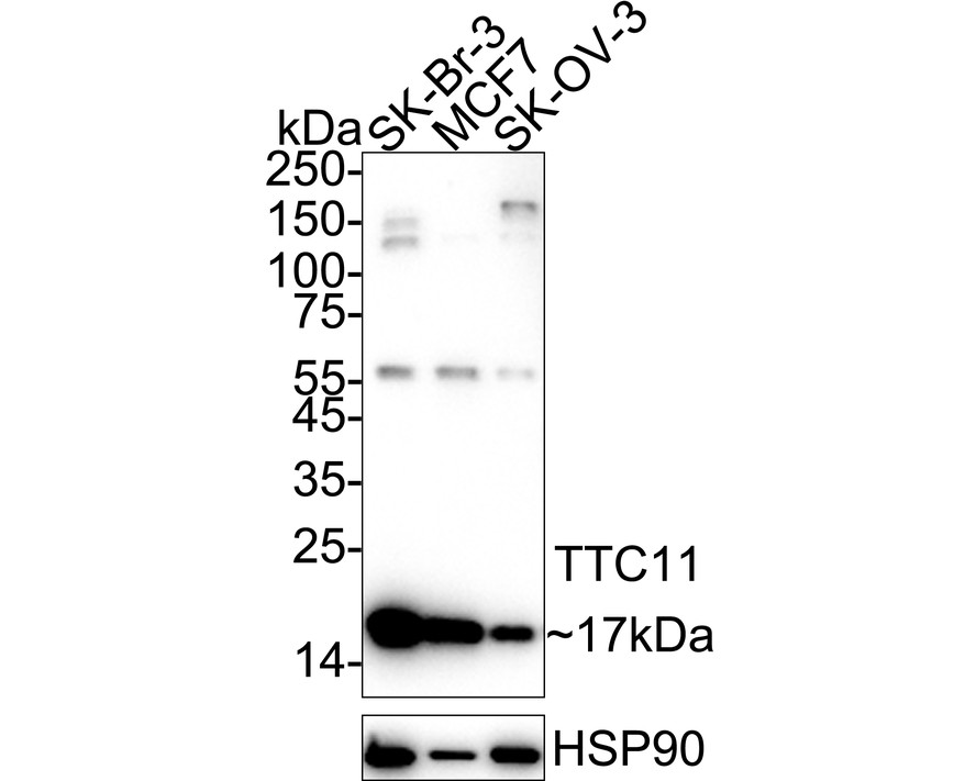
Western blot analysis of TTC11/FIS1 on different lysates with Rabbit anti-TTC11/FIS1 antibody at 1/2,000 dilution. Lane 1: SK-Br-3 cell lysate, Lane 2: MCF7 cell lysate, Lane 3: SK-OV-3 cell lysate, Lysates/proteins at 10 µg/Lane. Exposure time: 2 minutes; 4-20% SDS-PAGE gel. Proteins were transferred to a PVDF membrane and blocked with 5% NFDM/TBST for 1 hour at room temperature. The primary antibody at 1/2,000 dilution was used in 5% NFDM/TBST at 4℃ overnight. Goat Anti-Rabbit IgG - HRP Secondary Antibody at 1/50,000 dilution was used for 1 hour at room temperature.ICC/IF
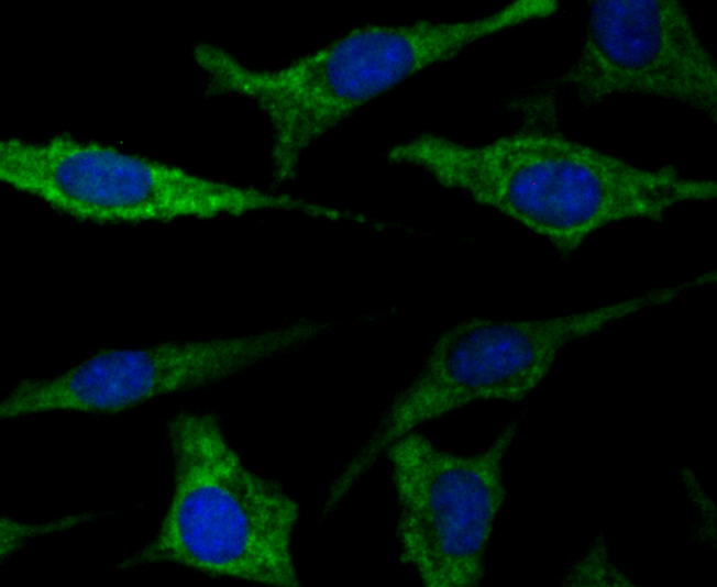
ICC staining TTC11 in SH-SY-5Y cells (green). The nuclear counter stain is DAPI (blue). Cells were fixed in paraformaldehyde, permeabilised with 0.25% Triton X100/PBS.ICC/IF
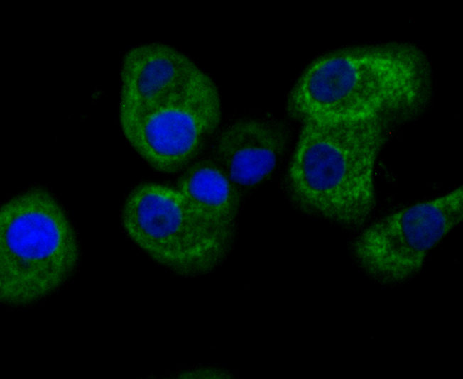
ICC staining TTC11 in SK-Br-3 cells (green). The nuclear counter stain is DAPI (blue). Cells were fixed in paraformaldehyde, permeabilised with 0.25% Triton X100/PBS.IHC
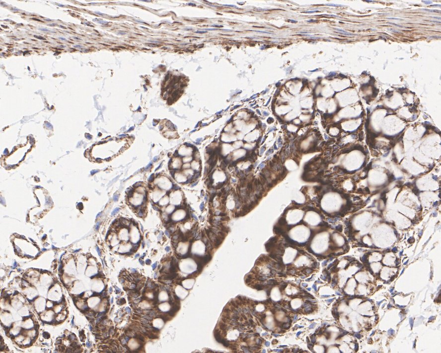
Immunohistochemical analysis of paraffin-embedded mouse colon tissue with Rabbit anti-TTC11/FIS1 antibody at 1/1,000 dilution. The section was pre-treated using heat mediated antigen retrieval with Tris-EDTA buffer (pH 9.0) for 20 minutes. The tissues were blocked in 1% BSA for 20 minutes at room temperature, washed with ddH2O and PBS, and then probed with the primary antibody at 1/1,000 dilution for 1 hour at room temperature. The detection was performed using an HRP conjugated compact polymer system. DAB was used as the chromogen. Tissues were counterstained with hematoxylin and mounted with DPX.IHC
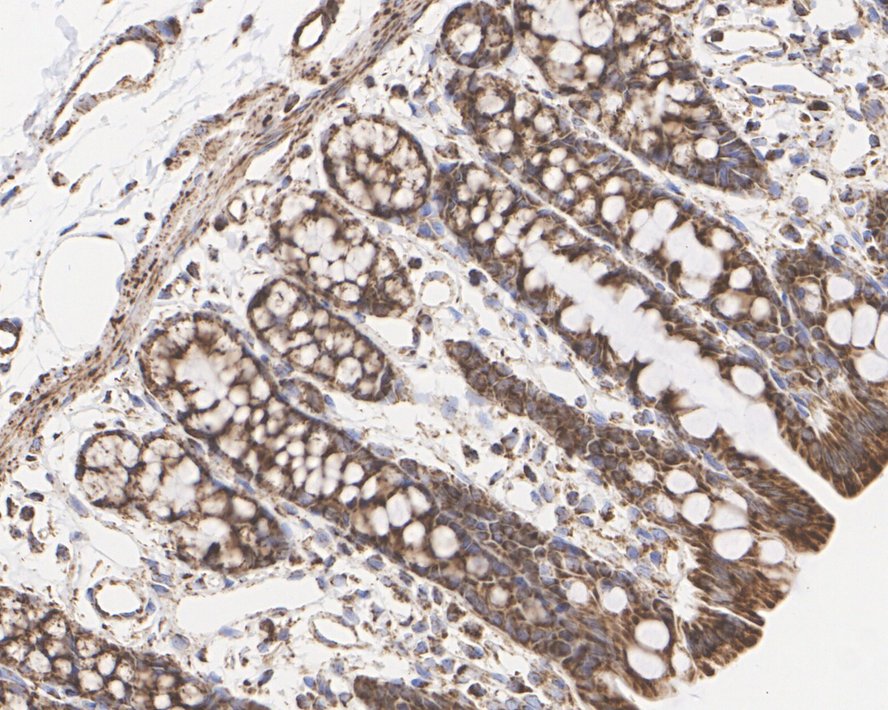
Immunohistochemical analysis of paraffin-embedded rat colon tissue with Rabbit anti-TTC11/FIS1 antibody at 1/1,000 dilution. The section was pre-treated using heat mediated antigen retrieval with Tris-EDTA buffer (pH 9.0) for 20 minutes. The tissues were blocked in 1% BSA for 20 minutes at room temperature, washed with ddH2O and PBS, and then probed with the primary antibody at 1/1,000 dilution for 1 hour at room temperature. The detection was performed using an HRP conjugated compact polymer system. DAB was used as the chromogen. Tissues were counterstained with hematoxylin and mounted with DPX.| Product Name | TTC11/FIS1 Recombinant Rabbit Monoclonal Antibody |
|---|---|
| Antibody Type | Primary Antibodies |
| Product description | Fis1 localizes to the outer mitochondrial membrane and, along with dynamin-related protein (Drp1), participates in mitochondrial fission. Fission and fusion mechanisms regulate mitochondrial morphology within the cell. Fission frequency is determined by the level of Fis1 molecules at the mitochondrial surface. Fis1 contains a C-terminal domain, which is required for mitochondrial localization, and an N-terminal domain, which is necessary for mitochondrial fission. Fragmentation of the mitochondrial network by Fis1 leads to cytochrome c release and apoptosis. The mitochondrial fission mechanisms may be involved in positively and negatively regulating apoptosis. |
| Immunogen | Recombinant protein within Human TTC11 aa 1-125 / 152. |
| Clonality | Monoclonal |
|---|---|
| Isotype | IgG |
| Host Species | Recombinant rabbit |
| Tested Applications | ICC/IFIHCWB |
| WB:1:500-1:2000 ICC/IF:1:50-1:200 IHC:1:1000 |
|
| Species Reactivity | HumanMouseRat |
| Concentration | 1mg/ml |
| Purification | Protein A |
| Alternative Names | 2010003O14Rik antibody
CGI 135 antibody
CGI135 antibody
FIS 1 antibody
FIS1 antibody
Fis1 homolog antibody
FIS1 S. cerevisiae homolog of antibody FIS1_HUMAN antibody Fission 1 (mitochondrial outer membrane) homolog (S. cerevisiae) antibody Fission 1 (mitochondrial outer membrane) homolog (yeast) antibody Fission 1 (mitochondrial outer membrane) homolog antibody Fission 1 homolog antibody H NH0132A01.6 antibody hFis 1 antibody hFis1 antibody Mitochondrial fission 1 protein antibody mitochondrial fission molecule antibody Tetratricopeptide repeat domain 11 antibody Tetratricopeptide repeat protein 11 antibody TPR repeat protein 11 antibody TTC 11 antibody |
|---|---|
| Molecular Weight(MW) | 17 kDa/15 kDa |
| Cellular Localization | Peroxisome. Mitochondrion. |
| SwissProt ID | Q9Y3D6 |
|---|
WB
Western blot analysis of TTC11/FIS1 on different lysates with Rabbit anti-TTC11/FIS1 antibody at 1/1,000 dilution. Lane 1: Mouse spleen tissue lysate, Lane 2: Mouse brain tissue lysate, Lane 3: Rat spleen tissue lysate, Lane 4: Rat brain tissue lysate, Lysates/proteins at 40 µg/Lane. Exposure time: 25 seconds; 4-20% SDS-PAGE gel. Proteins were transferred to a PVDF membrane and blocked with 5% NFDM/TBST for 1 hour at room temperature. The primary antibody at 1/1,000 dilution was used in 5% NFDM/TBST at 4℃ overnight. Goat Anti-Rabbit IgG - HRP Secondary Antibody at 1/50,000 dilution was used for 1 hour at room temperature.
WB
Western blot analysis of TTC11/FIS1 on different lysates with Rabbit anti-TTC11/FIS1 antibody at 1/2,000 dilution. Lane 1: SK-Br-3 cell lysate, Lane 2: MCF7 cell lysate, Lane 3: SK-OV-3 cell lysate, Lysates/proteins at 10 µg/Lane. Exposure time: 2 minutes; 4-20% SDS-PAGE gel. Proteins were transferred to a PVDF membrane and blocked with 5% NFDM/TBST for 1 hour at room temperature. The primary antibody at 1/2,000 dilution was used in 5% NFDM/TBST at 4℃ overnight. Goat Anti-Rabbit IgG - HRP Secondary Antibody at 1/50,000 dilution was used for 1 hour at room temperature.
ICC/IF
ICC staining TTC11 in SH-SY-5Y cells (green). The nuclear counter stain is DAPI (blue). Cells were fixed in paraformaldehyde, permeabilised with 0.25% Triton X100/PBS.
ICC/IF
ICC staining TTC11 in SK-Br-3 cells (green). The nuclear counter stain is DAPI (blue). Cells were fixed in paraformaldehyde, permeabilised with 0.25% Triton X100/PBS.
IHC
Immunohistochemical analysis of paraffin-embedded mouse colon tissue with Rabbit anti-TTC11/FIS1 antibody at 1/1,000 dilution. The section was pre-treated using heat mediated antigen retrieval with Tris-EDTA buffer (pH 9.0) for 20 minutes. The tissues were blocked in 1% BSA for 20 minutes at room temperature, washed with ddH2O and PBS, and then probed with the primary antibody at 1/1,000 dilution for 1 hour at room temperature. The detection was performed using an HRP conjugated compact polymer system. DAB was used as the chromogen. Tissues were counterstained with hematoxylin and mounted with DPX.
IHC
Immunohistochemical analysis of paraffin-embedded rat colon tissue with Rabbit anti-TTC11/FIS1 antibody at 1/1,000 dilution. The section was pre-treated using heat mediated antigen retrieval with Tris-EDTA buffer (pH 9.0) for 20 minutes. The tissues were blocked in 1% BSA for 20 minutes at room temperature, washed with ddH2O and PBS, and then probed with the primary antibody at 1/1,000 dilution for 1 hour at room temperature. The detection was performed using an HRP conjugated compact polymer system. DAB was used as the chromogen. Tissues were counterstained with hematoxylin and mounted with DPX.| Application Notes | WB:1:500-1:2000 ICC/IF:1:50-1:200 IHC:1:1000 |
|---|
| Form | Liquid |
|---|---|
| Storage Instructions | Store at +4℃ after thawing. Aliquot store at -20℃. Avoid repeated freeze / thaw cycles. |
| Storage Buffer | 1*TBS (pH7.4), 1%BSA, 40%Glycerol. Preservative: 0.05% Sodium Azide. |


