Application
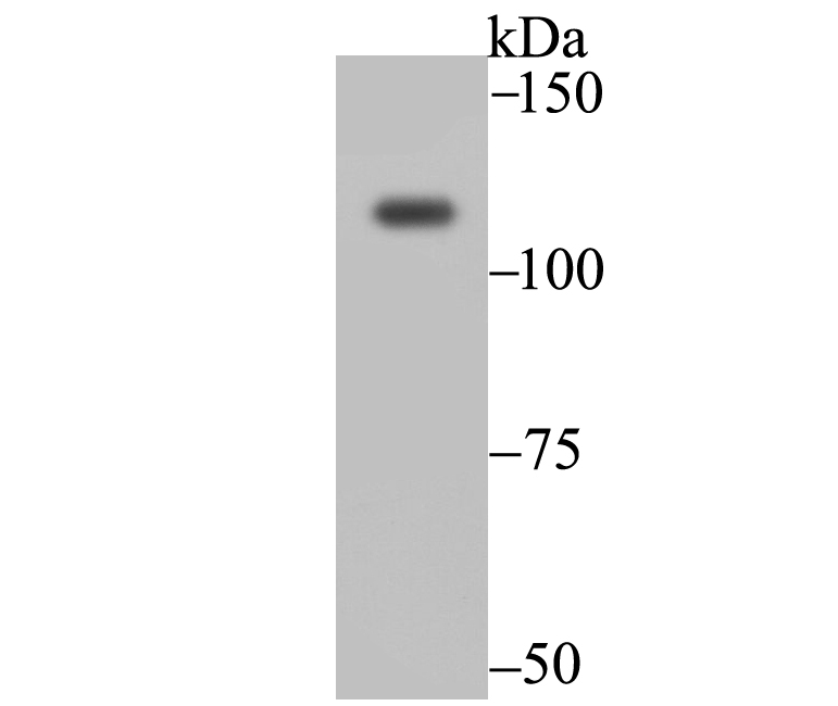
Fig1: Western blot analysis of Exportin-5 on 293 cell using anti-Exportin-5 antibody at 1/2,000 dilution.Application
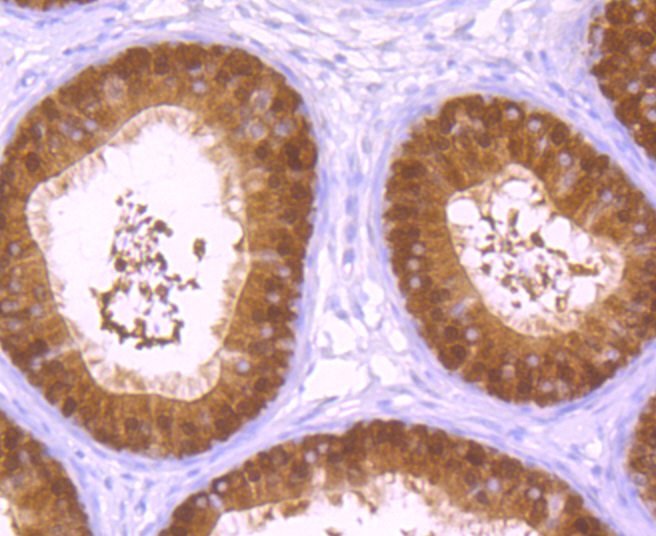
Fig2: Immunohistochemical analysis of paraffin-embedded rat epididymis tissue using anti-Exportin-5 antibody. Counter stained with hematoxylin.Application
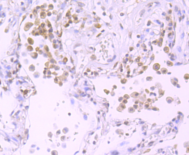
Fig3: Immunohistochemical analysis of paraffin-embedded human lung cancer tissue using anti-Exportin-5 antibody. Counter stained with hematoxylin.Application
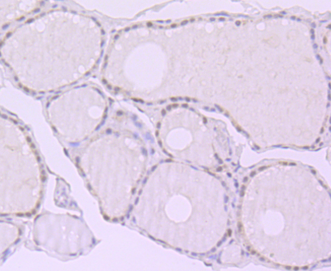
Fig4: Immunohistochemical analysis of paraffin-embedded human thyroid gland tissue using anti-Exportin-5 antibody. Counter stained with hematoxylin.Application
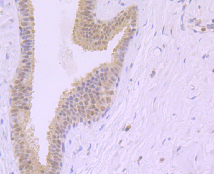
Fig5: Immunohistochemical analysis of paraffin-embedded human breast tissue using anti-Exportin-5 antibody. Counter stained with hematoxylin.Application
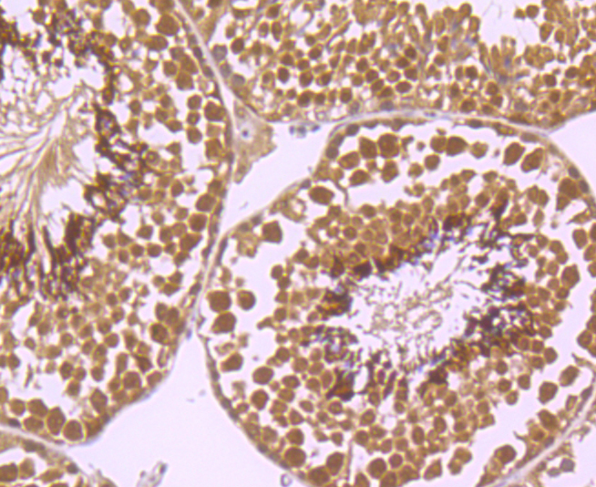
Fig6: Immunohistochemical analysis of paraffin-embedded mouse testis tissue using anti-Exportin-5 antibody. Counter stained with hematoxylin.Application
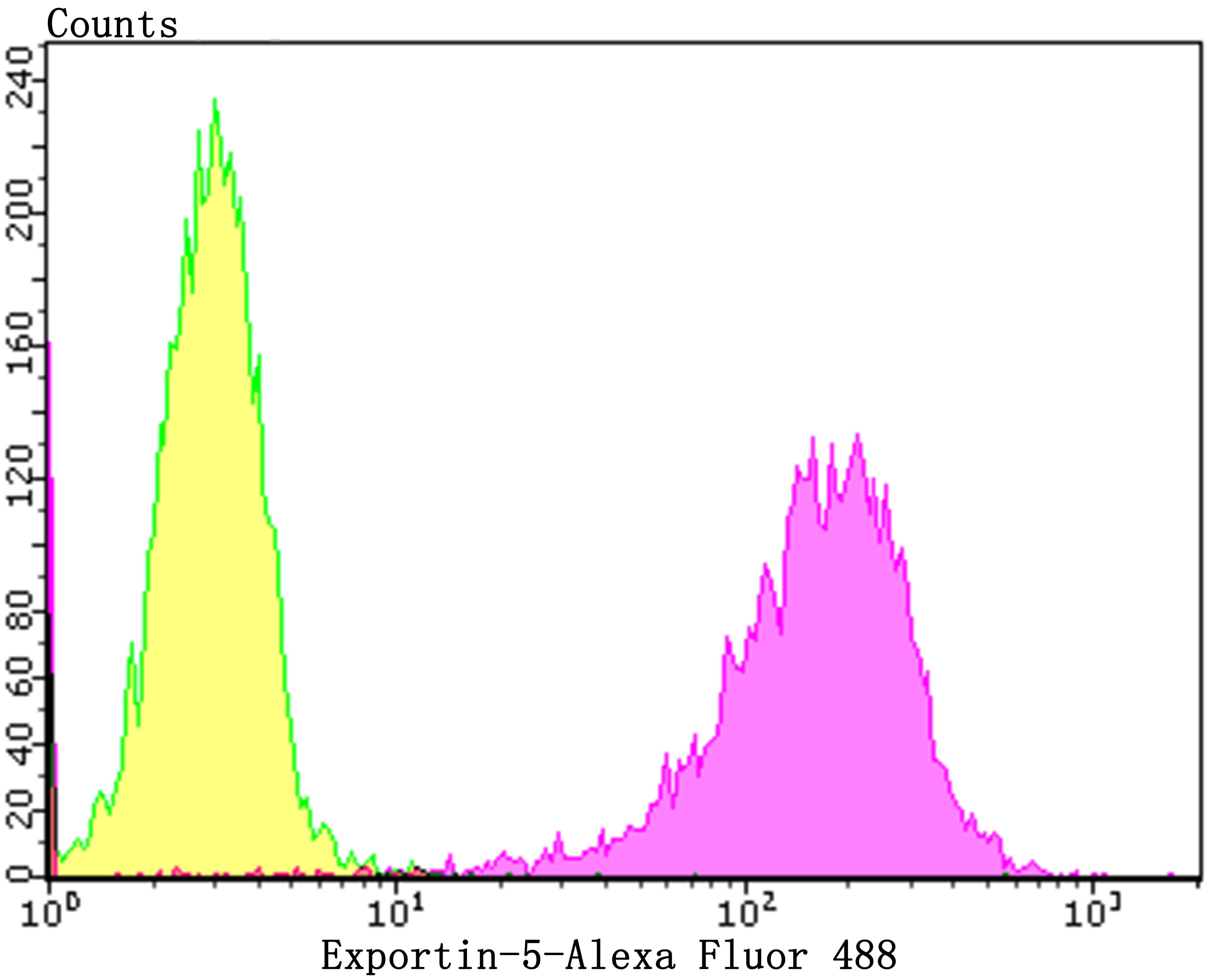
Fig7: Flow cytometric analysis of K562 cells with Exportin-5 antibody at 1/100 dilution (purple) compared with an unlabelled control (cells without incubation with primary antibody; yellow). Alexa Fluor 488-conjugated goat anti-rabbit IgG was used as the secondary antibody.| Product Name | Exportin-5 [JE40-91] |
|---|---|
| Antibody Type | Primary Antibodies |
| Antigen Alias | Exp 5 antibody Exp5 antibody Exportin 5 antibody Exportin-5 antibody Exportin5 antibody FLJ14239 antibody FLJ32057 antibody FLJ45606 antibody KIAA1291 antibody OTTHUMP00000016472 antibody Ran binding protein 21 antibody Ran-binding protein 21 antibody Ranbp 21 antibody Ranbp21 antibody Xpo 5 antibody xpo5 antibody XPO5_HUMAN antibody |
| Product description | The karyopherin-related nuclear transport factor Exportin 5, also known as Exp5, preferentially recognizes and transports RNAs containing minihelix motifs, structural cis-acting export elements that comprise a double-stranded stem (14 nucleotides) with a base-paired 5' end and a 3-8-nucleotide protruding 3' end. Exportin 5 also mediates protein transport between the nuclear and cytoplasmic compartment. Exportin 5 belongs to a large family of karyopherins and stimulates nuclear export of dsRNA binding proteins eEF1A and tRNA. |
| Immunogen | Recombinant protein within human Exportin-5 aa 1-200. |
| Clonality | Monoclonal |
|---|---|
| Isotype | IgG |
| Host Species | Recombinant rabbit |
| Tested Applications | WBIHCFCICC/IF |
| WB:1:500-1:2,000 IHC:1:100-1:400 FC:1:50-1:100 ICC/IF:1:100 |
|
| Species Reactivity | HumanMouseRat |
| Concentration | 1mg/ml |
| Alternative Names | Exp 5 antibody Exp5 antibody Exportin 5 antibody Exportin-5 antibody Exportin5 antibody FLJ14239 antibody FLJ32057 antibody FLJ45606 antibody KIAA1291 antibody OTTHUMP00000016472 antibody Ran binding protein 21 antibody Ran-binding protein 21 antibody Ranbp 21 antibody Ranbp21 antibody Xpo 5 antibody xpo5 antibody XPO5_HUMAN antibody |
|---|---|
| Molecular Weight(MW) | 136 kDa |
| Cellular Localization | Cytoplasm. Nucleus. |
| SwissProt ID | Q9HAV4 |
|---|
Application
Fig1: Western blot analysis of Exportin-5 on 293 cell using anti-Exportin-5 antibody at 1/2,000 dilution.
Application
Fig2: Immunohistochemical analysis of paraffin-embedded rat epididymis tissue using anti-Exportin-5 antibody. Counter stained with hematoxylin.
Application
Fig3: Immunohistochemical analysis of paraffin-embedded human lung cancer tissue using anti-Exportin-5 antibody. Counter stained with hematoxylin.
Application
Fig4: Immunohistochemical analysis of paraffin-embedded human thyroid gland tissue using anti-Exportin-5 antibody. Counter stained with hematoxylin.
Application
Fig5: Immunohistochemical analysis of paraffin-embedded human breast tissue using anti-Exportin-5 antibody. Counter stained with hematoxylin.
Application
Fig6: Immunohistochemical analysis of paraffin-embedded mouse testis tissue using anti-Exportin-5 antibody. Counter stained with hematoxylin.
Application
Fig7: Flow cytometric analysis of K562 cells with Exportin-5 antibody at 1/100 dilution (purple) compared with an unlabelled control (cells without incubation with primary antibody; yellow). Alexa Fluor 488-conjugated goat anti-rabbit IgG was used as the secondary antibody.| Positive Control | 293, rat epididymis tissue, human lung cancer tissue, human thyroid gland tissue, human breast tissue, mouse testis tissue, K562. |
|---|---|
| Application Notes | WB:1:500-1:2,000 IHC:1:100-1:400 FC:1:50-1:100 ICC/IF:1:100 |
| Form | Liquid |
|---|---|
| Storage Instructions | Store at +4℃ after thawing. Aliquot store at -20℃. Avoid repeated freeze / thaw cycles. |
| Storage Buffer | 1*TBS (pH7.4), 1%BSA, 40%Glycerol. Preservative: 0.05% Sodium Azide. |


