Application
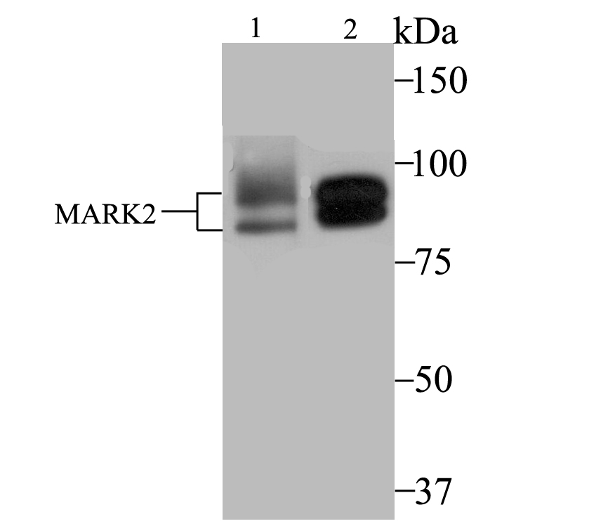
Fig1: Western blot analysis of MARK2 on MCF-7 (1) and SK-Br-3 (2) cell lysate using anti-MARK2 antibody at 1/2,000 dilution.Application
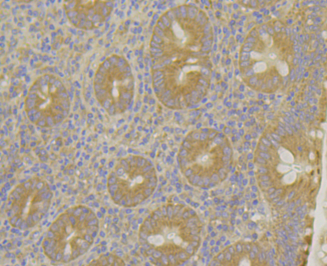
Fig2: Immunohistochemical analysis of paraffin-embedded human appendix tissue using anti-MARK2 antibody. Counter stained with hematoxylin.Application
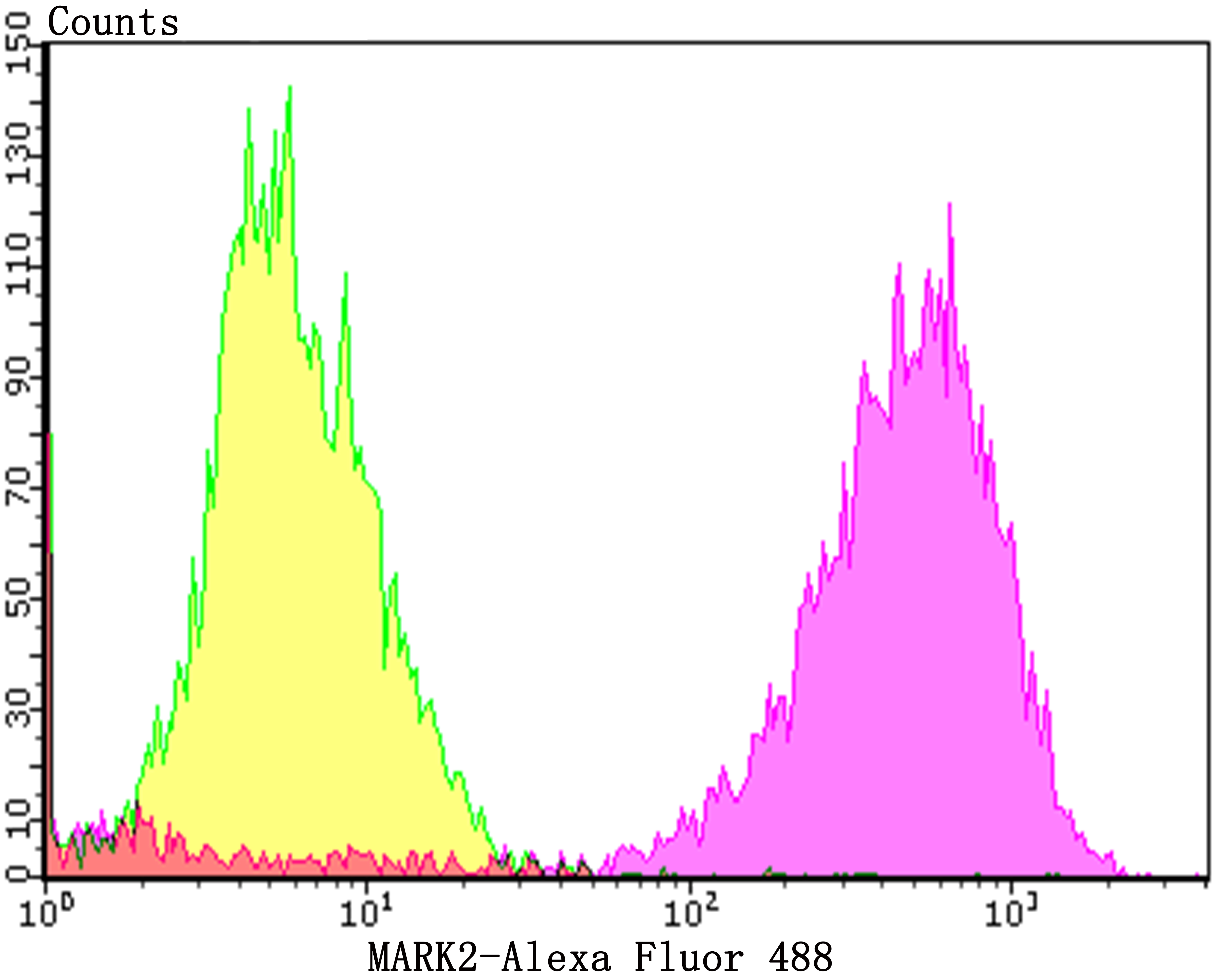
Fig3: Flow cytometric analysis of MCF-7 cells with MARK2 antibody at 1/100 dilution (purple) compared with an unlabelled control (cells without incubation with primary antibody; yellow). Alexa Fluor 488-conjugated goat anti-rabbit IgG was used as the secondary antibody.| Product Name | MARK2 [JE40-97] |
|---|---|
| Antibody Type | Primary Antibodies |
| Antigen Alias | ELKL motif kinase 1 antibody ELKL motif kinase antibody EMK-1 antibody EMK1 antibody MAP/microtubule affinity regulating kinase 2 antibody MAP/microtubule affinity-regulating kinase 2 antibody Mark2 antibody MARK2_HUMAN antibody MGC99619 antibody PAR 1 antibody Par 1b antibody PAR1 homolog antibody Par1b antibody Ser/Thr protein kinase PAR 1B antibody Serine/threonine protein kinase EMK antibody Serine/threonine protein kinase MARK2 antibody Serine/threonine-protein kinase MARK2 antibody |
| Product description | Microtubule affinity-regulating kinase 2 (MARK2), also known as EMK1 (ELKL motif kinase 1) or Par1b, is a 788 amino acid protein that is a member of the protein kinase superfamily, MARK subfamily. Highly expressed in heart, brain, skeletal muscle and pancreas, MARK2 is essential for the asymmetric development of membrane domains around polarized epithelial cells. Activation of MARK2 by phosphorylation on Thr 208 allows the protein to modulate the building of a columnar versus a hepatic epithelial cell. MARK2 contains one KA1 (kinase-associated) domain, one protein kinase domain and one UBA domain. MARK2 is expressed as 14 isoforms produced by alternative splicing events. Some of these isoforms may function in graft rejection. |
| Immunogen | Recombinant protein near the C-terminus of human MARK2. |
| Clonality | Monoclonal |
|---|---|
| Isotype | IgG |
| Host Species | Recombinant rabbit |
| Tested Applications | WBIHCFC |
| WB:1:500-1:2,000 IHC:1:50-1:200 FC:1:50-1:100 |
|
| Species Reactivity | HumanMouseRat |
| Concentration | 1mg/ml |
| Alternative Names | ELKL motif kinase 1 antibody ELKL motif kinase antibody EMK-1 antibody EMK1 antibody MAP/microtubule affinity regulating kinase 2 antibody MAP/microtubule affinity-regulating kinase 2 antibody Mark2 antibody MARK2_HUMAN antibody MGC99619 antibody PAR 1 antibody Par 1b antibody PAR1 homolog antibody Par1b antibody Ser/Thr protein kinase PAR 1B antibody Serine/threonine protein kinase EMK antibody Serine/threonine protein kinase MARK2 antibody Serine/threonine-protein kinase MARK2 antibody |
|---|---|
| Molecular Weight(MW) | 77/88 kDa |
| Cellular Localization | Cell membrane. Cytoplasm. Cytoskeleton. |
| SwissProt ID | Q7KZI7 |
|---|
Application
Fig1: Western blot analysis of MARK2 on MCF-7 (1) and SK-Br-3 (2) cell lysate using anti-MARK2 antibody at 1/2,000 dilution.
Application
Fig2: Immunohistochemical analysis of paraffin-embedded human appendix tissue using anti-MARK2 antibody. Counter stained with hematoxylin.
Application
Fig3: Flow cytometric analysis of MCF-7 cells with MARK2 antibody at 1/100 dilution (purple) compared with an unlabelled control (cells without incubation with primary antibody; yellow). Alexa Fluor 488-conjugated goat anti-rabbit IgG was used as the secondary antibody.| Positive Control | MCF-7, SK-Br-3, human appendix tissue. |
|---|---|
| Application Notes | WB:1:500-1:2,000 IHC:1:50-1:200 FC:1:50-1:100 |
| Form | Liquid |
|---|---|
| Storage Instructions | Store at +4℃ after thawing. Aliquot store at -20℃. Avoid repeated freeze / thaw cycles. |
| Storage Buffer | 1*TBS (pH7.4), 1%BSA, 40%Glycerol. Preservative: 0.05% Sodium Azide. |


