Application
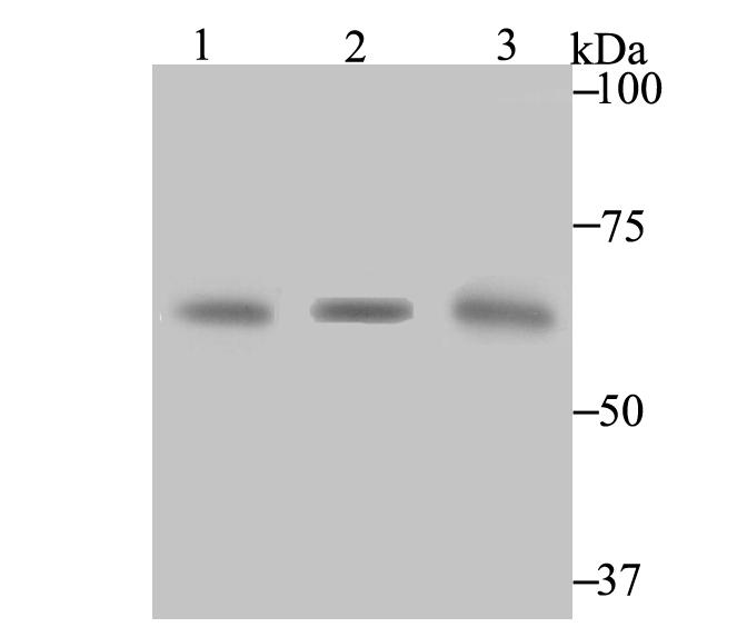
Fig1: Western blot analysis of CBS on different lysates using anti-CBS antibody at 1/1,000 dilution. Positive control: Lane 1: 293 Lane 2: 293 Lane 3: human liver tissueApplication
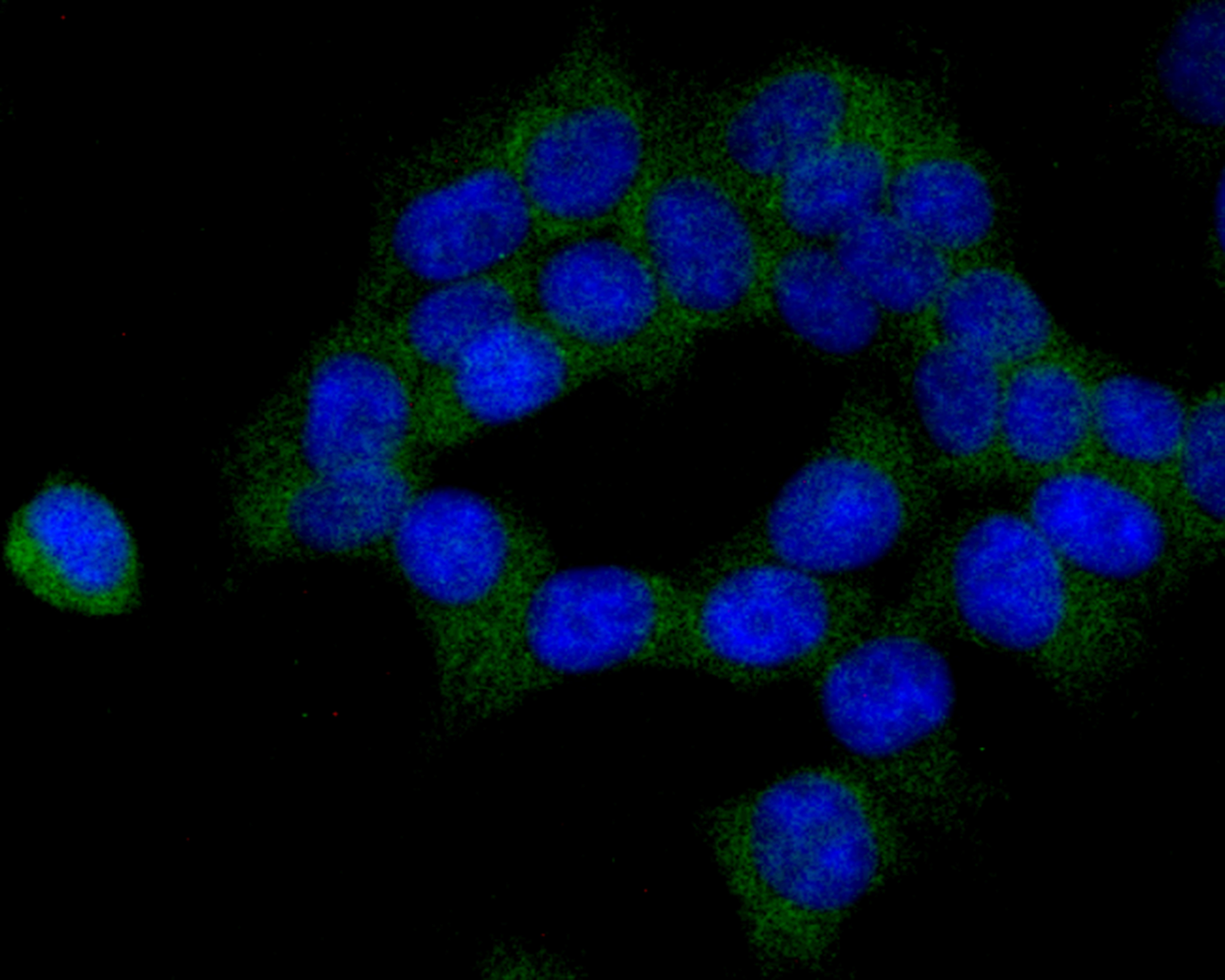
Fig2: ICC staining CBS in 293T cells (green). The nuclear counter stain is DAPI (blue). Cells were fixed in paraformaldehyde, permeabilised with 0.25% Triton X100/PBS.| Product Name | CBS [JE40-99] |
|---|---|
| Antibody Type | Primary Antibodies |
| Antigen Alias | AI047524 antibody AI303044 antibody Beta thionase antibody Beta-thionase antibody Cbs antibody Cbs cystathionine beta-synthase antibody CBS_HUMAN antibody Cystathionine beta synthase antibody Cystathionine beta-synthase antibody EC 4.2.1.22 antibody HIP 4 antibody HIP4 antibody Methylcysteine synthase antibody MGC18856 antibody MGC18895 antibody MGC37300 antibody OTTHUMP00000109416 antibody OTTHUMP00000109418 antibody Serine sulfhydrase antibody |
| Product description | Strongly expressed in human liver and pancreas, with weaker expression in heart and brain, the cytoplasmic protein cystathionine b-synthase (CBS) operates in the first step of homocysteine transulfuration. CBS, which belongs to the cysteine synthase/cystathionine b-synthase family of proteins, catalyzes the formation of cystathionine from the thrombogenic amino acid homocysteine using pyridoxal phosphate cofactor. Allosteric activation by adenosyl-methionine regulates CBS activity. Deficiencies in CBS are assoc-iated with homocystinuria, a recessively inherited error in sulfur amino acid metabolism that affects many organs and tissues. Symptoms of homocytinuria include arteriosclerosis, thrombosis, dislocated optic lenses, mental retard-ation and skeletal abnormalities. |
| Immunogen | Recombinant protein within human CBS aa 400-550. |
| Clonality | Monoclonal |
|---|---|
| Isotype | IgG |
| Host Species | Recombinant rabbit |
| Tested Applications | WBICC |
| WB:1:500-1:2,000 ICC:1:50-1:100 |
|
| Species Reactivity | Human |
| Concentration | 1mg/ml |
| Alternative Names | AI047524 antibody AI303044 antibody Beta thionase antibody Beta-thionase antibody Cbs antibody Cbs cystathionine beta-synthase antibody CBS_HUMAN antibody Cystathionine beta synthase antibody Cystathionine beta-synthase antibody EC 4.2.1.22 antibody HIP 4 antibody HIP4 antibody Methylcysteine synthase antibody MGC18856 antibody MGC18895 antibody MGC37300 antibody OTTHUMP00000109416 antibody OTTHUMP00000109418 antibody Serine sulfhydrase antibody |
|---|---|
| Molecular Weight(MW) | 61 kDa |
| Cellular Localization | Cytoplasm. Nucleus. |
| SwissProt ID | P35520 |
|---|
Application
Fig1: Western blot analysis of CBS on different lysates using anti-CBS antibody at 1/1,000 dilution. Positive control: Lane 1: 293 Lane 2: 293 Lane 3: human liver tissue
Application
Fig2: ICC staining CBS in 293T cells (green). The nuclear counter stain is DAPI (blue). Cells were fixed in paraformaldehyde, permeabilised with 0.25% Triton X100/PBS.| Positive Control | 293, human liver tissue lysate, 293T. |
|---|---|
| Application Notes | WB:1:500-1:2,000 ICC:1:50-1:100 |
| Form | Liquid |
|---|---|
| Storage Instructions | Store at +4℃ after thawing. Aliquot store at -20℃. Avoid repeated freeze / thaw cycles. |
| Storage Buffer | 1*TBS (pH7.4), 1%BSA, 40%Glycerol. Preservative: 0.05% Sodium Azide. |


