Application
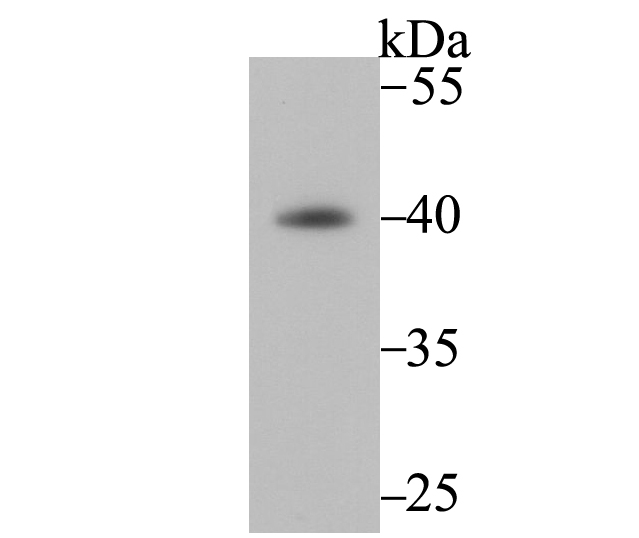
Fig1: Western blot analysis of Fbx32 on mouse skeletal muscle tissue using anti-Fbx32 antibody at 1/1,000 dilution.Application
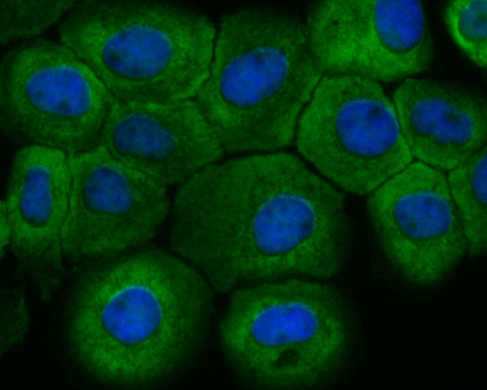
Fig2: ICC staining Fbx32 in A431 cells (green). The nuclear counter stain is DAPI (blue). Cells were fixed in paraformaldehyde, permeabilised with 0.25% Triton X100/PBS.Application
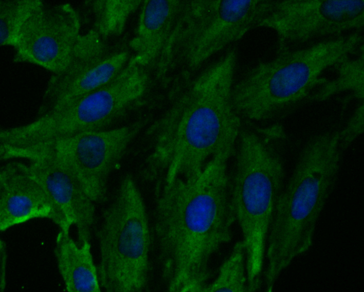
Fig3: ICC staining Fbx32 in L6 cells (green). The nuclear counter stain is DAPI (blue). Cells were fixed in paraformaldehyde, permeabilised with 0.25% Triton X100/PBS.| Product Name | Fbx32 [JE41-27] |
|---|---|
| Antibody Type | Primary Antibodies |
| Antigen Alias | 4833442G10Rik antibody AI430017 antibody Atrogin 1 antibody Atrogin-1 antibody ATROGIN1 antibody Atrophy gene 1 antibody F box only protein 32 antibody F-box only protein 32 antibody F-box protein 32 antibody FBX32_HUMAN antibody fbxo25 antibody FBXO32 antibody FLJ32424 antibody MAFbx antibody MGC108443 antibody MGC137646 antibody MGC33610 antibody Muscle atrophy F box antibody Muscle atrophy F box protein antibody Muscle atrophy F-box protein antibody |
| Product description | Muscle atrophy F-box (MAFbx) is an E3 ubiquitin ligase that initiates ATP-dependent ubiquitin-mediated proteolysis and promotes muscle atrophy. MAFbx transcript is abundant in cardiac and skeletal muscle undergoing atrophy. MAFbx -/- mice are resistant to muscle atrophy. MAFbx is thought to recognize and bind to some phosphorylated proteins and promote their ubiquitination and degradation during skeletal muscle atrophy. It interacts with MyoD by ubiquitination via a sequence found in transcriptional coactivators and therefore may play an important role in the course of muscle differentiation by determining the abundance of MyoD. MAFbx is specifically expressed in cardiac and skeletal muscle. |
| Immunogen | Recombinant protein corresponding to human Fbx32 1-200aa. |
| Clonality | Monoclonal |
|---|---|
| Isotype | IgG |
| Host Species | Recombinant rabbit |
| Tested Applications | WBICC |
| WB:1:500-1:1,000 ICC:1:50-1:200 |
|
| Species Reactivity | HumanMouseRat |
| Concentration | 1mg/ml |
| Alternative Names | 4833442G10Rik antibody AI430017 antibody Atrogin 1 antibody Atrogin-1 antibody ATROGIN1 antibody Atrophy gene 1 antibody F box only protein 32 antibody F-box only protein 32 antibody F-box protein 32 antibody FBX32_HUMAN antibody fbxo25 antibody FBXO32 antibody FLJ32424 antibody MAFbx antibody MGC108443 antibody MGC137646 antibody MGC33610 antibody Muscle atrophy F box antibody Muscle atrophy F box protein antibody Muscle atrophy F-box protein antibody |
|---|---|
| Molecular Weight(MW) | 41 kDa |
| Cellular Localization | Cytoplasm. Nucleus. |
Application
Fig1: Western blot analysis of Fbx32 on mouse skeletal muscle tissue using anti-Fbx32 antibody at 1/1,000 dilution.
Application
Fig2: ICC staining Fbx32 in A431 cells (green). The nuclear counter stain is DAPI (blue). Cells were fixed in paraformaldehyde, permeabilised with 0.25% Triton X100/PBS.
Application
Fig3: ICC staining Fbx32 in L6 cells (green). The nuclear counter stain is DAPI (blue). Cells were fixed in paraformaldehyde, permeabilised with 0.25% Triton X100/PBS.| Positive Control | Mouse skeletal muscle tissue lysate, A431, L6. |
|---|---|
| Application Notes | WB:1:500-1:1,000 ICC:1:50-1:200 |
| Form | Liquid |
|---|---|
| Storage Instructions | Store at +4℃ after thawing. Aliquot store at -20℃. Avoid repeated freeze / thaw cycles. |
| Storage Buffer | 1*TBS (pH7.4), 1%BSA, 40%Glycerol. Preservative: 0.05% Sodium Azide. |


