Application
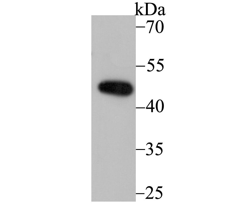
Fig1: Western blot analysis of DDB2 on Daudi cell using anti-DDB2 antibody at 1/1,000 dilution.Application
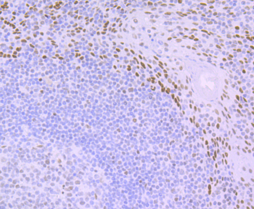
Fig2: Immunohistochemical analysis of paraffin-embedded human tonsil tissue using anti-DDB2 antibody. Counter stained with hematoxylin.Application
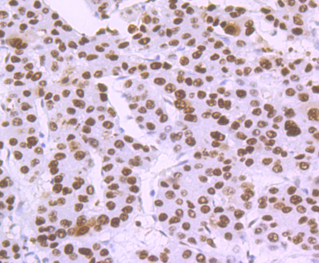
Fig3: Immunohistochemical analysis of paraffin-embedded human liver cancer tissue using anti-DDB2 antibody. Counter stained with hematoxylin.Application
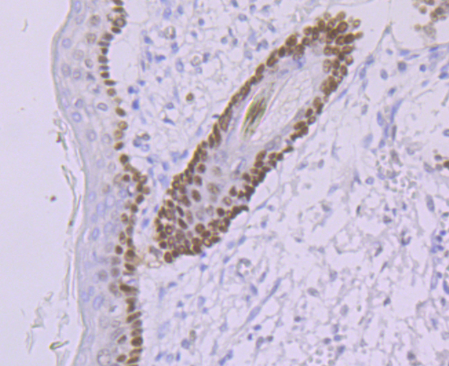
Fig4: Immunohistochemical analysis of paraffin-embedded human skin tissue using anti-DDB2 antibody. Counter stained with hematoxylin.Application
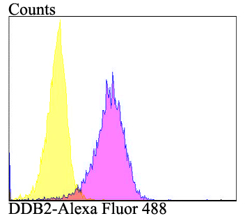
Fig5: Flow cytometric analysis of A549 cells with DDB2 antibody at 1/100 dilution (purple) compared with an unlabelled control (cells without incubation with primary antibody; yellow). Alexa Fluor 488-conjugated goat anti-rabbit IgG was used as the secondary antibody.| Product Name | DDB2 [JE16-41] |
|---|---|
| Antibody Type | Primary Antibodies |
| Antigen Alias | damage-specific DNA binding protein 2 antibody Damage-specific DNA-binding protein 2 antibody DDB p48 subunit antibody Ddb2 antibody DDB2_HUMAN antibody DDBb antibody DNA damage-binding protein 2 antibody UV-damaged DNA-binding protein 2 antibody UV-DDB 2 antibody Xeroderma pigmentosum group E protei antibody |
| Product description | Damaged DNA binding protein (DDB) is a heterodimer composed of two subunits, p127 and p48, which are designated DDB1 and DDB2, respectively. The DDB heterodimer is involved in repairing DNA damaged by ultraviolet light. Specifically, DDB, also designated UV-damaged DNA binding protein (UV-DDB), xeroderma pigmentosum group E binding factor (XPE-BF) and hepatitis B virus X-associated protein 1 (XAP-1), binds to damaged cyclobutane pyrimidine dimers (CPDs). Mutations in the DDB2 gene are implicated as causes of xeroderma pigmentosum group E, an autosomal recessive disease in which patients are defective in nucleotide excision DNA repair. XPE is characterized by hypersensitivity of the skin to sunlight with a high frequency of skin cancer as well as neurologic abnormalities. The hepatitis B virus (HBV) X protein interacts with DDB1, which may mediate HBx transactivation. |
| Immunogen | Recombinant protein with C-terminal human DDB2. |
| Clonality | Monoclonal |
|---|---|
| Isotype | IgG |
| Host Species | Recombinant rabbit |
| Tested Applications | WBIHCFC |
| WB:1:500-1:2,000 IHC:1:50-1:200 FC:1:50-1:100 |
|
| Species Reactivity | Human |
| Concentration | 1mg/ml |
| Alternative Names | damage-specific DNA binding protein 2 antibody Damage-specific DNA-binding protein 2 antibody DDB p48 subunit antibody Ddb2 antibody DDB2_HUMAN antibody DDBb antibody DNA damage-binding protein 2 antibody UV-damaged DNA-binding protein 2 antibody UV-DDB 2 antibody Xeroderma pigmentosum group E protei antibody |
|---|---|
| Molecular Weight(MW) | 48 kDa |
| Cellular Localization | Nucleus. |
| SwissProt ID | Q92466 |
|---|
Application
Fig1: Western blot analysis of DDB2 on Daudi cell using anti-DDB2 antibody at 1/1,000 dilution.
Application
Fig2: Immunohistochemical analysis of paraffin-embedded human tonsil tissue using anti-DDB2 antibody. Counter stained with hematoxylin.
Application
Fig3: Immunohistochemical analysis of paraffin-embedded human liver cancer tissue using anti-DDB2 antibody. Counter stained with hematoxylin.
Application
Fig4: Immunohistochemical analysis of paraffin-embedded human skin tissue using anti-DDB2 antibody. Counter stained with hematoxylin.
Application
Fig5: Flow cytometric analysis of A549 cells with DDB2 antibody at 1/100 dilution (purple) compared with an unlabelled control (cells without incubation with primary antibody; yellow). Alexa Fluor 488-conjugated goat anti-rabbit IgG was used as the secondary antibody.| Positive Control | Daudi, human tonsil tissue, human liver cancer tissue, human skin tissue, A549. |
|---|---|
| Application Notes | WB:1:500-1:2,000 IHC:1:50-1:200 FC:1:50-1:100 |
| Form | Liquid |
|---|---|
| Storage Instructions | Store at +4℃ after thawing. Aliquot store at -20℃. Avoid repeated freeze / thaw cycles. |
| Storage Buffer | 1*TBS (pH7.4), 1%BSA, 40%Glycerol. Preservative: 0.05% Sodium Azide. |


