Application
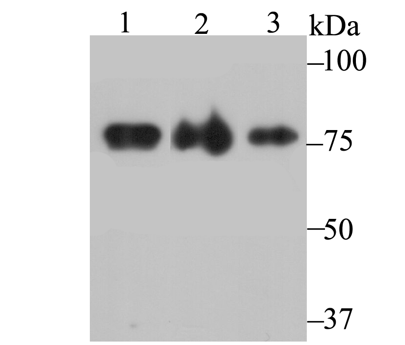
Fig1: Western blot analysis of KHSRP on different lysates using anti-KHSRP antibody at 1/1,000 dilution. Positive control: Lane 1: 293 Lane 2: mouse brain Lane 3: rat brainApplication
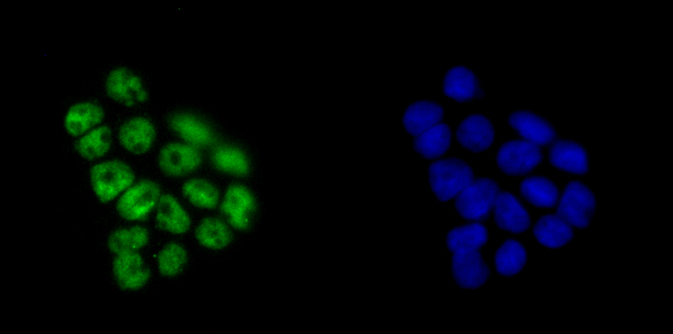
Fig2: ICC staining KHSRP in 293T cells (green). The nuclear counter stain is DAPI (blue). Cells were fixed in paraformaldehyde, permeabilised with 0.25% Triton X100/PBS.Application
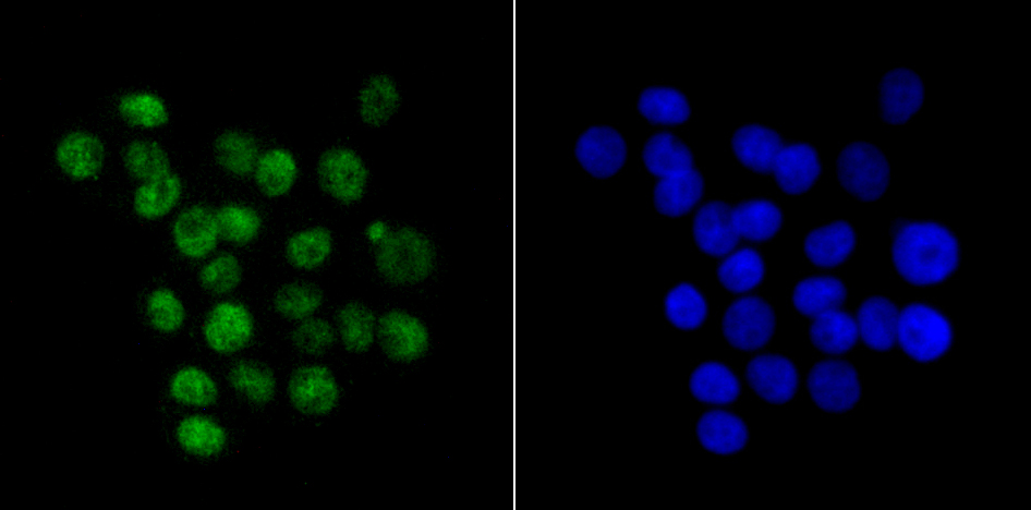
Fig3: ICC staining KHSRP in LOVO cells (green). The nuclear counter stain is DAPI (blue). Cells were fixed in paraformaldehyde, permeabilised with 0.25% Triton X100/PBS.Application
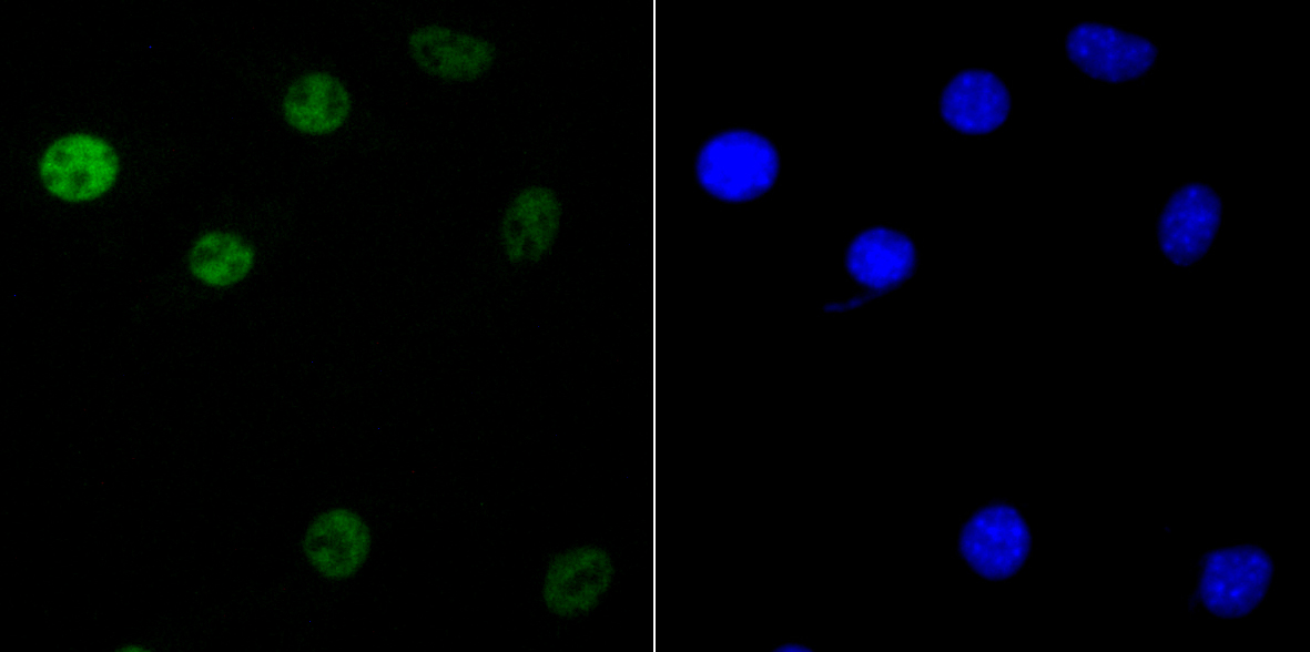
Fig4: ICC staining KHSRP in SH-SY-5Y cells (green). The nuclear counter stain is DAPI (blue). Cells were fixed in paraformaldehyde, permeabilised with 0.25% Triton X100/PBS.Application
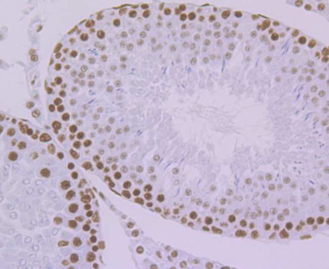
Fig5: Immunohistochemical analysis of paraffin-embedded rat testis tissue using anti-KHSRP antibody. Counter stained with hematoxylin.Application
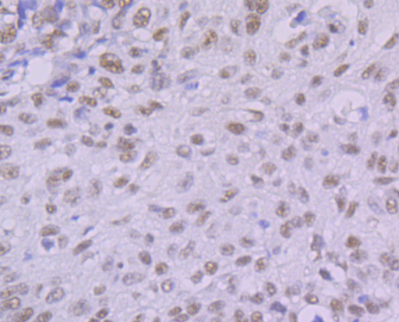
Fig6: Immunohistochemical analysis of paraffin-embedded human lung cancer tissue using anti-KHSRP antibody. Counter stained with hematoxylin.Application
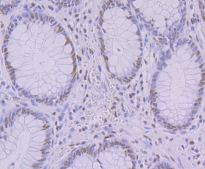
Fig7: Immunohistochemical analysis of paraffin-embedded human colon tissue using anti-KHSRP antibody. Counter stained with hematoxylin.Application
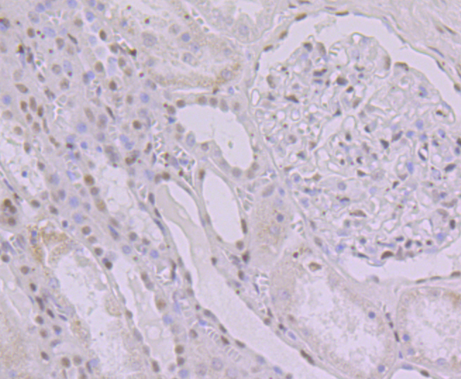
Fig8: Immunohistochemical analysis of paraffin-embedded human kidney tissue using anti-KHSRP antibody. Counter stained with hematoxylin.Application
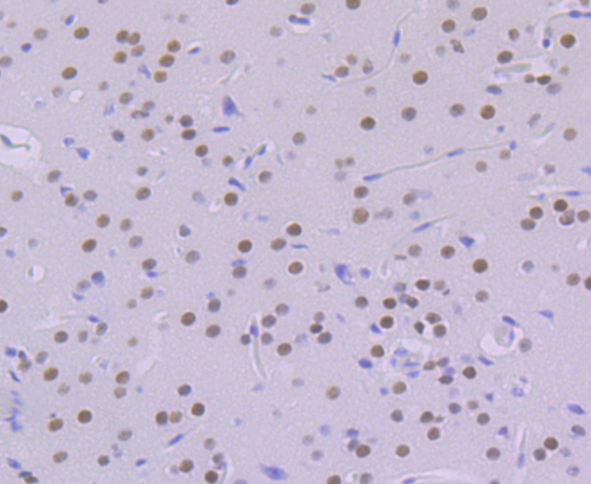
Fig9: Immunohistochemical analysis of paraffin-embedded mouse brain tissue using anti-KHSRP antibody. Counter stained with hematoxylin.| Product Name | KHSRP [JE41-64] |
|---|---|
| Antibody Type | Primary Antibodies |
| Antigen Alias | Far upstream element-binding protein 2 antibody FBP2 antibody FUBP2 antibody FUBP2_HUMAN antibody FUSE binding protein 2 antibody FUSE-binding protein 2 antibody KH type splicing regulatory protein (FUSE binding protein 2) antibody KH type splicing regulatory protein antibody KH type-splicing regulatory protein antibody KHSRP antibody KSRP antibody p75 antibody |
| Product description | Binds to the dendritic targeting element and may play a role in mRNA trafficking (By similarity). Part of a ternary complex that binds to the downstream control sequence (DCS) of the pre-mRNA. Mediates exon inclusion in transcripts that are subject to tissue-specific alternative splicing. May interact with single-stranded DNA from the far-upstream element (FUSE). May activate gene expression. Also involved in degradation of inherently unstable mRNAs that contain AU-rich elements (AREs) in their 3'-UTR, possibly by recruiting degradation machinery to ARE-containing mRNAs. |
| Immunogen | Recombinant protein within human KHSRP aa 1-150. |
| Clonality | Monoclonal |
|---|---|
| Isotype | IgG |
| Host Species | Recombinant rabbit |
| Tested Applications | WBICCIFIHCIP |
| WB:1:500-1:2,000 ICC/IF:1:50-1:200 IHC:1:50-1:200 IP:1:10-1:50 |
|
| Species Reactivity | HumanMouseRat |
| Concentration | 1mg/ml |
| Alternative Names | Far upstream element-binding protein 2 antibody FBP2 antibody FUBP2 antibody FUBP2_HUMAN antibody FUSE binding protein 2 antibody FUSE-binding protein 2 antibody KH type splicing regulatory protein (FUSE binding protein 2) antibody KH type splicing regulatory protein antibody KH type-splicing regulatory protein antibody KHSRP antibody KSRP antibody p75 antibody |
|---|---|
| Molecular Weight(MW) | 73 kDa |
| Cellular Localization | Nucleus. Cytoplasm. |
Application
Fig1: Western blot analysis of KHSRP on different lysates using anti-KHSRP antibody at 1/1,000 dilution. Positive control: Lane 1: 293 Lane 2: mouse brain Lane 3: rat brain
Application
Fig2: ICC staining KHSRP in 293T cells (green). The nuclear counter stain is DAPI (blue). Cells were fixed in paraformaldehyde, permeabilised with 0.25% Triton X100/PBS.
Application
Fig3: ICC staining KHSRP in LOVO cells (green). The nuclear counter stain is DAPI (blue). Cells were fixed in paraformaldehyde, permeabilised with 0.25% Triton X100/PBS.
Application
Fig4: ICC staining KHSRP in SH-SY-5Y cells (green). The nuclear counter stain is DAPI (blue). Cells were fixed in paraformaldehyde, permeabilised with 0.25% Triton X100/PBS.
Application
Fig5: Immunohistochemical analysis of paraffin-embedded rat testis tissue using anti-KHSRP antibody. Counter stained with hematoxylin.
Application
Fig6: Immunohistochemical analysis of paraffin-embedded human lung cancer tissue using anti-KHSRP antibody. Counter stained with hematoxylin.
Application
Fig7: Immunohistochemical analysis of paraffin-embedded human colon tissue using anti-KHSRP antibody. Counter stained with hematoxylin.
Application
Fig8: Immunohistochemical analysis of paraffin-embedded human kidney tissue using anti-KHSRP antibody. Counter stained with hematoxylin.
Application
Fig9: Immunohistochemical analysis of paraffin-embedded mouse brain tissue using anti-KHSRP antibody. Counter stained with hematoxylin.| Positive Control | 293, mouse brain tissue, rat brain tissue, 293T, LOVO, SH-SY-5Y, rat testis tissue, human lung cancer tissue, human colon tissue, human kidney tissue. |
|---|---|
| Application Notes | WB:1:500-1:2,000 ICC/IF:1:50-1:200 IHC:1:50-1:200 IP:1:10-1:50 |
| Form | Liquid |
|---|---|
| Storage Instructions | Store at +4℃ after thawing. Aliquot store at -20℃. Avoid repeated freeze / thaw cycles. |
| Storage Buffer | 1*TBS (pH7.4), 1%BSA, 40%Glycerol. Preservative: 0.05% Sodium Azide. |


