Application
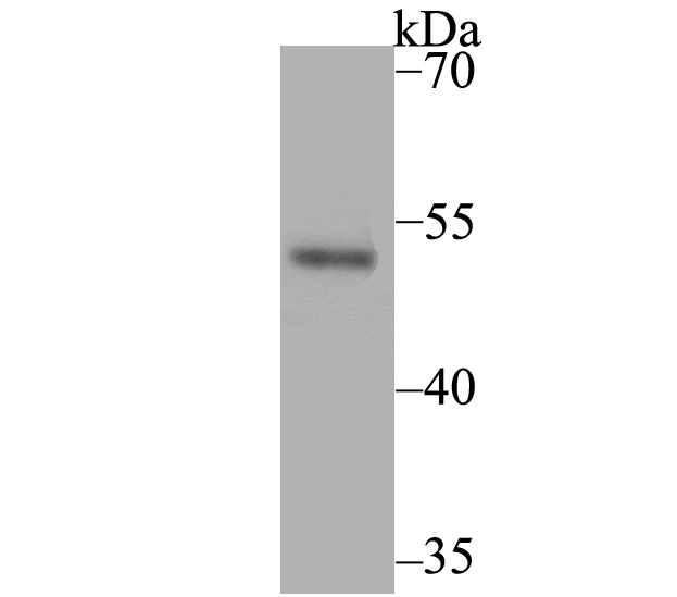
Fig1: Western blot analysis of Brachyury / Bry on SH-SY-5Y lysates. Proteins were transferred to a PVDF membrane and blocked with 5% BSA in PBS for 1 hour at room temperature. The primary antibody was used at a 1:500 dilution in 5% BSA at room temperature for 2 hours. Goat Anti-Rabbit IgG - HRP Secondary Antibody (HA1001) at 1:5,000 dilution was used for 1 hour at room temperature.Application
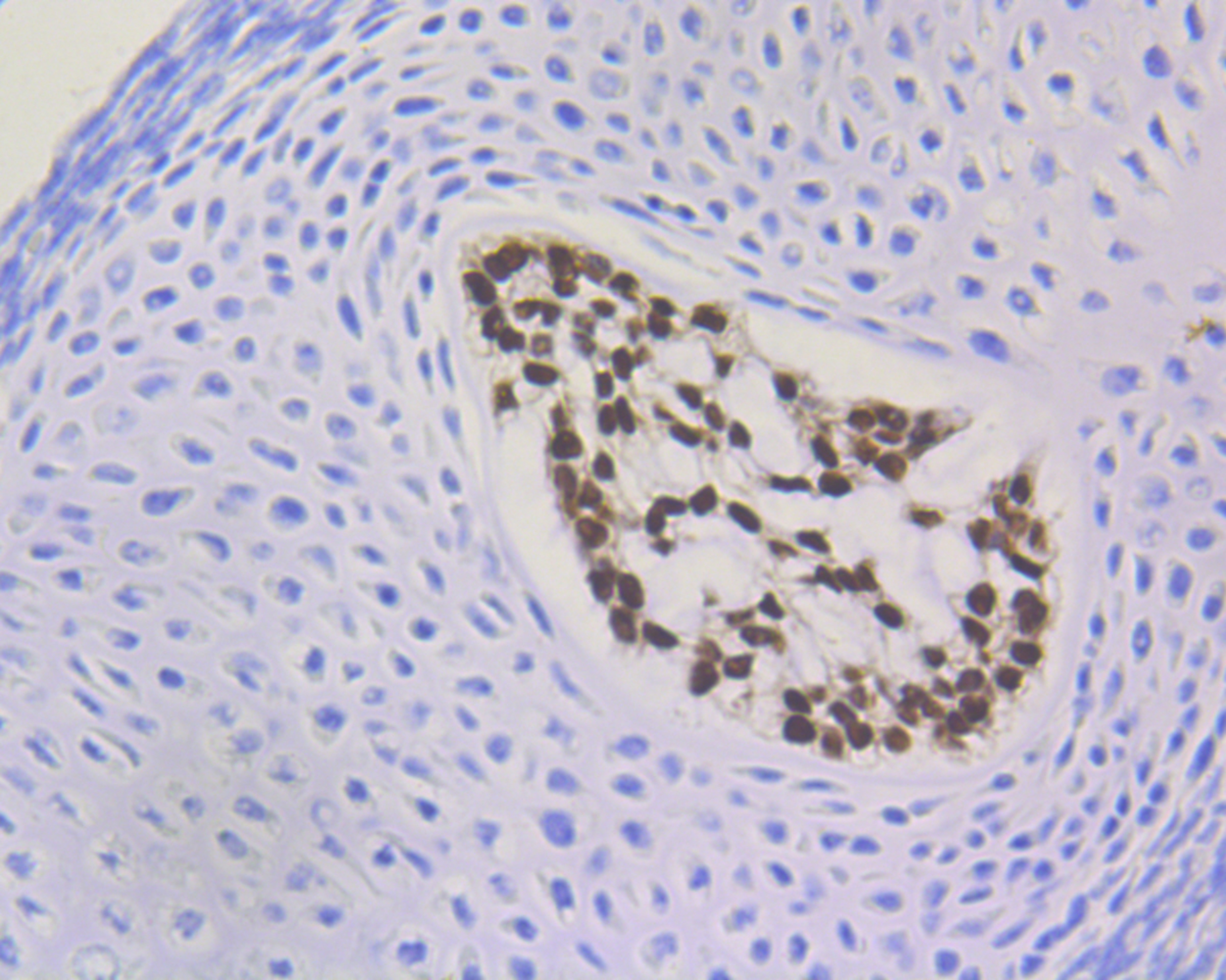
Fig2: Immunohistochemical analysis of paraffin-embedded mouse E14.5 embryo tissue using anti-Brachyury antibody. The section was pre-treated using heat mediated antigen retrieval with sodium citrate buffer (pH 6.0) for 20 mins. The tissues were blocked in 5% BSA for 30 minutes at room temperature, washed with ddH2O and PBS, and then probed with the antibody (ET7109-35) at 1/200 dilution, for 30 minutes at room temperature and detected using an HRP conjugated compact polymer system. DAB was used as the chrogen. Counter stained with hematoxylin and mounted with DPX.Application
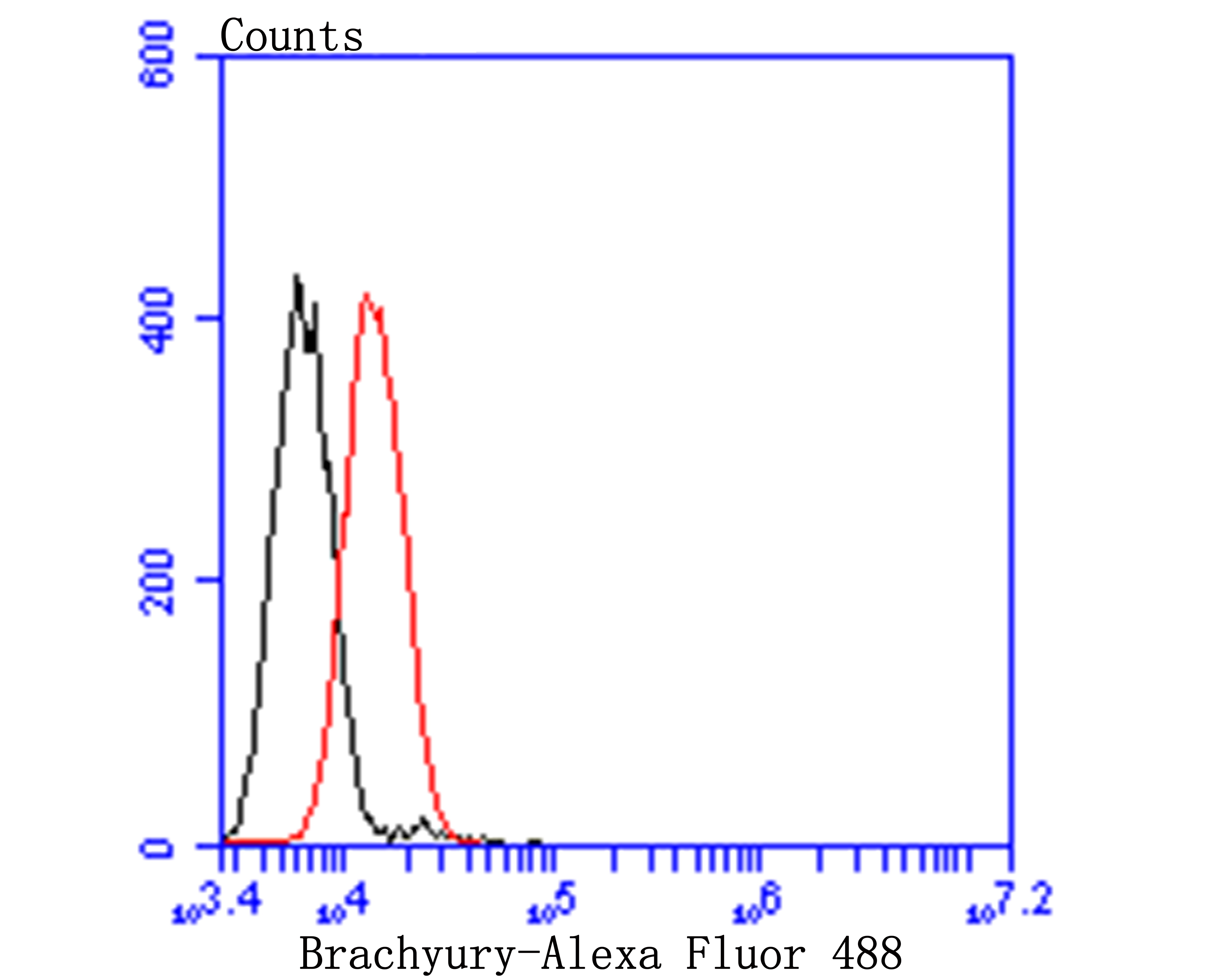
Fig3: Flow cytometric analysis of Brachyury was done on A549 cells. The cells were fixed, permeabilized and stained with Brachyury antibody at 1/100 dilution (red) compared with an unlabelled control (cells without incubation with primary antibody; black). After incubation of the primary antibody on room temperature for an hour, the cells was stained with a Alexa Fluor™ 488-conjugated goat anti-rabbit IgG Secondary antibody at 1/500 dilution for 30 minutes.| Product Name | Brachyury / Bry [JE44-11] |
|---|---|
| Antibody Type | Primary Antibodies |
| Antigen Alias | BRAC_HUMAN antibody Brachyury homolog antibody Brachyury protein antibody Bry antibody MGC104817 antibody Protein T antibody SAVA antibody T antibody T brachyury homolog antibody T brachyury transcription factor antibody T Protein antibody T, brachyury homolog (mouse) antibody TFT antibody Transcription factor T antibody |
| Product description | The protein encoded by this gene is an embryonic nuclear transcription factor that binds to a specific DNA element, the palindromic T-site. It binds through a region in its N-terminus, called the T-box, and effects transcription of genes required for mesoderm formation and differentiation. The protein is localized to notochord-derived cells. Two transcript variants encoding different isoforms have been found for this gene. Genetic variations in T are associated with susceptibility to neural tube defects (NTD). NTD are common congenital malformations. Spina bifida, which results from malformations in the caudal region of the neural tube, is compatible with life but associated with significant morbidity, including lower limb paralysis. |
| Immunogen | Recombinant protein within human Brachyury/ Bry aa 200-400. |
| Clonality | Monoclonal |
|---|---|
| Isotype | IgG |
| Host Species | Recombinant rabbit |
| Tested Applications | WBIHCFCIP |
| WB:1:500 IHC:1:50-1:200 FC:1:50-1:100 IP:1:10-1:50 |
|
| Species Reactivity | HumanMouse |
| Concentration | 1mg/ml |
| Alternative Names | BRAC_HUMAN antibody
Brachyury homolog antibody
Brachyury protein antibody
Bry antibody
MGC104817 antibody
Protein T antibody
SAVA antibody
T antibody
T brachyury homolog antibody
T brachyury transcription factor antibody
T Protein antibody
T brachyury homolog (mouse) antibody TFT antibody Transcription factor T antibody |
|---|---|
| Molecular Weight(MW) | 47 kDa |
| Cellular Localization | Nucleus. |
Application
Fig1: Western blot analysis of Brachyury / Bry on SH-SY-5Y lysates. Proteins were transferred to a PVDF membrane and blocked with 5% BSA in PBS for 1 hour at room temperature. The primary antibody was used at a 1:500 dilution in 5% BSA at room temperature for 2 hours. Goat Anti-Rabbit IgG - HRP Secondary Antibody (HA1001) at 1:5,000 dilution was used for 1 hour at room temperature.
Application
Fig2: Immunohistochemical analysis of paraffin-embedded mouse E14.5 embryo tissue using anti-Brachyury antibody. The section was pre-treated using heat mediated antigen retrieval with sodium citrate buffer (pH 6.0) for 20 mins. The tissues were blocked in 5% BSA for 30 minutes at room temperature, washed with ddH2O and PBS, and then probed with the antibody (ET7109-35) at 1/200 dilution, for 30 minutes at room temperature and detected using an HRP conjugated compact polymer system. DAB was used as the chrogen. Counter stained with hematoxylin and mounted with DPX.
Application
Fig3: Flow cytometric analysis of Brachyury was done on A549 cells. The cells were fixed, permeabilized and stained with Brachyury antibody at 1/100 dilution (red) compared with an unlabelled control (cells without incubation with primary antibody; black). After incubation of the primary antibody on room temperature for an hour, the cells was stained with a Alexa Fluor™ 488-conjugated goat anti-rabbit IgG Secondary antibody at 1/500 dilution for 30 minutes.| Positive Control | SH-SY-5Y, Mouse E14.5 embryo tissue. |
|---|---|
| Application Notes | WB:1:500 IHC:1:50-1:200 FC:1:50-1:100 IP:1:10-1:50 |
| Form | Liquid |
|---|---|
| Storage Instructions | Store at +4℃ after thawing. Aliquot store at -20℃. Avoid repeated freeze / thaw cycles. |
| Storage Buffer | 1*TBS (pH7.4), 1%BSA, 40%Glycerol. Preservative: 0.05% Sodium Azide. |


