Application
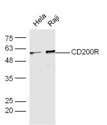
Fig1: Sample:hela(Human) Cell Lysate at 30 ug Raji(Human) Cell Lysate at 30 ug Primary: Anti-CD200R at 1/300 dilution Secondary: IRDye800CW Goat Anti-Rabbit IgG at 1/20000 dilution Predicted band size: 33 kD Observed band size: 58 kDApplication
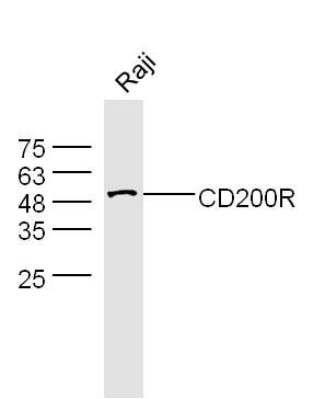
Fig2: Sample: Raji(Human) Cell Lysate at 30 ug Primary: Anti-CD200R at 1/300 dilution Secondary: IRDye800CW Goat Anti-Rabbit IgG at 1/20000 dilution Predicted band size: 33 kD Observed band size: 53 kDApplication
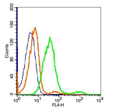
Fig3: Blank control:Hepg2 Cells(blue). Primary Antibody: Rabbit Anti-CD200R/AF647 Conjugated antibody , Dilution: 5μg in 100 μL 1X PBS containing 0.5% BSA; Isotype Control Antibody: Rabbit IgG/AF647(orange) ,used under the same conditions.Application
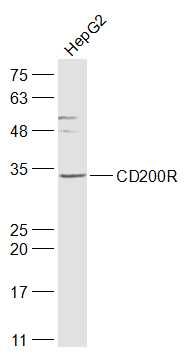
Fig4: Sample: HepG2(Human) Cell Lysate at 30 ug Primary: Anti-CD200R at 1/1000 dilution Secondary: IRDye800CW Goat Anti-Rabbit IgG at 1/20000 dilution Predicted band size: 34 kD Observed band size: 34 kDApplication
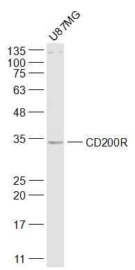
Fig5: Sample: U87MG(Human) Cell Lysate at 30 ug Primary: Anti-CD200R at 1/1000 dilution Secondary: IRDye800CW Goat Anti-Rabbit IgG at 1/20000 dilution Predicted band size: 34 kD Observed band size: 34 kDApplication
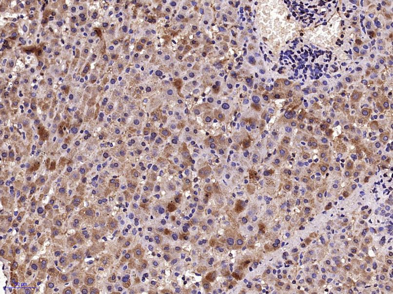
Fig6: Paraformaldehyde-fixed, paraffin embedded (Human liver); Antigen retrieval by microwave in sodium citrate buffer (pH6.0) ; Block endogenous peroxidase by 3% hydrogen peroxide for 30 minutes; Blocking buffer (3% BSA) at RT for 30min; Antibody incubation with (CD200R) Polyclonal Antibody, Unconjugated at 1:400 overnight at 4℃, followed by conjugation to the secondary antibody (labeled with HRP)and DAB staining.| Product Name | CD200R |
|---|---|
| Antibody Type | Primary Antibodies |
| Product description | Receptor for the CD200/OX2 cell surface glycoprotein. Also binds to HHV-8 K14 viral CD200 homolog with identical affinity and kinetics as the host CD200. |
| Immunogen | KLH conjugated synthetic peptide derived from human CD200R 151-250/325 |
| Clonality | Polyclonal |
|---|---|
| Isotype | IgG |
| Host Species | Rabbit |
| Tested Applications | FCIHC-PWB |
| WB:1:500-1:2000 IHC:1:100-1:500 FC:2μg/Test |
|
| Species Reactivity | HumanRat |
| Concentration | 1mg/ml |
| Alternative Names | CD 200R CD200 cell surface glycoprotein receptor CD200 receptor 1 Cd200r1 Cell surface glycoprotein CD200 receptor 1 Cell surface glycoprotein OX2 receptor 1 Cell surface glycoprotein OX2 receptor cell surface glycoprotein receptor CD200 CRTR 2 CRTR2 HCRTR2 MO2R1_HUMAN MOX2 receptor MOX2R OX2R. |
|---|---|
| Molecular Weight(MW) | 34 KDa |
| Cellular Localization | Secreted and Cell membrane. |
| SwissProt ID | Q6Q8B3 |
|---|
Application
Fig1: Sample:hela(Human) Cell Lysate at 30 ug Raji(Human) Cell Lysate at 30 ug Primary: Anti-CD200R at 1/300 dilution Secondary: IRDye800CW Goat Anti-Rabbit IgG at 1/20000 dilution Predicted band size: 33 kD Observed band size: 58 kD
Application
Fig2: Sample: Raji(Human) Cell Lysate at 30 ug Primary: Anti-CD200R at 1/300 dilution Secondary: IRDye800CW Goat Anti-Rabbit IgG at 1/20000 dilution Predicted band size: 33 kD Observed band size: 53 kD
Application
Fig3: Blank control:Hepg2 Cells(blue). Primary Antibody: Rabbit Anti-CD200R/AF647 Conjugated antibody , Dilution: 5μg in 100 μL 1X PBS containing 0.5% BSA; Isotype Control Antibody: Rabbit IgG/AF647(orange) ,used under the same conditions.
Application
Fig4: Sample: HepG2(Human) Cell Lysate at 30 ug Primary: Anti-CD200R at 1/1000 dilution Secondary: IRDye800CW Goat Anti-Rabbit IgG at 1/20000 dilution Predicted band size: 34 kD Observed band size: 34 kD
Application
Fig5: Sample: U87MG(Human) Cell Lysate at 30 ug Primary: Anti-CD200R at 1/1000 dilution Secondary: IRDye800CW Goat Anti-Rabbit IgG at 1/20000 dilution Predicted band size: 34 kD Observed band size: 34 kD
Application
Fig6: Paraformaldehyde-fixed, paraffin embedded (Human liver); Antigen retrieval by microwave in sodium citrate buffer (pH6.0) ; Block endogenous peroxidase by 3% hydrogen peroxide for 30 minutes; Blocking buffer (3% BSA) at RT for 30min; Antibody incubation with (CD200R) Polyclonal Antibody, Unconjugated at 1:400 overnight at 4℃, followed by conjugation to the secondary antibody (labeled with HRP)and DAB staining.| Positive Control | Expressed in granulocytes, monocytes, most T cells, neutrophils, basophils and a subset of NK, NKT and B cells (at protein level). Expressed in bone marrow, lymph nodes, spleen, lung, liver, spinal cord, kidney. Expressed in monocyte-derived dendritic and mast cells. |
|---|---|
| Application Notes | WB:1:500-1:2000 IHC:1:100-1:500 FC:2μg/Test |
| Form | Liquid |
|---|---|
| Storage Instructions | Store at -20℃ for one year. Avoid repeated freeze/thaw cycles. The lyophilized antibody is stable at room temperature for at least one month and for greater than a year when kept at -20℃. When reconstituted in sterile pH 7.4 0.01M PBS or diluent of antibody the antibody is stable for at least two weeks at 2-4℃. |
| Storage Buffer | 0.01M TBS(pH7.4) with 1% BSA, 0.03% Proclin300 and 50% Glycerol. |


