Application
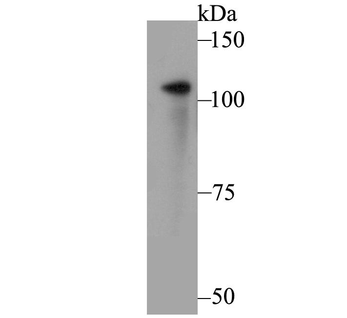
Fig1: Western blot analysis of FYB on U937 cell lysate. Proteins were transferred to a PVDF membrane and blocked with 5% BSA in PBS for 1 hour at room temperature. The primary antibody was used at a 1:500 dilution in 5% BSA at room temperature for 2 hours. Goat Anti-Rabbit IgG - HRP Secondary Antibody (HA1001) at 1:5,000 dilution was used for 1 hour at room temperature.Application
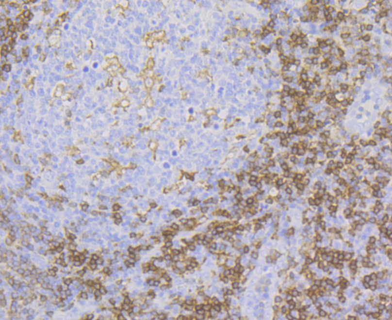
Fig2: Immunohistochemical analysis of paraffin-embedded human tonsil tissue using anti-FYB antibody. The section was pre-treated using heat mediated antigen retrieval with Tris-EDTA buffer (pH 8.0-8.4) for 20 minutes. The tissues were blocked in 5% BSA for 30 minutes at room temperature, washed with ddH2O and PBS, and then probed with the antibody (ET7109-38) at 1/200 dilution, for 30 minutes at room temperature and detected using an HRP conjugated compact polymer system. DAB was used as the chrogen. Counter stained with hematoxylin and mounted with DPX.Application
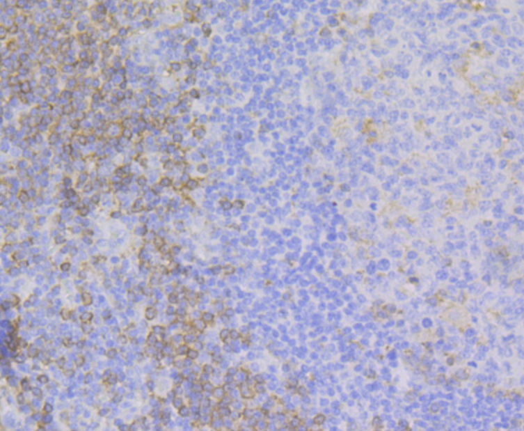
Fig3: Immunohistochemical analysis of paraffin-embedded human appendix tissue using anti-FYB antibody. The section was pre-treated using heat mediated antigen retrieval with Tris-EDTA buffer (pH 8.0-8.4) for 20 minutes. The tissues were blocked in 5% BSA for 30 minutes at room temperature, washed with ddH2O and PBS, and then probed with the antibody (ET7109-38) at 1/200 dilution, for 30 minutes at room temperature and detected using an HRP conjugated compact polymer system. DAB was used as the chrogen. Counter stained with hematoxylin and mounted with DPX.Application
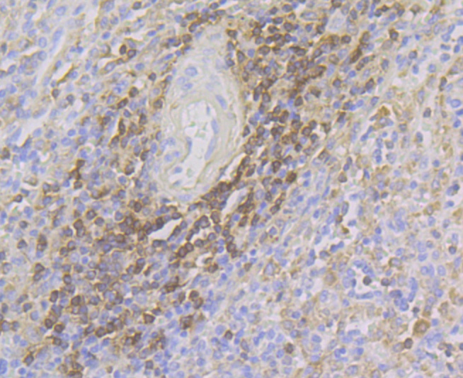
Fig4: Immunohistochemical analysis of paraffin-embedded human spleen tissue using anti-FYB antibody. The section was pre-treated using heat mediated antigen retrieval with Tris-EDTA buffer (pH 8.0-8.4) for 20 minutes. The tissues were blocked in 5% BSA for 30 minutes at room temperature, washed with ddH2O and PBS, and then probed with the antibody (ET7109-38) at 1/200 dilution, for 30 minutes at room temperature and detected using an HRP conjugated compact polymer system. DAB was used as the chrogen. Counter stained with hematoxylin and mounted with DPX.Application
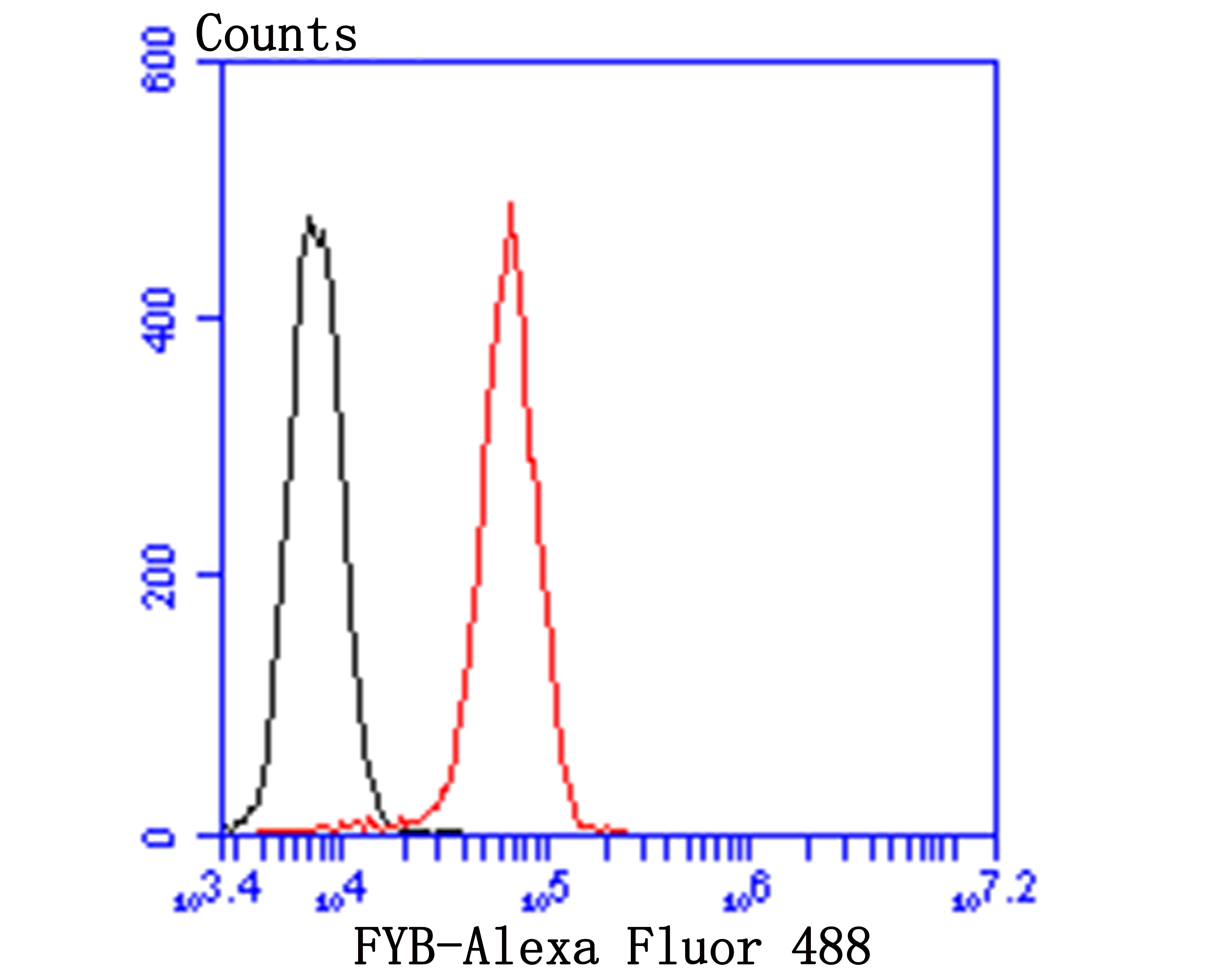
Fig5: Flow cytometric analysis of FYB was done on HepG2 cells. The cells were fixed, permeabilized and stained with FYB antibody at 1/100 dilution (red) compared with an unlabelled control (cells without incubation with primary antibody; black). After incubation of the primary antibody on room temperature for 1 hour, the cells was stained with a Alexa Fluor™ 488-conjugated goat anti-rabbit IgG Secondary antibody at 1/500 dilution for 30 minutes.| Product Name | FYB [JE44-80] |
|---|---|
| Antibody Type | Primary Antibodies |
| Antigen Alias | ADAP antibody Adhesion and degranulation promoting adaptor protein antibody FYB 120/130 antibody Fyb antibody FYB-120/130 antibody FYB_HUMAN antibody FYN binding protein antibody FYN T binding protein antibody FYN-binding protein antibody FYN-T-binding protein antibody p120/p130 antibody PRO0823 antibody SLAP 130 antibody SLAP-130 antibody SLAP130 antibody SLP 76 associated phosphoprotein antibody SLP-76-associated phosphoprotein antibody SLP76 associated phosphoprotein antibody |
| Product description | Acts as an adapter protein of the FYN and LCP2 signaling cascades in T-cells. Modulates the expression of interleukin-2 (IL-2). Involved in platelet activation. Prevents the degradation of SKAP1 and SKAP2. May play a role in linking T-cell signaling to remodeling of the actin cytoskeleton. Expressed in hematopoietic tissues such as myeloid and T-cells, spleen and thymus. Not expressed in B-cells, nor in non-lymphoid tissues. |
| Immunogen | Recombinant protein within C-terminal human FYB. |
| Clonality | Monoclonal |
|---|---|
| Isotype | IgG |
| Host Species | Recombinant rabbit |
| Tested Applications | WBIHCFC |
| WB:1:500-1:1,000 IHC:1:50-1:200 FC:1:50-1:100 |
|
| Species Reactivity | Human |
| Concentration | 1mg/ml |
| Alternative Names | ADAP antibody Adhesion and degranulation promoting adaptor protein antibody FYB 120/130 antibody Fyb antibody FYB-120/130 antibody FYB_HUMAN antibody FYN binding protein antibody FYN T binding protein antibody FYN-binding protein antibody FYN-T-binding protein antibody p120/p130 antibody PRO0823 antibody SLAP 130 antibody SLAP-130 antibody SLAP130 antibody SLP 76 associated phosphoprotein antibody SLP-76-associated phosphoprotein antibody SLP76 associated phosphoprotein antibody |
|---|---|
| Molecular Weight(MW) | 120 kDa, predicted band size 85 kDa |
| Cellular Localization | Cell junction, Cytoplasm, Nucleus. |
| SwissProt ID | O15117 |
|---|
Application
Fig1: Western blot analysis of FYB on U937 cell lysate. Proteins were transferred to a PVDF membrane and blocked with 5% BSA in PBS for 1 hour at room temperature. The primary antibody was used at a 1:500 dilution in 5% BSA at room temperature for 2 hours. Goat Anti-Rabbit IgG - HRP Secondary Antibody (HA1001) at 1:5,000 dilution was used for 1 hour at room temperature.
Application
Fig2: Immunohistochemical analysis of paraffin-embedded human tonsil tissue using anti-FYB antibody. The section was pre-treated using heat mediated antigen retrieval with Tris-EDTA buffer (pH 8.0-8.4) for 20 minutes. The tissues were blocked in 5% BSA for 30 minutes at room temperature, washed with ddH2O and PBS, and then probed with the antibody (ET7109-38) at 1/200 dilution, for 30 minutes at room temperature and detected using an HRP conjugated compact polymer system. DAB was used as the chrogen. Counter stained with hematoxylin and mounted with DPX.
Application
Fig3: Immunohistochemical analysis of paraffin-embedded human appendix tissue using anti-FYB antibody. The section was pre-treated using heat mediated antigen retrieval with Tris-EDTA buffer (pH 8.0-8.4) for 20 minutes. The tissues were blocked in 5% BSA for 30 minutes at room temperature, washed with ddH2O and PBS, and then probed with the antibody (ET7109-38) at 1/200 dilution, for 30 minutes at room temperature and detected using an HRP conjugated compact polymer system. DAB was used as the chrogen. Counter stained with hematoxylin and mounted with DPX.
Application
Fig4: Immunohistochemical analysis of paraffin-embedded human spleen tissue using anti-FYB antibody. The section was pre-treated using heat mediated antigen retrieval with Tris-EDTA buffer (pH 8.0-8.4) for 20 minutes. The tissues were blocked in 5% BSA for 30 minutes at room temperature, washed with ddH2O and PBS, and then probed with the antibody (ET7109-38) at 1/200 dilution, for 30 minutes at room temperature and detected using an HRP conjugated compact polymer system. DAB was used as the chrogen. Counter stained with hematoxylin and mounted with DPX.
Application
Fig5: Flow cytometric analysis of FYB was done on HepG2 cells. The cells were fixed, permeabilized and stained with FYB antibody at 1/100 dilution (red) compared with an unlabelled control (cells without incubation with primary antibody; black). After incubation of the primary antibody on room temperature for 1 hour, the cells was stained with a Alexa Fluor™ 488-conjugated goat anti-rabbit IgG Secondary antibody at 1/500 dilution for 30 minutes.| Positive Control | U937, human tonsil tissue, human appendix tissue, human spleen tissue. |
|---|---|
| Application Notes | WB:1:500-1:1,000 IHC:1:50-1:200 FC:1:50-1:100 |
| Form | Liquid |
|---|---|
| Storage Instructions | Store at +4℃ after thawing. Aliquot store at -20℃. Avoid repeated freeze / thaw cycles. |
| Storage Buffer | 1*TBS (pH7.4), 1%BSA, 40%Glycerol. Preservative: 0.05% Sodium Azide. |


