Application
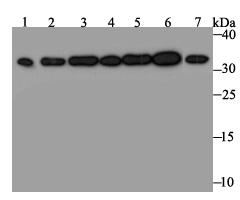
Fig1: Western blot analysis of RPA32/RPA2 on different lysates. Proteins were transferred to a PVDF membrane and blocked with 5% BSA in PBS for 1 hour at room temperature. The primary antibody was used at a 1:500 dilution in 5% BSA at room temperature for 2 hours. Goat Anti-Rabbit IgG - HRP Secondary Antibody (HA1001) at 1:5,000 dilution was used for 1 hour at room temperature. Positive control: Lane 1: Mouse testis tissue lysate Lane 2: A431 cell lysate Lane 3: SH-SY-5Y cell lysate Lane 4: SiHa cell lysate Lane 5: 293 cell lysate Lane 6: Hela cell lysate Lane 7: HepG2 cell lysateApplication
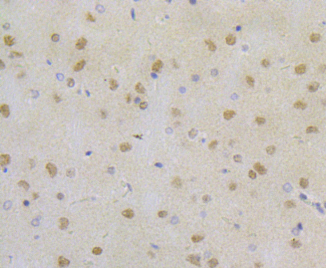
Fig2: Immunohistochemical analysis of paraffin-embedded rat brain tissue using anti-RPA32/RPA2 antibody. The section was pre-treated using heat mediated antigen retrieval with Tris-EDTA buffer (pH 8.0-8.4) for 20 minutes. The tissues were blocked in 5% BSA for 30 minutes at room temperature, washed with ddH2O and PBS, and then probed with the antibody (ET7109-41) at 1/50 dilution, for 30 minutes at room temperature and detected using an HRP conjugated compact polymer system. DAB was used as the chrogen. Counter stained with hematoxylin and mounted with DPX.Application
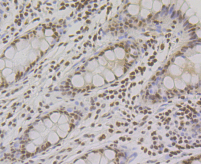
Fig3: Immunohistochemical analysis of paraffin-embedded human colon tissue using anti-RPA32/RPA2 antibody. The section was pre-treated using heat mediated antigen retrieval with Tris-EDTA buffer (pH 8.0-8.4) for 20 minutes. The tissues were blocked in 5% BSA for 30 minutes at room temperature, washed with ddH2O and PBS, and then probed with the antibody (ET7109-41) at 1/200 dilution, for 30 minutes at room temperature and detected using an HRP conjugated compact polymer system. DAB was used as the chrogen. Counter stained with hematoxylin and mounted with DPX.Application
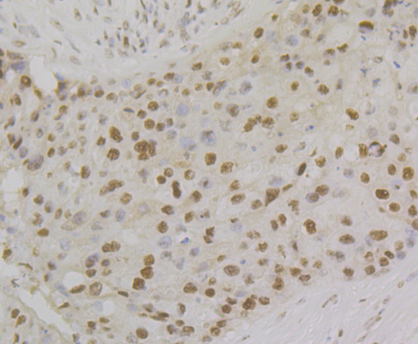
Fig4: Immunohistochemical analysis of paraffin-embedded human breast cancer tissue using anti-RPA32/RPA2 antibody. The section was pre-treated using heat mediated antigen retrieval with Tris-EDTA buffer (pH 8.0-8.4) for 20 minutes. The tissues were blocked in 5% BSA for 30 minutes at room temperature, washed with ddH2O and PBS, and then probed with the antibody (ET7109-41) at 1/200 dilution, for 30 minutes at room temperature and detected using an HRP conjugated compact polymer system. DAB was used as the chrogen. Counter stained with hematoxylin and mounted with DPX.Application
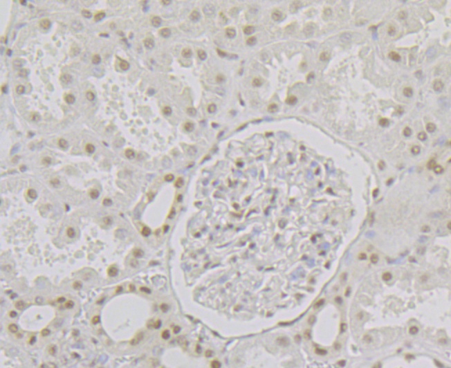
Fig5: Immunohistochemical analysis of paraffin-embedded human kidney tissue using anti-RPA32/RPA2 antibody. The section was pre-treated using heat mediated antigen retrieval with Tris-EDTA buffer (pH 8.0-8.4) for 20 minutes. The tissues were blocked in 5% BSA for 30 minutes at room temperature, washed with ddH2O and PBS, and then probed with the antibody (ET7109-41) at 1/200 dilution, for 30 minutes at room temperature and detected using an HRP conjugated compact polymer system. DAB was used as the chrogen. Counter stained with hematoxylin and mounted with DPX.Application
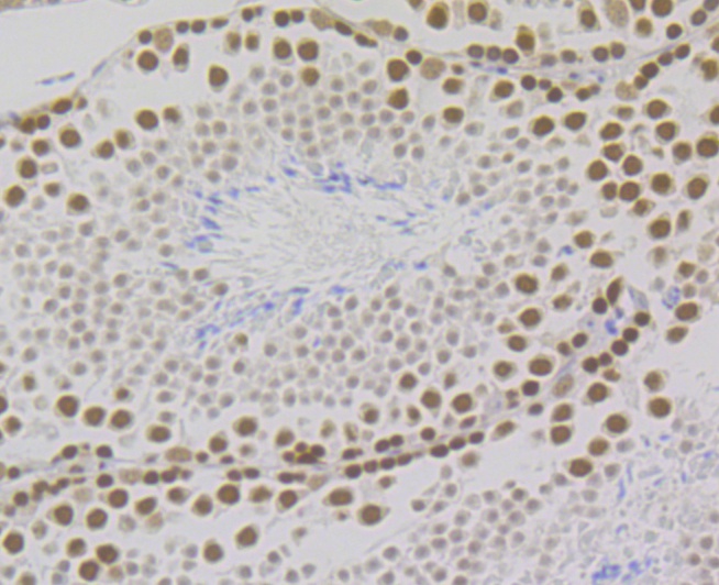
Fig6: Immunohistochemical analysis of paraffin-embedded mouse testis tissue using anti-RPA32/RPA2 antibody. The section was pre-treated using heat mediated antigen retrieval with Tris-EDTA buffer (pH 8.0-8.4) for 20 minutes. The tissues were blocked in 5% BSA for 30 minutes at room temperature, washed with ddH2O and PBS, and then probed with the antibody (ET7109-41) at 1/200 dilution, for 30 minutes at room temperature and detected using an HRP conjugated compact polymer system. DAB was used as the chrogen. Counter stained with hematoxylin and mounted with DPX.| Product Name | RPA32/RPA2 [JE45-59] |
|---|---|
| Antibody Type | Primary Antibodies |
| Antigen Alias | 60S acidic ribosomal protein P1 antibody AA409079 antibody AI325195 antibody AU020965 antibody ik:tdsubc_2g1 antibody M(2)21C antibody MGC137236 antibody OTTHUMP00000004008 antibody p32 antibody p34 antibody RCJMB04_6d17 replication protein A2, 32kDa antibody REPA2 antibody Replication factor A protein 2 antibody Replication protein A 32 kDa subunit antibody Replication protein A 32kDa subunit antibody Replication protein A 34 kDa subunit antibody Replication protein A antibody Replication Protein A2 (32kDa) antibody Replication protein A2 antibody Replication protein A2, 32kDa antibody RF-A protein 2 antibody Rf-A2 antibody RFA antibody RFA2_HUMAN antibody RP-A p32 antibody RP-A p34 antibody RP21C antibody RPA 2 antibody RPA 32 antibody RPA antibody Rpa2 antibody RPA32 antibody RPA34 antibody RpLP1 antibody RpP2 antibody xx:tdsubc_2g1 antibody zgc:109822 antibody |
| Product description | As part of the heterotrimeric replication protein A complex (RPA/RP-A), binds and stabilizes single-stranded DNA intermediates, that form during DNA replication or upon DNA stress. It prevents their reannealing and in parallel, recruits and activates different proteins and complexes involved in DNA metabolism. Thereby, it plays an essential role both in DNA replication and the cellular response to DNA damage. In the cellular response to DNA damage, the RPA complex controls DNA repair and DNA damage checkpoint activation. Through recruitment of ATRIP activates the ATR kinase a master regulator of the DNA damage response. It is required for the recruitment of the DNA double-strand break repair factors RAD51 and RAD52 to chromatin in response to DNA damage. Also recruits to sites of DNA damage proteins like XPA and XPG that are involved in nucleotide excision repair and is required for this mechanism of DNA repair. Plays also a role in base excision repair (BER) probably through interaction with UNG. Also recruits SMARCAL1/HARP, which is involved in replication fork restart, to sites of DNA damage. May also play a role in telomere maintenance. |
| Immunogen | Recombinant protein within C-terminal human RPA32/RPA2. |
| Clonality | Monoclonal |
|---|---|
| Isotype | IgG |
| Host Species | Recombinant rabbit |
| Tested Applications | WBIPIHC |
| WB:1:500-1:2,000 IP:1:10-1:50 IHC:1:50-1:200 |
|
| Species Reactivity | HumanMouseRat |
| Concentration | 1mg/ml |
| Alternative Names | 60S acidic ribosomal protein P1 antibody
AA409079 antibody
AI325195 antibody
AU020965 antibody
ik:tdsubc_2g1 antibody
M(2)21C antibody
MGC137236 antibody
OTTHUMP00000004008 antibody
p32 antibody
p34 antibody
RCJMB04_6d17 replication protein A2 32kDa antibody REPA2 antibody Replication factor A protein 2 antibody Replication protein A 32 kDa subunit antibody Replication protein A 32kDa subunit antibody Replication protein A 34 kDa subunit antibody Replication protein A antibody Replication Protein A2 (32kDa) antibody Replication protein A2 antibody Replication protein A2 32kDa antibody RF-A protein 2 antibody Rf-A2 antibody RFA antibody RFA2_HUMAN antibody RP-A p32 antibody RP-A p34 antibody RP21C antibody RPA 2 antibody RPA 32 antibody RPA antibody Rpa2 antibody RPA32 antibody RPA34 antibody RpLP1 antibody RpP2 antibody xx:tdsubc_2g1 antibody zgc:109822 antibody |
|---|---|
| Molecular Weight(MW) | 32 kDa |
| Cellular Localization | Nucleus. |
Application
Fig1: Western blot analysis of RPA32/RPA2 on different lysates. Proteins were transferred to a PVDF membrane and blocked with 5% BSA in PBS for 1 hour at room temperature. The primary antibody was used at a 1:500 dilution in 5% BSA at room temperature for 2 hours. Goat Anti-Rabbit IgG - HRP Secondary Antibody (HA1001) at 1:5,000 dilution was used for 1 hour at room temperature. Positive control: Lane 1: Mouse testis tissue lysate Lane 2: A431 cell lysate Lane 3: SH-SY-5Y cell lysate Lane 4: SiHa cell lysate Lane 5: 293 cell lysate Lane 6: Hela cell lysate Lane 7: HepG2 cell lysate
Application
Fig2: Immunohistochemical analysis of paraffin-embedded rat brain tissue using anti-RPA32/RPA2 antibody. The section was pre-treated using heat mediated antigen retrieval with Tris-EDTA buffer (pH 8.0-8.4) for 20 minutes. The tissues were blocked in 5% BSA for 30 minutes at room temperature, washed with ddH2O and PBS, and then probed with the antibody (ET7109-41) at 1/50 dilution, for 30 minutes at room temperature and detected using an HRP conjugated compact polymer system. DAB was used as the chrogen. Counter stained with hematoxylin and mounted with DPX.
Application
Fig3: Immunohistochemical analysis of paraffin-embedded human colon tissue using anti-RPA32/RPA2 antibody. The section was pre-treated using heat mediated antigen retrieval with Tris-EDTA buffer (pH 8.0-8.4) for 20 minutes. The tissues were blocked in 5% BSA for 30 minutes at room temperature, washed with ddH2O and PBS, and then probed with the antibody (ET7109-41) at 1/200 dilution, for 30 minutes at room temperature and detected using an HRP conjugated compact polymer system. DAB was used as the chrogen. Counter stained with hematoxylin and mounted with DPX.
Application
Fig4: Immunohistochemical analysis of paraffin-embedded human breast cancer tissue using anti-RPA32/RPA2 antibody. The section was pre-treated using heat mediated antigen retrieval with Tris-EDTA buffer (pH 8.0-8.4) for 20 minutes. The tissues were blocked in 5% BSA for 30 minutes at room temperature, washed with ddH2O and PBS, and then probed with the antibody (ET7109-41) at 1/200 dilution, for 30 minutes at room temperature and detected using an HRP conjugated compact polymer system. DAB was used as the chrogen. Counter stained with hematoxylin and mounted with DPX.
Application
Fig5: Immunohistochemical analysis of paraffin-embedded human kidney tissue using anti-RPA32/RPA2 antibody. The section was pre-treated using heat mediated antigen retrieval with Tris-EDTA buffer (pH 8.0-8.4) for 20 minutes. The tissues were blocked in 5% BSA for 30 minutes at room temperature, washed with ddH2O and PBS, and then probed with the antibody (ET7109-41) at 1/200 dilution, for 30 minutes at room temperature and detected using an HRP conjugated compact polymer system. DAB was used as the chrogen. Counter stained with hematoxylin and mounted with DPX.
Application
Fig6: Immunohistochemical analysis of paraffin-embedded mouse testis tissue using anti-RPA32/RPA2 antibody. The section was pre-treated using heat mediated antigen retrieval with Tris-EDTA buffer (pH 8.0-8.4) for 20 minutes. The tissues were blocked in 5% BSA for 30 minutes at room temperature, washed with ddH2O and PBS, and then probed with the antibody (ET7109-41) at 1/200 dilution, for 30 minutes at room temperature and detected using an HRP conjugated compact polymer system. DAB was used as the chrogen. Counter stained with hematoxylin and mounted with DPX.| Positive Control | Mouse testis tissue, A431, SH-SY-5Y, SiHa, 293, Hela, HepG2, rat brain tissue, human colon tissue, human breast cancer tissue, human kidney tissue. |
|---|---|
| Application Notes | WB:1:500-1:2,000 IP:1:10-1:50 IHC:1:50-1:200 |
| Form | Liquid |
|---|---|
| Storage Instructions | Store at +4℃ after thawing. Aliquot store at -20℃. Avoid repeated freeze / thaw cycles. |
| Storage Buffer | 1*TBS (pH7.4), 1%BSA, 40%Glycerol. Preservative: 0.05% Sodium Azide. |


