Application
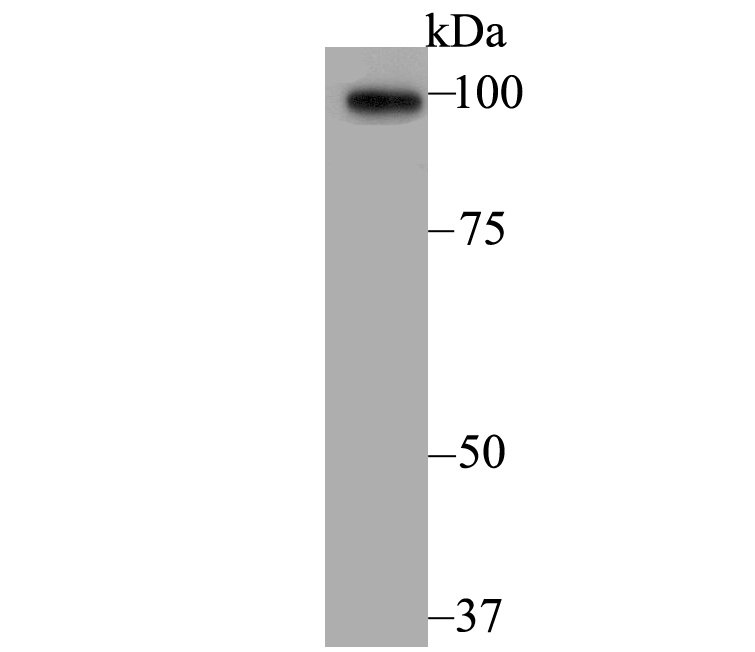
Fig1: Western blot analysis of Sarcomeric Alpha Actinin on mouse heart tissue lysate. Proteins were transferred to a PVDF membrane and blocked with 5% BSA in PBS for 1 hour at room temperature. The primary antibody was used at a 1:500 dilution in 5% BSA at room temperature for 2 hours. Goat Anti-Rabbit IgG - HRP Secondary Antibody (HA1001) at 1:5,000 dilution was used for 1 hour at room temperature.Application
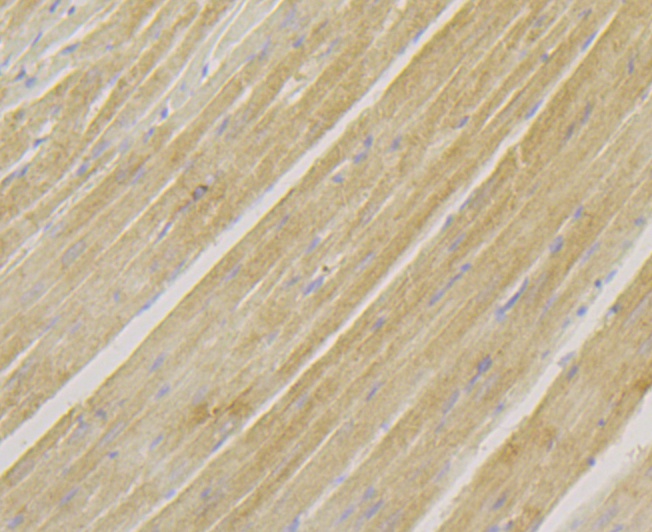
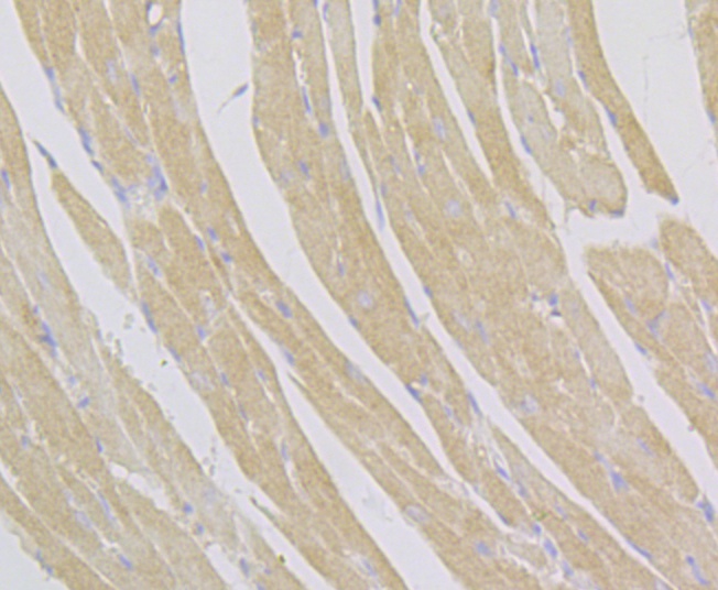
Fig2: Immunohistochemical analysis of paraffin-embedded rat heart tissue using anti-Sarcomeric Alpha Actinin antibody. The section was pre-treated using heat mediated antigen retrieval with Tris-EDTA buffer (pH 8.0-8.4) for 20 minutes. The tissues were blocked in 5% BSA for 30 minutes at room temperature, washed with ddH2O and PBS, and then probed with the antibody (ET7109-42) at 1/200 dilution, for 30 minutes at room temperature and detected using an HRP conjugated compact polymer system. DAB was used as the chrogen. Counter stained with hematoxylin and mounted with DPX.Application
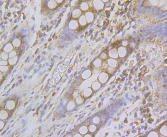
Fig3: Immunohistochemical analysis of paraffin-embedded human colon tissue using anti-Sarcomeric Alpha Actinin antibody. The section was pre-treated using heat mediated antigen retrieval with Tris-EDTA buffer (pH 8.0-8.4) for 20 minutes. The tissues were blocked in 5% BSA for 30 minutes at room temperature, washed with ddH2O and PBS, and then probed with the antibody (ET7109-42) at 1/200 dilution, for 30 minutes at room temperature and detected using an HRP conjugated compact polymer system. DAB was used as the chrogen. Counter stained with hematoxylin and mounted with DPX.Application
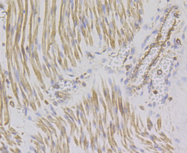
Fig4: Immunohistochemical analysis of paraffin-embedded human fetal skeletal muscle tissue using anti-Sarcomeric Alpha Actinin antibody. The section was pre-treated using heat mediated antigen retrieval with Tris-EDTA buffer (pH 8.0-8.4) for 20 minutes. The tissues were blocked in 5% BSA for 30 minutes at room temperature, washed with ddH2O and PBS, and then probed with the antibody (ET7109-42) at 1/200 dilution, for 30 minutes at room temperature and detected using an HRP conjugated compact polymer system. DAB was used as the chrogen. Counter stained with hematoxylin and mounted with DPX.Application
Fig5: Immunohistochemical analysis of paraffin-embedded mouse heart tissue using anti-Sarcomeric Alpha Actinin antibody. The section was pre-treated using heat mediated antigen retrieval with Tris-EDTA buffer (pH 8.0-8.4) for 20 minutes. The tissues were blocked in 5% BSA for 30 minutes at room temperature, washed with ddH2O and PBS, and then probed with the antibody (ET7109-42) at 1/200 dilution, for 30 minutes at room temperature and detected using an HRP conjugated compact polymer system. DAB was used as the chrogen. Counter stained with hematoxylin and mounted with DPX.| Product Name | Sarcomeric Alpha Actinin [JE45-22] |
|---|---|
| Antibody Type | Primary Antibodies |
| Antigen Alias | Actin binding protein antibody Actinin alpha 2 antibody ACTN 2 antibody ACTN2 antibody ACTN2_HUMAN antibody Alpha actinin 2 antibody Alpha actinin skeletal muscle antibody Alpha actinin skeletal muscle isoform 2 antibody Alpha-actinin skeletal muscle isoform 2 antibody Alpha-actinin-2 antibody CMD1AA antibody F actin cross linking protein antibody F-actin cross-linking protein antibody |
| Product description | This gene encodes a member of the alpha-actin binding protein gene family. The encoded protein is primarily expressed in skeletal muscle and functions as a structural component of sarcomeric Z line. This protein is involved in crosslinking actin containing thin filaments. An allelic polymorphism in this gene results in both coding and non-coding variants; the reference genome represents the coding allele. The non-functional allele of this gene is associated with elite athlete status. F-actin cross-linking protein which is thought to anchor actin to a variety of intracellular structures. This is a bundling protein. |
| Immunogen | Recombinant protein within human Sarcomeric Alpha Actinin aa 1-150. |
| Clonality | Monoclonal |
|---|---|
| Isotype | IgG |
| Host Species | Recombinant rabbit |
| Tested Applications | WBIPIHC |
| WB:1:500: IP:1:10-1:50: IHC:1:50-1:200: |
|
| Species Reactivity | HumanMouseRat |
| Concentration | 1mg/ml |
| Alternative Names | Actin binding protein antibody Actinin alpha 2 antibody ACTN 2 antibody ACTN2 antibody ACTN2_HUMAN antibody Alpha actinin 2 antibody Alpha actinin skeletal muscle antibody Alpha actinin skeletal muscle isoform 2 antibody Alpha-actinin skeletal muscle isoform 2 antibody Alpha-actinin-2 antibody CMD1AA antibody F actin cross linking protein antibody F-actin cross-linking protein antibody |
|---|---|
| Molecular Weight(MW) | 104 kDa |
| Cellular Localization | Cytoplasm. |
Application
Fig1: Western blot analysis of Sarcomeric Alpha Actinin on mouse heart tissue lysate. Proteins were transferred to a PVDF membrane and blocked with 5% BSA in PBS for 1 hour at room temperature. The primary antibody was used at a 1:500 dilution in 5% BSA at room temperature for 2 hours. Goat Anti-Rabbit IgG - HRP Secondary Antibody (HA1001) at 1:5,000 dilution was used for 1 hour at room temperature.
Application
Fig2: Immunohistochemical analysis of paraffin-embedded rat heart tissue using anti-Sarcomeric Alpha Actinin antibody. The section was pre-treated using heat mediated antigen retrieval with Tris-EDTA buffer (pH 8.0-8.4) for 20 minutes. The tissues were blocked in 5% BSA for 30 minutes at room temperature, washed with ddH2O and PBS, and then probed with the antibody (ET7109-42) at 1/200 dilution, for 30 minutes at room temperature and detected using an HRP conjugated compact polymer system. DAB was used as the chrogen. Counter stained with hematoxylin and mounted with DPX.
Application
Fig3: Immunohistochemical analysis of paraffin-embedded human colon tissue using anti-Sarcomeric Alpha Actinin antibody. The section was pre-treated using heat mediated antigen retrieval with Tris-EDTA buffer (pH 8.0-8.4) for 20 minutes. The tissues were blocked in 5% BSA for 30 minutes at room temperature, washed with ddH2O and PBS, and then probed with the antibody (ET7109-42) at 1/200 dilution, for 30 minutes at room temperature and detected using an HRP conjugated compact polymer system. DAB was used as the chrogen. Counter stained with hematoxylin and mounted with DPX.
Application
Fig4: Immunohistochemical analysis of paraffin-embedded human fetal skeletal muscle tissue using anti-Sarcomeric Alpha Actinin antibody. The section was pre-treated using heat mediated antigen retrieval with Tris-EDTA buffer (pH 8.0-8.4) for 20 minutes. The tissues were blocked in 5% BSA for 30 minutes at room temperature, washed with ddH2O and PBS, and then probed with the antibody (ET7109-42) at 1/200 dilution, for 30 minutes at room temperature and detected using an HRP conjugated compact polymer system. DAB was used as the chrogen. Counter stained with hematoxylin and mounted with DPX.
Application
Fig5: Immunohistochemical analysis of paraffin-embedded mouse heart tissue using anti-Sarcomeric Alpha Actinin antibody. The section was pre-treated using heat mediated antigen retrieval with Tris-EDTA buffer (pH 8.0-8.4) for 20 minutes. The tissues were blocked in 5% BSA for 30 minutes at room temperature, washed with ddH2O and PBS, and then probed with the antibody (ET7109-42) at 1/200 dilution, for 30 minutes at room temperature and detected using an HRP conjugated compact polymer system. DAB was used as the chrogen. Counter stained with hematoxylin and mounted with DPX.| Positive Control | Mouse heart tissue, rat heart tissue, human colon tissue, human fetal skeletal muscle tissue. |
|---|---|
| Application Notes | WB:1:500: IP:1:10-1:50: IHC:1:50-1:200: |
| Form | Liquid |
|---|---|
| Storage Instructions | Store at +4℃ after thawing. Aliquot store at -20℃. Avoid repeated freeze / thaw cycles. |
| Storage Buffer | 1*TBS (pH7.4), 1%BSA, 40%Glycerol. Preservative: 0.05% Sodium Azide. |


