Application
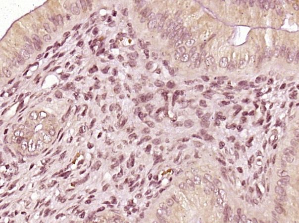
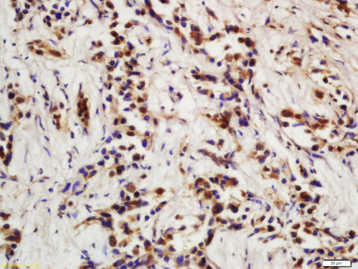
Fig1: Paraformaldehyde-fixed, paraffin embedded (Rat uterus); Antigen retrieval by boiling in sodium citrate buffer (pH6.0) for 15min; Block endogenous peroxidase by 3% hydrogen peroxide for 20 minutes; Blocking buffer (normal goat serum) at 37℃ for 30min; Antibody incubation with (P21) Polyclonal Antibody, Unconjugated (ER1906-07) at 1:400 overnight at 4℃, followed by operating according to SP Kit(Rabbit) (sp-0023) instructionsand DAB staining.Application
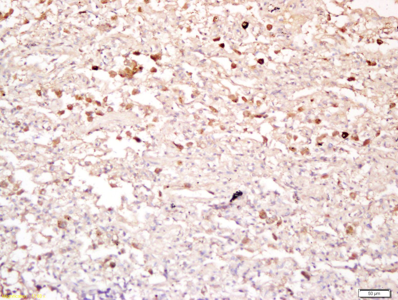
Fig2: Tissue/cell: human lung carcinoma; 4% Paraformaldehyde-fixed and paraffin-embedded; Antigen retrieval: citrate buffer ( 0.01M, pH 6.0 ), Boiling bathing for 15min; Block endogenous peroxidase by 3% Hydrogen peroxide for 30min; Blocking buffer (normal goat serum,C-0005) at 37℃ for 20 min; Incubation: Anti-P21 Polyclonal Antibody, Unconjugated(ER1906-07) 1:200, overnight at 4℃, followed by conjugation to the secondary antibody(SP-0023) and DAB(C-0010) stainingApplication
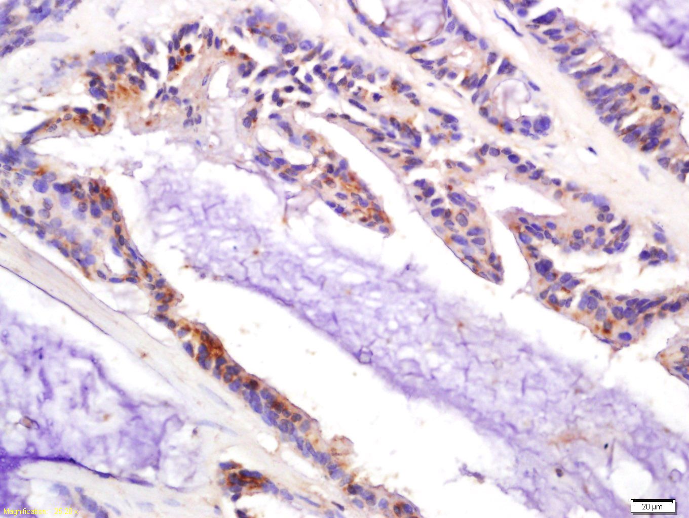
Fig3: Tissue/cell: human colon carcinoma; 4% Paraformaldehyde-fixed and paraffin-embedded; Antigen retrieval: citrate buffer ( 0.01M, pH 6.0 ), Boiling bathing for 15min; Block endogenous peroxidase by 3% Hydrogen peroxide for 30min; Blocking buffer (normal goat serum,C-0005) at 37℃ for 20 min; Incubation: Anti-P21 Polyclonal Antibody, Unconjugated(ER1906-07) 1:200, overnight at 4℃, followed by conjugation to the secondary antibody(SP-0023) and DAB(C-0010) stainingApplication
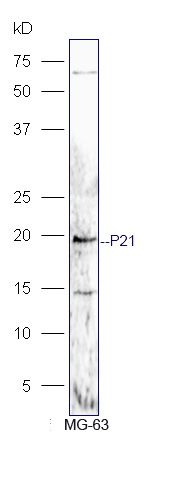
Fig4: Protein: MG-63(human) lysate at 40ug; Primary: rabbit Anti-P21 (ER1906-07) at 1:300; Secondary: HRP conjugated Goat-Anti-rabbit IgG(bs-0295G-HRP) at 1: 5000; Predicted band size: 18 kD Observed band size: 18 kDApplication
Fig5: Tissue/cell: human hepatoma tissue; 4% Paraformaldehyde-fixed and paraffin-embedded; Antigen retrieval: citrate buffer ( 0.01M, pH 6.0 ), Boiling bathing for 15min; Block endogenous peroxidase by 3% Hydrogen peroxide for 30min; Blocking buffer (normal goat serum,C-0005) at 37℃ for 20 min; Incubation: Anti-P21 Polyclonal Antibody, Unconjugated(ER1906-07) 1:200, overnight at 4℃, followed by conjugation to the secondary antibody(SP-0023) and DAB(C-0010) stainingApplication
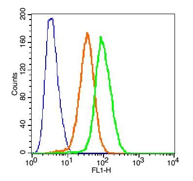
Fig6: Blank control(blue): Hep G2 Cells(fixed with 2% paraformaldehyde (10 min) and then permeabilized with ice-cold 90% methanol for 30 min on ice). Primary Antibody: Rabbit Anti-TNFR1/FITC Conjugated antibody (ER1906-07 /FITC), Dilution: 1μg in 100 μL 1X PBS containing 0.5% BSA; Isotype Control Antibody: Rabbit IgG/FITC(orange) ,used under the same conditions.| Product Name | CDKN1A/P21 |
|---|---|
| Antibody Type | Primary Antibodies |
| Antigen Alias | Activating Fragment 1 CAP20 Cation chloride cotransporter-interacting protein 1 CDK Interacting Protein 1 CDK-interacting protein 1 CDKI CDKN 1 CDKN1 CDKN1A CIP1 Cyclin Dependent Kinase Inhibitor 1A Cyclin-dependent kinase inhibitor 1 Cyclin-dependent kinase inhibitor 1A (P21) Cyclin-dependent kinase inhibitor 1A (p21, Cip1) DNA Synthesis Inhibitor MDA 6 MDA-6 MDA6 Melanoma Differentiation Associated Protein 6 Melanoma differentiation-associated protein 6 Melanoma differentiation-associated protein p21 P21 protein p21CIP1 p21WAF PIC1 SDI1 SLC12A9 WAF1 Wildtype p53 Activating Fragment 1 Wildtype p53-activated fragment 1 CDN1A_HUMAN. |
| Product description | May be the important intermediate by which p53/TP53 mediates its role as an inhibitor of cellular proliferation in response to DNA damage. Binds to and inhibits cyclin-dependent kinase activity, preventing phosphorylation of critical cyclin-dependent kinase substrates and blocking cell cycle progression. Functions in the nuclear localization and assembly of cyclin D-CDK4 complex and promotes its kinase activity towards RB1. At higher stoichiometric ratios, inhibits the kinase activity of the cyclin D-CDK4 complex. |
| Immunogen | KLH conjugated synthetic peptide derived from human P21 21-100/164 |
| Clonality | Polyclonal |
|---|---|
| Isotype | IgG |
| Host Species | Rabbit |
| Tested Applications | WBIHC-PFC |
| WB:1:500-2000: IHC-P:1:400-800: FC:1μg/Test: |
|
| Species Reactivity | HumanMouseRatDogRabbit |
| Concentration | 1mg/ml |
| Alternative Names | Activating Fragment 1
CAP20
Cation chloride cotransporter-interacting protein 1
CDK Interacting Protein 1
CDK-interacting protein 1
CDKI
CDKN 1
CDKN1
CDKN1A
CIP1
Cyclin Dependent Kinase Inhibitor 1A
Cyclin-dependent kinase inhibitor 1
Cyclin-dependent kinase inhibitor 1A (P21)
Cyclin-dependent kinase inhibitor 1A (p21 Cip1) DNA Synthesis Inhibitor MDA 6 MDA-6 MDA6 Melanoma Differentiation Associated Protein 6 Melanoma differentiation-associated protein 6 Melanoma differentiation-associated protein p21 P21 protein p21CIP1 p21WAF PIC1 SDI1 SLC12A9 WAF1 Wildtype p53 Activating Fragment 1 Wildtype p53-activated fragment 1 CDN1A_HUMAN. |
|---|---|
| Molecular Weight(MW) | 18 KDa |
| Cellular Localization | Cytoplasmic and Nuclear. |
| SwissProt ID | P38936 |
|---|
Application
Fig1: Paraformaldehyde-fixed, paraffin embedded (Rat uterus); Antigen retrieval by boiling in sodium citrate buffer (pH6.0) for 15min; Block endogenous peroxidase by 3% hydrogen peroxide for 20 minutes; Blocking buffer (normal goat serum) at 37℃ for 30min; Antibody incubation with (P21) Polyclonal Antibody, Unconjugated (ER1906-07) at 1:400 overnight at 4℃, followed by operating according to SP Kit(Rabbit) (sp-0023) instructionsand DAB staining.
Application
Fig2: Tissue/cell: human lung carcinoma; 4% Paraformaldehyde-fixed and paraffin-embedded; Antigen retrieval: citrate buffer ( 0.01M, pH 6.0 ), Boiling bathing for 15min; Block endogenous peroxidase by 3% Hydrogen peroxide for 30min; Blocking buffer (normal goat serum,C-0005) at 37℃ for 20 min; Incubation: Anti-P21 Polyclonal Antibody, Unconjugated(ER1906-07) 1:200, overnight at 4℃, followed by conjugation to the secondary antibody(SP-0023) and DAB(C-0010) staining
Application
Fig3: Tissue/cell: human colon carcinoma; 4% Paraformaldehyde-fixed and paraffin-embedded; Antigen retrieval: citrate buffer ( 0.01M, pH 6.0 ), Boiling bathing for 15min; Block endogenous peroxidase by 3% Hydrogen peroxide for 30min; Blocking buffer (normal goat serum,C-0005) at 37℃ for 20 min; Incubation: Anti-P21 Polyclonal Antibody, Unconjugated(ER1906-07) 1:200, overnight at 4℃, followed by conjugation to the secondary antibody(SP-0023) and DAB(C-0010) staining
Application
Fig4: Protein: MG-63(human) lysate at 40ug; Primary: rabbit Anti-P21 (ER1906-07) at 1:300; Secondary: HRP conjugated Goat-Anti-rabbit IgG(bs-0295G-HRP) at 1: 5000; Predicted band size: 18 kD Observed band size: 18 kD
Application
Fig5: Tissue/cell: human hepatoma tissue; 4% Paraformaldehyde-fixed and paraffin-embedded; Antigen retrieval: citrate buffer ( 0.01M, pH 6.0 ), Boiling bathing for 15min; Block endogenous peroxidase by 3% Hydrogen peroxide for 30min; Blocking buffer (normal goat serum,C-0005) at 37℃ for 20 min; Incubation: Anti-P21 Polyclonal Antibody, Unconjugated(ER1906-07) 1:200, overnight at 4℃, followed by conjugation to the secondary antibody(SP-0023) and DAB(C-0010) staining
Application
Fig6: Blank control(blue): Hep G2 Cells(fixed with 2% paraformaldehyde (10 min) and then permeabilized with ice-cold 90% methanol for 30 min on ice). Primary Antibody: Rabbit Anti-TNFR1/FITC Conjugated antibody (ER1906-07 /FITC), Dilution: 1μg in 100 μL 1X PBS containing 0.5% BSA; Isotype Control Antibody: Rabbit IgG/FITC(orange) ,used under the same conditions.| Positive Control | Expressed in all adult tissues, with 5-fold lower levels observed in the brain. |
|---|---|
| Application Notes | WB:1:500-2000: IHC-P:1:400-800: FC:1μg/Test: |
| Form | Liquid |
|---|---|
| Storage Instructions | Store at -20℃ for one year. Avoid repeated freeze/thaw cycles. The lyophilized antibody is stable at room temperature for at least one month and for greater than a year when kept at -20℃. When reconstituted in sterile pH 7.4 0.01M PBS or diluent of antibody the antibody is stable for at least two weeks at 2-4℃. |
| Storage Buffer | 0.01M TBS(pH7.4) with 1% BSA, 0.03% Proclin300 and 50% Glycerol. |


