Application
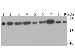
Fig1: Western blot analysis of SFRS3 on different lysates. Proteins were transferred to a PVDF membrane and blocked with 5% BSA in PBS for 1 hour at room temperature. The primary antibody was used at a 1:500 dilution in 5% BSA at room temperature for 2 hours. Goat Anti-Rabbit IgG - HRP Secondary Antibody (HA1001) at 1:5,000 dilution was used for 1 hour at room temperature. Positive control: Lane 1: Mouse testis tissue lysate Lane 2: Rat brain tissue lysate Lane 3: A431 cell lysate Lane 4: SH-SY-5Y cell lysate Lane 5: SiHa cell lysate Lane 6: 293 cell lysate Lane 7: Hela cell lysate Lane 8: HepG2 cell lysate Lane 9: A549 cell lysateApplication
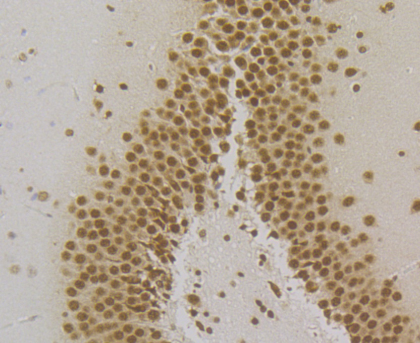
Fig2: Immunohistochemical analysis of paraffin-embedded rat brain tissue using anti-SFRS3 antibody. The section was pre-treated using heat mediated antigen retrieval with Tris-EDTA buffer (pH 8.0-8.4) for 20 minutes. The tissues were blocked in 5% BSA for 30 minutes at room temperature, washed with ddH2O and PBS, and then probed with the antibody (ET7109-47) at 1/200 dilution, for 30 minutes at room temperature and detected using an HRP conjugated compact polymer system. DAB was used as the chrogen. Counter stained with hematoxylin and mounted with DPX.Application
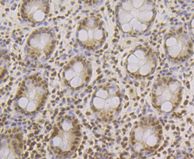
Fig3: Immunohistochemical analysis of paraffin-embedded human colon tissue using anti-SFRS3 antibody. The section was pre-treated using heat mediated antigen retrieval with Tris-EDTA buffer (pH 8.0-8.4) for 20 minutes. The tissues were blocked in 5% BSA for 30 minutes at room temperature, washed with ddH2O and PBS, and then probed with the antibody (ET7109-47) at 1/200 dilution, for 30 minutes at room temperature and detected using an HRP conjugated compact polymer system. DAB was used as the chrogen. Counter stained with hematoxylin and mounted with DPX.Application
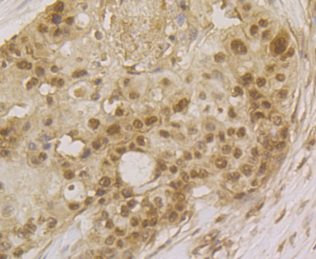
Fig4: Immunohistochemical analysis of paraffin-embedded human breast cancer tissue using anti-SFRS3 antibody. The section was pre-treated using heat mediated antigen retrieval with Tris-EDTA buffer (pH 8.0-8.4) for 20 minutes. The tissues were blocked in 5% BSA for 30 minutes at room temperature, washed with ddH2O and PBS, and then probed with the antibody (ET7109-47) at 1/200 dilution, for 30 minutes at room temperature and detected using an HRP conjugated compact polymer system. DAB was used as the chrogen. Counter stained with hematoxylin and mounted with DPX.Application
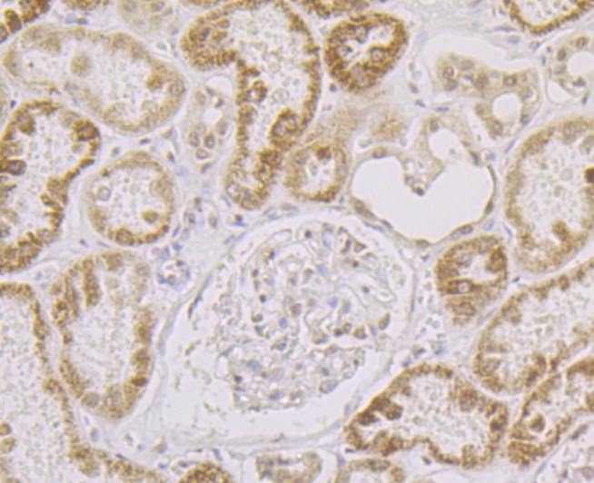
Fig5: Immunohistochemical analysis of paraffin-embedded human kidney tissue using anti-SFRS3 antibody. The section was pre-treated using heat mediated antigen retrieval with Tris-EDTA buffer (pH 8.0-8.4) for 20 minutes. The tissues were blocked in 5% BSA for 30 minutes at room temperature, washed with ddH2O and PBS, and then probed with the antibody (ET7109-47) at 1/200 dilution, for 30 minutes at room temperature and detected using an HRP conjugated compact polymer system. DAB was used as the chrogen. Counter stained with hematoxylin and mounted with DPX.Application
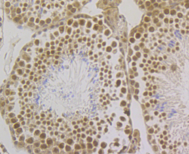
Fig6: Immunohistochemical analysis of paraffin-embedded mouse testis tissue using anti-SFRS3 antibody. The section was pre-treated using heat mediated antigen retrieval with Tris-EDTA buffer (pH 8.0-8.4) for 20 minutes. The tissues were blocked in 5% BSA for 30 minutes at room temperature, washed with ddH2O and PBS, and then probed with the antibody (ET7109-47) at 1/200 dilution, for 30 minutes at room temperature and detected using an HRP conjugated compact polymer system. DAB was used as the chrogen. Counter stained with hematoxylin and mounted with DPX.Application
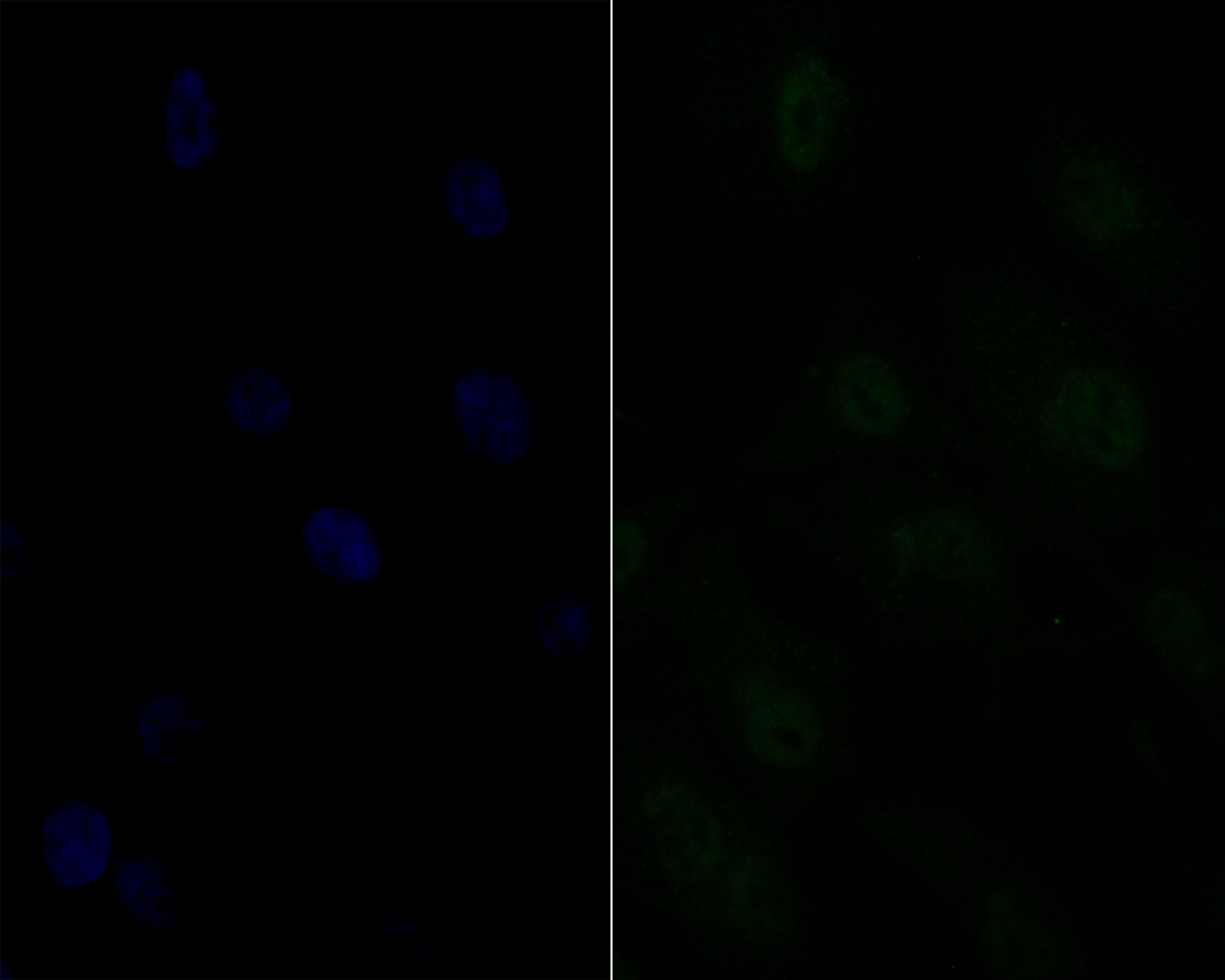
Fig7: Immunocytochemistry staining SFRS3 in MG-63 cells (green). Formalin fixed cells were permeabilized with 0.1% Triton X-100 in TBS for 10 minutes at room temperature and blocked with 1% Blocker BSA for 15 minutes at room temperature. Cells were probed with Ki67 polyclonal antibody at a dilution of 1:100 for 1 hour at room temperature, washed with PBS. Alexa Fluorc™ 488 Goat anti-Rabbit IgG was used as the secondary antibody at 1/100 dilution. The nuclear counter stain is DAPI (blue).| Product Name | SFRS3 [JE46-44] |
|---|---|
| Antibody Type | Primary Antibodies |
| Antigen Alias | arginine/serine-rich 3 antibody Pre mRNA splicing factor SRP20 antibody Pre-mRNA-splicing factor SRP20 antibody Serine/arginine-rich splicing factor 3 antibody Splicing factor antibody Splicing factor arginine/serine rich 20 kD antibody Splicing factor arginine/serine rich 3 antibody SRp20 antibody SRSF3 antibody SRSF3_HUMAN antibody X16 antibody |
| Product description | Splicing factor that specifically promotes exon-inclusion during alternative splicing. Interaction with YTHDC1, a RNA-binding protein that recognizes and binds N6-methyladenosine (m6A)-containing RNAs, promotes recruitment of SRSF3 to its mRNA-binding elements adjacent to m6A sites, leading to exon-inclusion during alternative splicing. Also functions as export adapter involved in mRNA nuclear export. Binds mRNA which is thought to be transferred to the NXF1-NXT1 heterodimer for export (TAP/NXF1 pathway); enhances NXF1-NXT1 RNA-binding activity. Involved in nuclear export of m6A-containing mRNAs via interaction with YTHDC1: interaction with YTHDC1 facilitates m6A-containing mRNA-binding to both SRSF3 and NXF1, promoting mRNA nuclear export (PubMed:28984244). RNA-binding is semi-sequence specific. |
| Immunogen | Full length recombinant protein. |
| Clonality | Monoclonal |
|---|---|
| Isotype | IgG |
| Host Species | Recombinant rabbit |
| Tested Applications | WBIHCICC |
| WB:1:500-1:2,000: ICC:1:50-1:200: IHC:1:100-1:500: |
|
| Species Reactivity | HumanMouseRat |
| Concentration | 1mg/ml |
| Alternative Names | arginine/serine-rich 3 antibody Pre mRNA splicing factor SRP20 antibody Pre-mRNA-splicing factor SRP20 antibody Serine/arginine-rich splicing factor 3 antibody Splicing factor antibody Splicing factor arginine/serine rich 20 kD antibody Splicing factor arginine/serine rich 3 antibody SRp20 antibody SRSF3 antibody SRSF3_HUMAN antibody X16 antibody |
|---|---|
| Molecular Weight(MW) | 19 kDa |
| Cellular Localization | Cytoplasm, Nucleus. |
Application
Fig1: Western blot analysis of SFRS3 on different lysates. Proteins were transferred to a PVDF membrane and blocked with 5% BSA in PBS for 1 hour at room temperature. The primary antibody was used at a 1:500 dilution in 5% BSA at room temperature for 2 hours. Goat Anti-Rabbit IgG - HRP Secondary Antibody (HA1001) at 1:5,000 dilution was used for 1 hour at room temperature. Positive control: Lane 1: Mouse testis tissue lysate Lane 2: Rat brain tissue lysate Lane 3: A431 cell lysate Lane 4: SH-SY-5Y cell lysate Lane 5: SiHa cell lysate Lane 6: 293 cell lysate Lane 7: Hela cell lysate Lane 8: HepG2 cell lysate Lane 9: A549 cell lysate
Application
Fig2: Immunohistochemical analysis of paraffin-embedded rat brain tissue using anti-SFRS3 antibody. The section was pre-treated using heat mediated antigen retrieval with Tris-EDTA buffer (pH 8.0-8.4) for 20 minutes. The tissues were blocked in 5% BSA for 30 minutes at room temperature, washed with ddH2O and PBS, and then probed with the antibody (ET7109-47) at 1/200 dilution, for 30 minutes at room temperature and detected using an HRP conjugated compact polymer system. DAB was used as the chrogen. Counter stained with hematoxylin and mounted with DPX.
Application
Fig3: Immunohistochemical analysis of paraffin-embedded human colon tissue using anti-SFRS3 antibody. The section was pre-treated using heat mediated antigen retrieval with Tris-EDTA buffer (pH 8.0-8.4) for 20 minutes. The tissues were blocked in 5% BSA for 30 minutes at room temperature, washed with ddH2O and PBS, and then probed with the antibody (ET7109-47) at 1/200 dilution, for 30 minutes at room temperature and detected using an HRP conjugated compact polymer system. DAB was used as the chrogen. Counter stained with hematoxylin and mounted with DPX.
Application
Fig4: Immunohistochemical analysis of paraffin-embedded human breast cancer tissue using anti-SFRS3 antibody. The section was pre-treated using heat mediated antigen retrieval with Tris-EDTA buffer (pH 8.0-8.4) for 20 minutes. The tissues were blocked in 5% BSA for 30 minutes at room temperature, washed with ddH2O and PBS, and then probed with the antibody (ET7109-47) at 1/200 dilution, for 30 minutes at room temperature and detected using an HRP conjugated compact polymer system. DAB was used as the chrogen. Counter stained with hematoxylin and mounted with DPX.
Application
Fig5: Immunohistochemical analysis of paraffin-embedded human kidney tissue using anti-SFRS3 antibody. The section was pre-treated using heat mediated antigen retrieval with Tris-EDTA buffer (pH 8.0-8.4) for 20 minutes. The tissues were blocked in 5% BSA for 30 minutes at room temperature, washed with ddH2O and PBS, and then probed with the antibody (ET7109-47) at 1/200 dilution, for 30 minutes at room temperature and detected using an HRP conjugated compact polymer system. DAB was used as the chrogen. Counter stained with hematoxylin and mounted with DPX.
Application
Fig6: Immunohistochemical analysis of paraffin-embedded mouse testis tissue using anti-SFRS3 antibody. The section was pre-treated using heat mediated antigen retrieval with Tris-EDTA buffer (pH 8.0-8.4) for 20 minutes. The tissues were blocked in 5% BSA for 30 minutes at room temperature, washed with ddH2O and PBS, and then probed with the antibody (ET7109-47) at 1/200 dilution, for 30 minutes at room temperature and detected using an HRP conjugated compact polymer system. DAB was used as the chrogen. Counter stained with hematoxylin and mounted with DPX.
Application
Fig7: Immunocytochemistry staining SFRS3 in MG-63 cells (green). Formalin fixed cells were permeabilized with 0.1% Triton X-100 in TBS for 10 minutes at room temperature and blocked with 1% Blocker BSA for 15 minutes at room temperature. Cells were probed with Ki67 polyclonal antibody at a dilution of 1:100 for 1 hour at room temperature, washed with PBS. Alexa Fluorc™ 488 Goat anti-Rabbit IgG was used as the secondary antibody at 1/100 dilution. The nuclear counter stain is DAPI (blue).| Positive Control | Mouse testis tissue, rat brain tissue, A431, SH-SY-5Y, SiHa, 293, Hela, HepG2, A549, MG-63. |
|---|---|
| Application Notes | WB:1:500-1:2,000: ICC:1:50-1:200: IHC:1:100-1:500: |
| Form | Liquid |
|---|---|
| Storage Instructions | Store at +4℃ after thawing. Aliquot store at -20℃. Avoid repeated freeze / thaw cycles. |
| Storage Buffer | 1*TBS (pH7.4), 1%BSA, 40%Glycerol. Preservative: 0.05% Sodium Azide. |


