Application
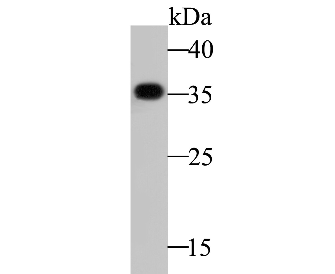
Fig1: Western blot analysis of GNB2 on MCF-7 cell lysate. Proteins were transferred to a PVDF membrane and blocked with 5% BSA in PBS for 1 hour at room temperature. The primary antibody (ET7109-53, 1/500) was used in 5% BSA at room temperature for 2 hours. Goat Anti-Rabbit IgG - HRP Secondary Antibody (HA1001) at 1:5,000 dilution was used for 1 hour at room temperature.Application
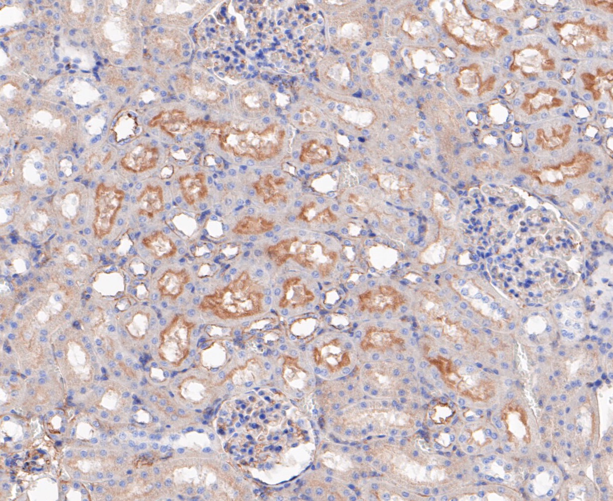
Fig2: Immunohistochemical analysis of paraffin-embedded rat kidney tissue using anti-GNB2 antibody. The section was pre-treated using heat mediated antigen retrieval with sodium citrate buffer (pH 6.0) for 20 minutes. The tissues were blocked in 5% BSA for 30 minutes at room temperature, washed with ddH2O and PBS, and then probed with the primary antibody (ET7109-53, 1/200) for 30 minutes at room temperature. The detection was performed using an HRP conjugated compact polymer system. DAB was used as the chromogen. Tissues were counterstained with hematoxylin and mounted with DPX.Application
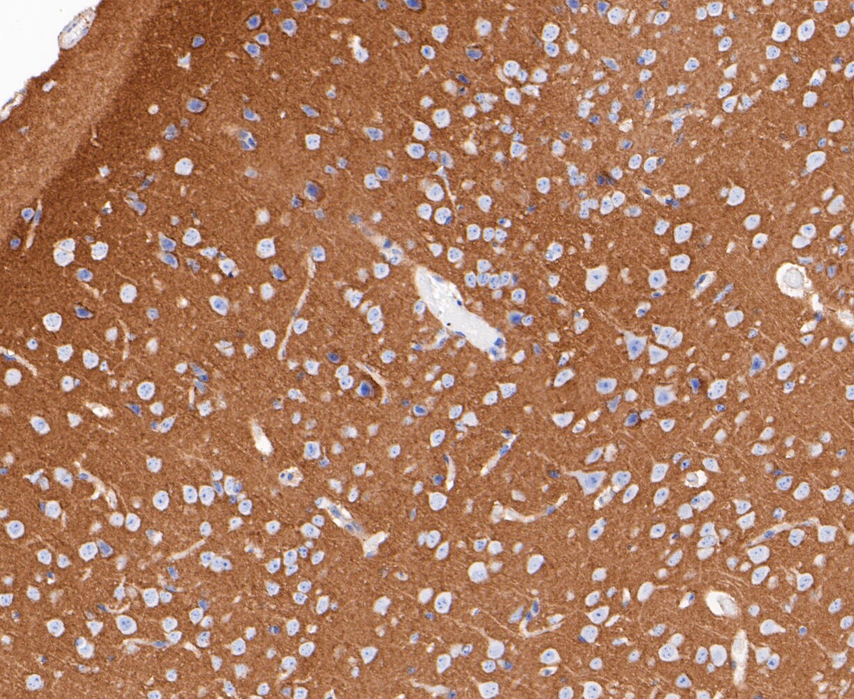
Fig3: Immunohistochemical analysis of paraffin-embedded mouse brain tissue using anti-GNB2 antibody. The section was pre-treated using heat mediated antigen retrieval with sodium citrate buffer (pH 6.0) for 20 minutes. The tissues were blocked in 5% BSA for 30 minutes at room temperature, washed with ddH2O and PBS, and then probed with the primary antibody (ET7109-53, 1/200) for 30 minutes at room temperature. The detection was performed using an HRP conjugated compact polymer system. DAB was used as the chromogen. Tissues were counterstained with hematoxylin and mounted with DPX.Application
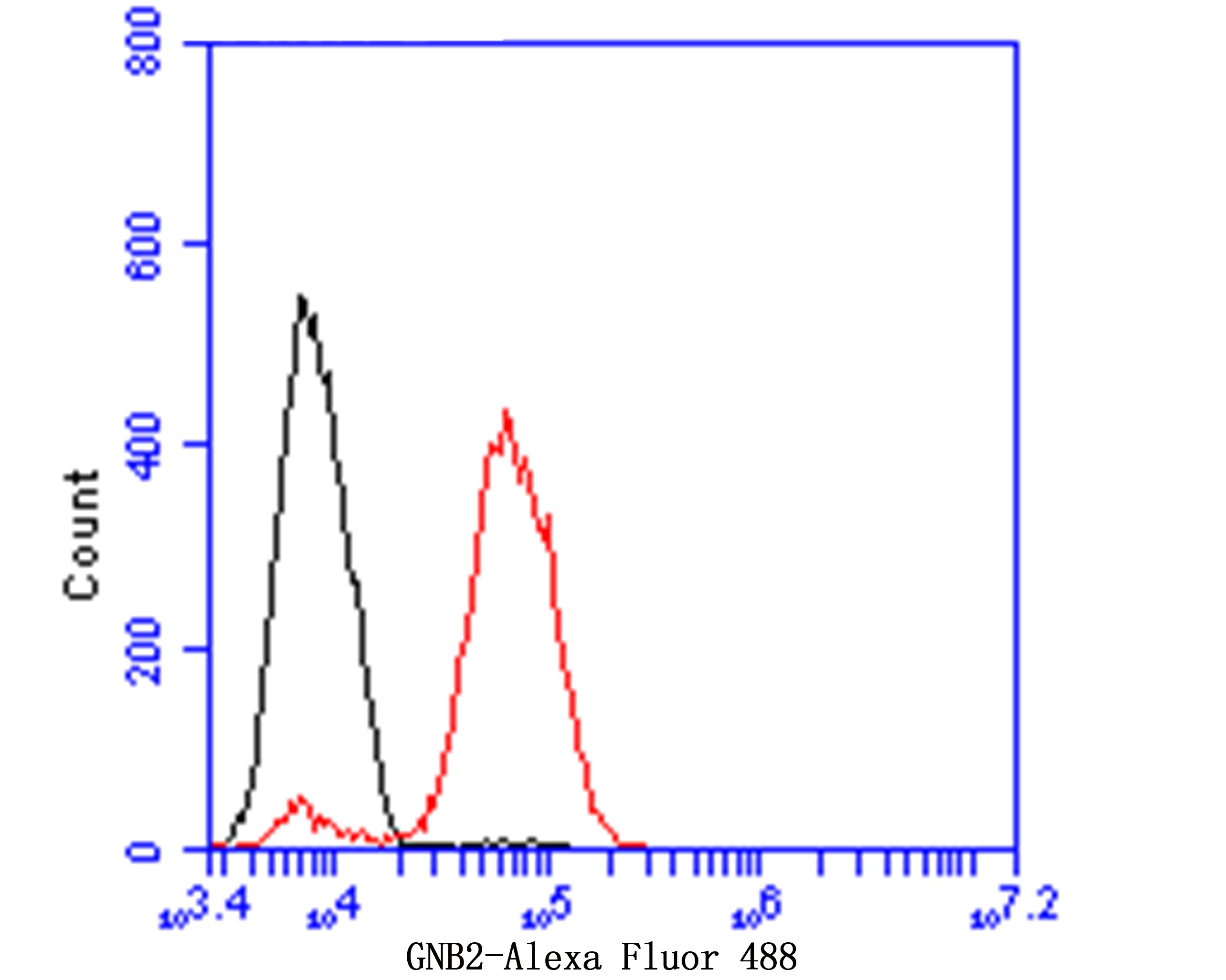
Fig4: Flow cytometric analysis of GNB2 was done on MCF-7 cells. The cells were fixed, permeabilized and stained with GNB2 antibody at 1/100 dilution (red) compared with an unlabelled control (cells without incubation with primary antibody; black). After incubation of the primary antibody on room temperature for an hour, the cells was stained with a Alexa Fluor™ 488-conjugated goat anti-rabbit IgG Secondary antibody at 1/500 dilution for 30 minutes.| Product Name | GNB2 [JE47-16] |
|---|---|
| Antibody Type | Primary Antibodies |
| Antigen Alias | G protein beta 2 subunit antibody G protein subunit beta 2 antibody G protein subunit beta-2 antibody GBB2_HUMAN antibody Gnb2 antibody Gnb2l1 antibody Guanine nucleotide binding protein beta 2 subunit antibody Guanine nucleotide binding protein G I G S G T beta 2 subunit 2 antibody Guanine nucleotide binding protein G protein beta polypeptide 2 antibody Guanine nucleotide-binding protein G(I)/G(S)/G(T) subunit beta-2 antibody OTTHUMP00000174601 antibody OTTHUMP00000174602 antibody RACK1 antibody Receptor for activated C kinase antibody Receptor of activated protein kinase C 1 antibody Signal transducing guanine nucleotide binding regulatory protein beta antibody Transducin beta chain 2 antibody |
| Product description | Heterotrimeric G proteins function to relay information from cell surface receptors to intracellular effectors. Each of a very broad range of receptors specifically detects an extracellular stimulus (i.e., a photon, pheromone, odorant, hormone or neurotransmitter), while the effectors (e.g., adenyl cyclase), which act to generate one or more intracellular messengers, are less numerous. Each subunit of the G protein complex is encoded by a member of one of three corresponding gene families (α, β, γ). In mammals, there are five different members of the β-subunit family. The β subunits of the G proteins are important regulators of G protein a subunits as well as of certain signal transduction receptors and effectors. In contrast to G β1-4, which are at least 83% homologous, G β5 is only 50% homologous to the other β subunits. Human G β5 is expressed at high levels in brain, pancreas, kidney, and heart. |
| Immunogen | Synthetic peptide within N terminal human GNB2. |
| Clonality | Monoclonal |
|---|---|
| Isotype | IgG |
| Host Species | Recombinant rabbit |
| Tested Applications | WBIHCFC |
| WB:1:500-1:1,000: IHC:1:50-1:200: FC:1:50-1:100: |
|
| Species Reactivity | HumanMouseRat |
| Concentration | 1mg/ml |
| Alternative Names | G protein beta 2 subunit antibody G protein subunit beta 2 antibody G protein subunit beta-2 antibody GBB2_HUMAN antibody Gnb2 antibody Gnb2l1 antibody Guanine nucleotide binding protein beta 2 subunit antibody Guanine nucleotide binding protein G I G S G T beta 2 subunit 2 antibody Guanine nucleotide binding protein G protein beta polypeptide 2 antibody Guanine nucleotide-binding protein G(I)/G(S)/G(T) subunit beta-2 antibody OTTHUMP00000174601 antibody OTTHUMP00000174602 antibody RACK1 antibody Receptor for activated C kinase antibody Receptor of activated protein kinase C 1 antibody Signal transducing guanine nucleotide binding regulatory protein beta antibody Transducin beta chain 2 antibody |
|---|---|
| Molecular Weight(MW) | 37 kDa |
| Cellular Localization | Perinuclear region. |
Application
Fig1: Western blot analysis of GNB2 on MCF-7 cell lysate. Proteins were transferred to a PVDF membrane and blocked with 5% BSA in PBS for 1 hour at room temperature. The primary antibody (ET7109-53, 1/500) was used in 5% BSA at room temperature for 2 hours. Goat Anti-Rabbit IgG - HRP Secondary Antibody (HA1001) at 1:5,000 dilution was used for 1 hour at room temperature.
Application
Fig2: Immunohistochemical analysis of paraffin-embedded rat kidney tissue using anti-GNB2 antibody. The section was pre-treated using heat mediated antigen retrieval with sodium citrate buffer (pH 6.0) for 20 minutes. The tissues were blocked in 5% BSA for 30 minutes at room temperature, washed with ddH2O and PBS, and then probed with the primary antibody (ET7109-53, 1/200) for 30 minutes at room temperature. The detection was performed using an HRP conjugated compact polymer system. DAB was used as the chromogen. Tissues were counterstained with hematoxylin and mounted with DPX.
Application
Fig3: Immunohistochemical analysis of paraffin-embedded mouse brain tissue using anti-GNB2 antibody. The section was pre-treated using heat mediated antigen retrieval with sodium citrate buffer (pH 6.0) for 20 minutes. The tissues were blocked in 5% BSA for 30 minutes at room temperature, washed with ddH2O and PBS, and then probed with the primary antibody (ET7109-53, 1/200) for 30 minutes at room temperature. The detection was performed using an HRP conjugated compact polymer system. DAB was used as the chromogen. Tissues were counterstained with hematoxylin and mounted with DPX.
Application
Fig4: Flow cytometric analysis of GNB2 was done on MCF-7 cells. The cells were fixed, permeabilized and stained with GNB2 antibody at 1/100 dilution (red) compared with an unlabelled control (cells without incubation with primary antibody; black). After incubation of the primary antibody on room temperature for an hour, the cells was stained with a Alexa Fluor™ 488-conjugated goat anti-rabbit IgG Secondary antibody at 1/500 dilution for 30 minutes.| Positive Control | MCF-7, rat kidney tissue, mouse brain tissue. |
|---|---|
| Application Notes | WB:1:500-1:1,000: IHC:1:50-1:200: FC:1:50-1:100: |
| Form | Liquid |
|---|---|
| Storage Instructions | Store at +4℃ after thawing. Aliquot store at -20℃. Avoid repeated freeze / thaw cycles. |
| Storage Buffer | 1*TBS (pH7.4), 1%BSA, 50%Glycerol. Preservative: 0.05% Sodium Azide. |


