Application
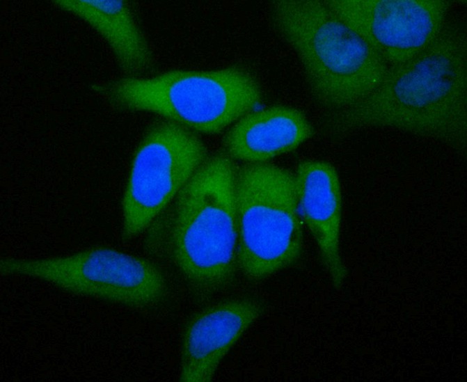
Fig1: ICC staining of GCDFP 15 in Hela cells (green). Formalin fixed cells were permeabilized with 0.1% Triton X-100 in TBS for 10 minutes at room temperature and blocked with 1% Blocker BSA for 15 minutes at room temperature. Cells were probed with the primary antibody (ET7109-80, 1/50) for 1 hour at room temperature, washed with PBS. Alexa Fluor®488 Goat anti-Rabbit IgG was used as the secondary antibody at 1/100 dilution. The nuclear counter stain is DAPI (blue).Application
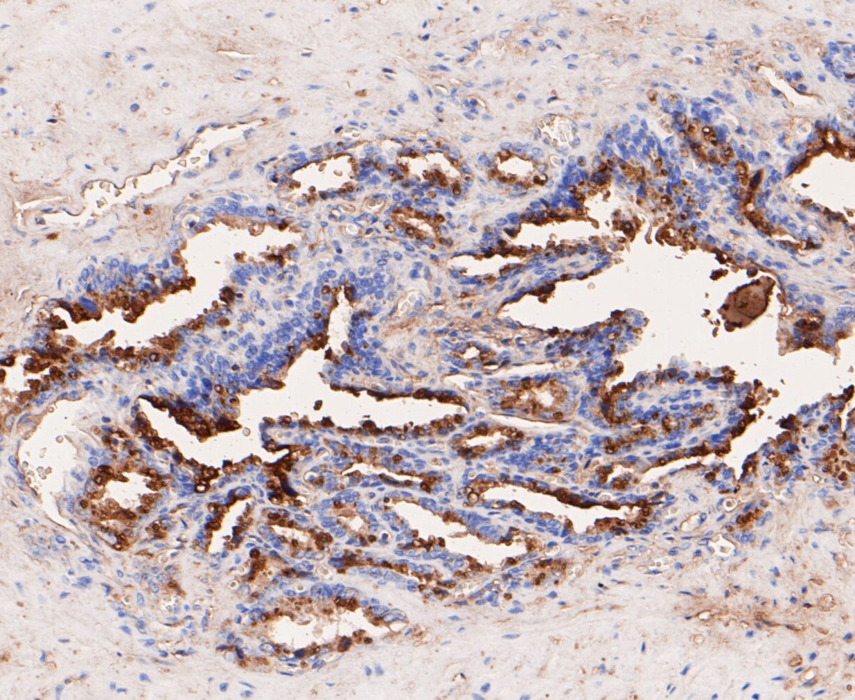
Fig2: Immunohistochemical analysis of paraffin-embedded human seminal vesicle tissue using anti-GCDFP 15 antibody. The section was pre-treated using heat mediated antigen retrieval with Tris-EDTA buffer (pH 8.0-8.4) for 20 minutes.The tissues were blocked in 5% BSA for 30 minutes at room temperature, washed with ddH2O and PBS, and then probed with the primary antibody (ET7109-80, 1/50) for 30 minutes at room temperature. The detection was performed using an HRP conjugated compact polymer system. DAB was used as the chromogen. Tissues were counterstained with hematoxylin and mounted with DPX.Application
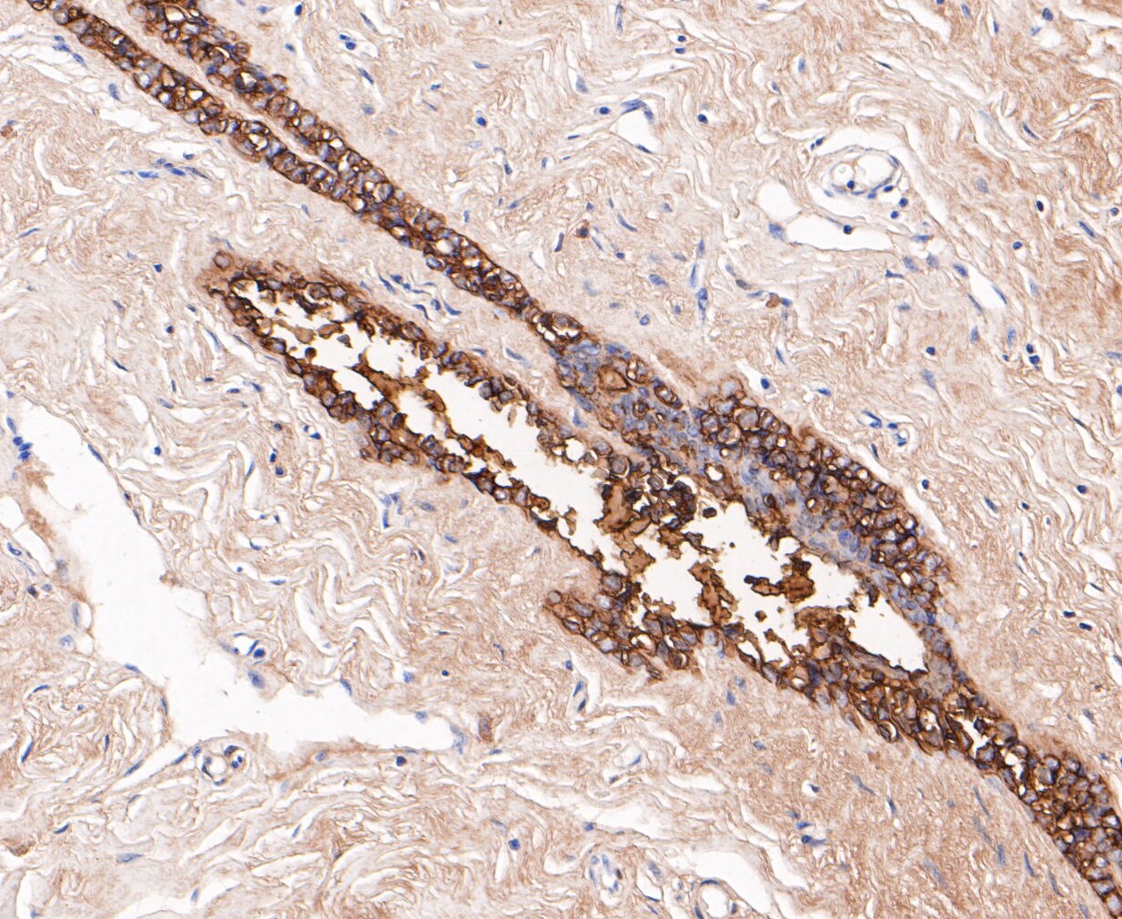
Fig3: Immunohistochemical analysis of paraffin-embedded human breast tissue using anti-GCDFP 15 antibody. The section was pre-treated using heat mediated antigen retrieval with Tris-EDTA buffer (pH 8.0-8.4) for 20 minutes.The tissues were blocked in 5% BSA for 30 minutes at room temperature, washed with ddH2O and PBS, and then probed with the primary antibody (ET7109-80, 1/200) for 30 minutes at room temperature. The detection was performed using an HRP conjugated compact polymer system. DAB was used as the chromogen. Tissues were counterstained with hematoxylin and mounted with DPX.Application
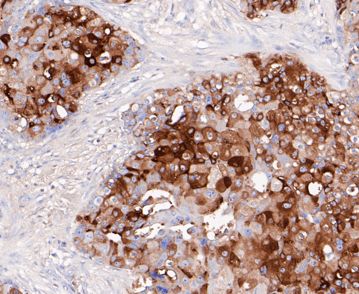
Fig4: Immunohistochemical analysis of paraffin-embedded human breast cancer tissue using anti-GCDFP 15 antibody. The section was pre-treated using heat mediated antigen retrieval with Tris-EDTA buffer (pH 8.0-8.4) for 20 minutes.The tissues were blocked in 5% BSA for 30 minutes at room temperature, washed with ddH2O and PBS, and then probed with the primary antibody (ET7109-80, 1/200) for 30 minutes at room temperature. The detection was performed using an HRP conjugated compact polymer system. DAB was used as the chromogen. Tissues were counterstained with hematoxylin and mounted with DPX.| Product Name | GCDFP 15 [JE48-53] |
|---|---|
| Antibody Type | Primary Antibodies |
| Antigen Alias | BRST-2 antibody BRST2 antibody GCDFP-15 antibody GCDGP15 antibody gp17 antibody GPIP4 antibody Gross cystic disease fluid protein 15 antibody PIP antibody PIP_HUMAN antibody Prolactin induced protein antibody Prolactin inducible protein precursor antibody Prolactin-induced protein antibody Prolactin-inducible protein antibody SABP antibody Secretory actin binding protein antibody Secretory actin-binding protein antibody |
| Product description | Gross cystic disease fluid protein 15 (GCDFP-15) is a major protein component of benign breast gross cysts. It is a known marker of breast cancer, as it is found in approximately 50% of all breast cancer specimens. GCDFP-15, also known as PIP, for prolactin inducible protein, is a prolactin and androgen controlled protein. It is detectable in saliva, tears, sweat, seminal plasma, submucosal glands of the lung and amniotic fluid. PIP, the gene encoding GCDFP-15 is expressed in exocrine glands and, in pathologic conditions, in breast cysts and breast cancers exhibiting apocrine features. The PIP gene maps to the long arm of chromosome 7, a region frequently altered in mammary tumors. |
| Immunogen | Synthetic peptide within N-terminal human GCDFP 15. |
| Clonality | Monoclonal |
|---|---|
| Isotype | IgG |
| Host Species | Recombinant rabbit |
| Tested Applications | ICCIHCWB |
| WB:1:500: ICC:1:50: IHC:1:50-1:200: |
|
| Species Reactivity | Human |
| Concentration | 1mg/ml |
| Alternative Names | BRST-2 antibody
BRST2 antibody
GCDFP-15 antibody GCDGP15 antibody gp17 antibody GPIP4 antibody Gross cystic disease fluid protein 15 antibody PIP antibody PIP_HUMAN antibody Prolactin induced protein antibody Prolactin inducible protein precursor antibody Prolactin-induced protein antibody Prolactin-inducible protein antibody SABP antibody Secretory actin binding protein antibody Secretory actin-binding protein antibody |
|---|---|
| Molecular Weight(MW) | 17 kDa |
| Cellular Localization | Secreted. |
| SwissProt ID | P12273 |
|---|
Application
Fig1: ICC staining of GCDFP 15 in Hela cells (green). Formalin fixed cells were permeabilized with 0.1% Triton X-100 in TBS for 10 minutes at room temperature and blocked with 1% Blocker BSA for 15 minutes at room temperature. Cells were probed with the primary antibody (ET7109-80, 1/50) for 1 hour at room temperature, washed with PBS. Alexa Fluor®488 Goat anti-Rabbit IgG was used as the secondary antibody at 1/100 dilution. The nuclear counter stain is DAPI (blue).
Application
Fig2: Immunohistochemical analysis of paraffin-embedded human seminal vesicle tissue using anti-GCDFP 15 antibody. The section was pre-treated using heat mediated antigen retrieval with Tris-EDTA buffer (pH 8.0-8.4) for 20 minutes.The tissues were blocked in 5% BSA for 30 minutes at room temperature, washed with ddH2O and PBS, and then probed with the primary antibody (ET7109-80, 1/50) for 30 minutes at room temperature. The detection was performed using an HRP conjugated compact polymer system. DAB was used as the chromogen. Tissues were counterstained with hematoxylin and mounted with DPX.
Application
Fig3: Immunohistochemical analysis of paraffin-embedded human breast tissue using anti-GCDFP 15 antibody. The section was pre-treated using heat mediated antigen retrieval with Tris-EDTA buffer (pH 8.0-8.4) for 20 minutes.The tissues were blocked in 5% BSA for 30 minutes at room temperature, washed with ddH2O and PBS, and then probed with the primary antibody (ET7109-80, 1/200) for 30 minutes at room temperature. The detection was performed using an HRP conjugated compact polymer system. DAB was used as the chromogen. Tissues were counterstained with hematoxylin and mounted with DPX.
Application
Fig4: Immunohistochemical analysis of paraffin-embedded human breast cancer tissue using anti-GCDFP 15 antibody. The section was pre-treated using heat mediated antigen retrieval with Tris-EDTA buffer (pH 8.0-8.4) for 20 minutes.The tissues were blocked in 5% BSA for 30 minutes at room temperature, washed with ddH2O and PBS, and then probed with the primary antibody (ET7109-80, 1/200) for 30 minutes at room temperature. The detection was performed using an HRP conjugated compact polymer system. DAB was used as the chromogen. Tissues were counterstained with hematoxylin and mounted with DPX.| Positive Control | Hela, human seminal vesicle tissue, human breast tissue, human breast cancer tissue. |
|---|---|
| Application Notes | WB:1:500: ICC:1:50: IHC:1:50-1:200: |
| Form | Liquid |
|---|---|
| Storage Instructions | Store at +4℃ after thawing. Aliquot store at -20℃. Avoid repeated freeze / thaw cycles. |
| Storage Buffer | 1*TBS (pH7.4), 1%BSA, 50%Glycerol. Preservative: 0.05% Sodium Azide. |


