Application
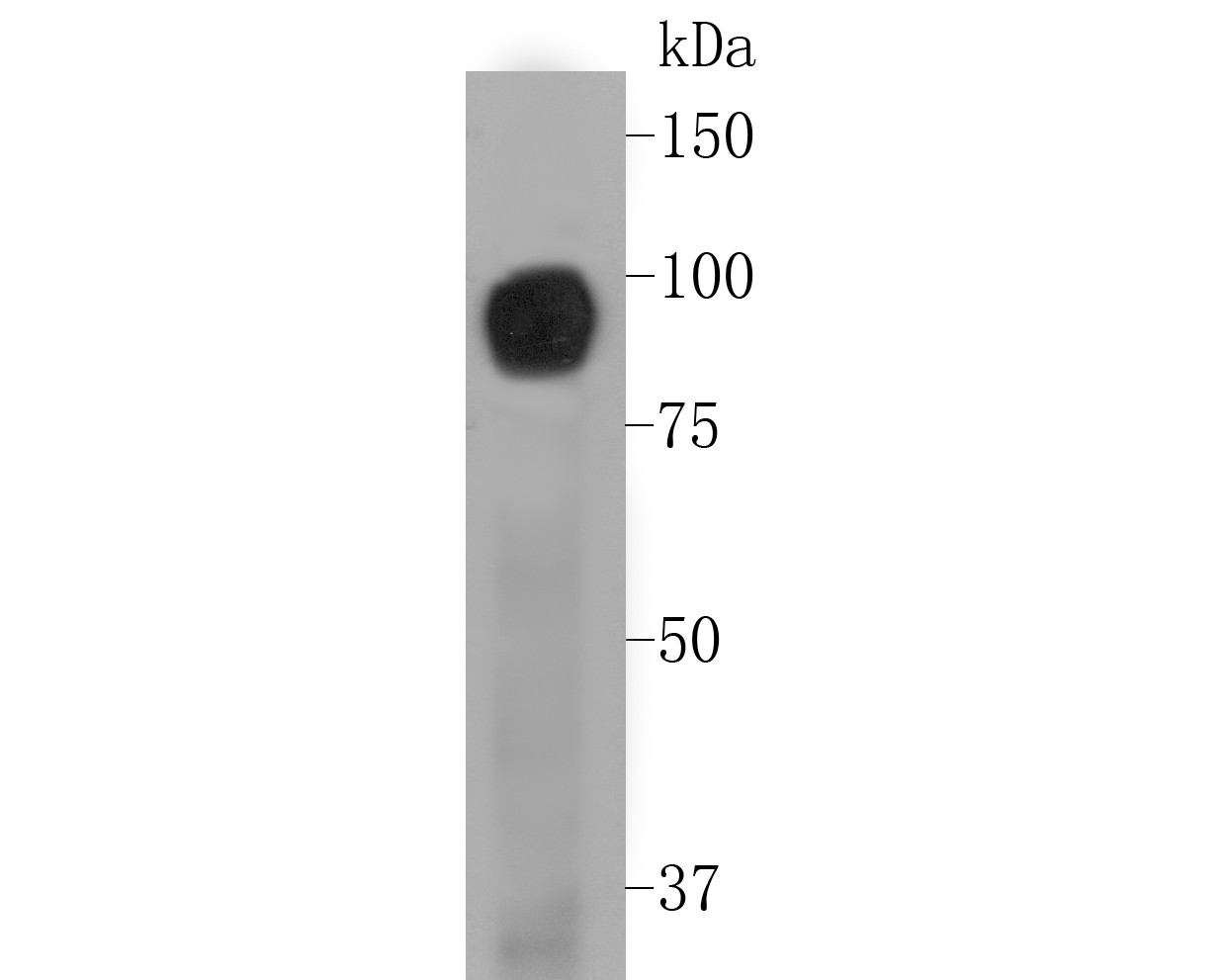
Fig1: Western blot analysis of CD239 on A431 cell lysate. Proteins were transferred to a PVDF membrane and blocked with 5% BSA in PBS for 1 hour at room temperature. The primary antibody (ET7110-04, 1/500) was used in 5% BSA at room temperature for 2 hours. Goat Anti-Rabbit IgG - HRP Secondary Antibody (HA1001) at 1:5,000 dilution was used for 1 hour at room temperature.Application
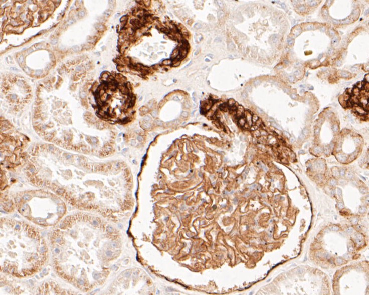
Fig2: Immunohistochemical analysis of paraffin-embedded human kidney tissue using anti-CD239 antibody. The section was pre-treated using heat mediated antigen retrieval with Tris-EDTA buffer (pH 8.0-8.4) for 20 minutes.The tissues were blocked in 5% BSA for 30 minutes at room temperature, washed with ddH2O and PBS, and then probed with the primary antibody (ET7110-04, 1/200) for 30 minutes at room temperature. The detection was performed using an HRP conjugated compact polymer system. DAB was used as the chromogen. Tissues were counterstained with hematoxylin and mounted with DPX.Application
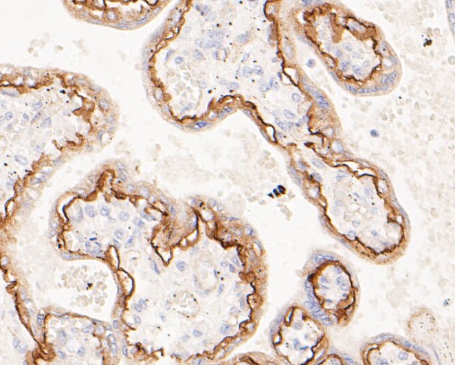
Fig3: Immunohistochemical analysis of paraffin-embedded human placenta tissue using anti-CD239 antibody. The section was pre-treated using heat mediated antigen retrieval with Tris-EDTA buffer (pH 8.0-8.4) for 20 minutes.The tissues were blocked in 5% BSA for 30 minutes at room temperature, washed with ddH2O and PBS, and then probed with the primary antibody (ET7110-04, 1/200) for 30 minutes at room temperature. The detection was performed using an HRP conjugated compact polymer system. DAB was used as the chromogen. Tissues were counterstained with hematoxylin and mounted with DPX.Application
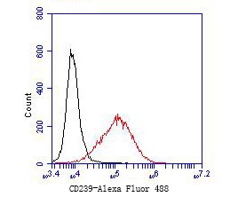
Fig4: Flow cytometric analysis of CD239 was done on JAR cells. The cells were fixed, permeabilized and stained with the primary antibody (ET7110-04, 1/50) (red). After incubation of the primary antibody at room temperature for an hour, the cells were stained with a Alexa Fluor 488-conjugated Goat anti-Rabbit IgG Secondary antibody at 1/1000 dilution for 30 minutes.Unlabelled sample was used as a control (cells without incubation with primary antibody; black).| Product Name | CD239 [JE51-07] |
|---|---|
| Antibody Type | Primary Antibodies |
| Antigen Alias | Antigen identified by monoclonal antibody F8 antibody AU antibody Auberger B antigen antibody B CAM cell surface glycoprotein antibody B cell adhesion molecule antibody B-CAM cell surface glycoprotein antibody Basal cell adhesion molecule (Lu and Au blood groups) antibody Basal cell adhesion molecule (Lutheran blood group) antibody Basal cell adhesion molecule antibody Basal cell adhesion molecule Lu and Au blood groups antibody Basal cell adhesion molecule Lutheran blood group antibody Bcam antibody BCAM_HUMAN antibody CD239 antibody CD239 antigen antibody F8/G253 antigen antibody Glycoprotein 95kDa antibody LU antibody Lutheran antibody Lutheran antigen antibody Lutheran blood group (Auberger b antigen included) antibody Lutheran blood group Auberger b antigen included antibody Lutheran blood group glycoprotein antibody MSK19 antibody |
| Product description | Basal cell adhesion molecule, also known as Lutheran antigen, is a plasma membrane glycoprotein that in humans is encoded by the BCAM gene. BCAM has also recently been designated CD239 (cluster of differentiation 239). Lutheran blood group glycoprotein is a member of the immunoglobulin superfamily and a receptor for the extracellular matrix protein, laminin. The protein contains five, N-terminus, extracellular immunoglobulin domains, a single transmembrane domain, and a short, C-terminal cytoplasmic tail. This protein may play a role in epithelial cell cancer and in vaso-occlusion of red blood cells in sickle cell disease. Polymorphisms in this gene define some of the antigens in the Lutheran system and also the Auberger system. Inactivating variants of this gene result in the recessive Lutheran null phenotype, Lu(a-b-), of the Lutheran blood group. Two transcript variants encoding different isoforms have been found for this gene. BCAM has been shown to interact with Laminin, alpha 5. |
| Immunogen | Recombinant protein corresponding to C-terminal human CD239 |
| Clonality | Monoclonal |
|---|---|
| Isotype | IgG |
| Host Species | Recombinant rabbit |
| Tested Applications | WBIHC-PFC |
| WB:1:500-1:2,000: IHC-P:1:100-1:500: FC:1:50-1:100: |
|
| Species Reactivity | Human |
| Concentration | 1mg/ml |
| Alternative Names | Antigen identified by monoclonal antibody F8 antibody
AU antibody
Auberger B antigen antibody B CAM cell surface glycoprotein antibody B cell adhesion molecule antibody B-CAM cell surface glycoprotein antibody Basal cell adhesion molecule (Lu and Au blood groups) antibody Basal cell adhesion molecule (Lutheran blood group) antibody Basal cell adhesion molecule antibody Basal cell adhesion molecule Lu and Au blood groups antibody Basal cell adhesion molecule Lutheran blood group antibody Bcam antibody BCAM_HUMAN antibody CD239 antibody CD239 antigen antibody F8/G253 antigen antibody Glycoprotein 95kDa antibody LU antibody Lutheran antibody Lutheran antigen antibody Lutheran blood group (Auberger b antigen included) antibody Lutheran blood group Auberger b antigen included antibody Lutheran blood group glycoprotein antibody MSK19 antibody |
|---|---|
| Molecular Weight(MW) | 67 kDa (Predicted band size) |
| Cellular Localization | Membrane. |
| SwissProt ID | P50895 |
|---|
Application
Fig1: Western blot analysis of CD239 on A431 cell lysate. Proteins were transferred to a PVDF membrane and blocked with 5% BSA in PBS for 1 hour at room temperature. The primary antibody (ET7110-04, 1/500) was used in 5% BSA at room temperature for 2 hours. Goat Anti-Rabbit IgG - HRP Secondary Antibody (HA1001) at 1:5,000 dilution was used for 1 hour at room temperature.
Application
Fig2: Immunohistochemical analysis of paraffin-embedded human kidney tissue using anti-CD239 antibody. The section was pre-treated using heat mediated antigen retrieval with Tris-EDTA buffer (pH 8.0-8.4) for 20 minutes.The tissues were blocked in 5% BSA for 30 minutes at room temperature, washed with ddH2O and PBS, and then probed with the primary antibody (ET7110-04, 1/200) for 30 minutes at room temperature. The detection was performed using an HRP conjugated compact polymer system. DAB was used as the chromogen. Tissues were counterstained with hematoxylin and mounted with DPX.
Application
Fig3: Immunohistochemical analysis of paraffin-embedded human placenta tissue using anti-CD239 antibody. The section was pre-treated using heat mediated antigen retrieval with Tris-EDTA buffer (pH 8.0-8.4) for 20 minutes.The tissues were blocked in 5% BSA for 30 minutes at room temperature, washed with ddH2O and PBS, and then probed with the primary antibody (ET7110-04, 1/200) for 30 minutes at room temperature. The detection was performed using an HRP conjugated compact polymer system. DAB was used as the chromogen. Tissues were counterstained with hematoxylin and mounted with DPX.
Application
Fig4: Flow cytometric analysis of CD239 was done on JAR cells. The cells were fixed, permeabilized and stained with the primary antibody (ET7110-04, 1/50) (red). After incubation of the primary antibody at room temperature for an hour, the cells were stained with a Alexa Fluor 488-conjugated Goat anti-Rabbit IgG Secondary antibody at 1/1000 dilution for 30 minutes.Unlabelled sample was used as a control (cells without incubation with primary antibody; black).| Positive Control | A431 cell, human kidney tissue, human placenta tissue, JAR. |
|---|---|
| Application Notes | WB:1:500-1:2,000: IHC-P:1:100-1:500: FC:1:50-1:100: |
| Form | Liquid |
|---|---|
| Storage Instructions | Store at +4℃ after thawing. Aliquot store at -20℃. Avoid repeated freeze / thaw cycles. |
| Storage Buffer | 1*TBS (pH7.4), 1%BSA, 50%Glycerol. Preservative: 0.05% Sodium Azide. |


