Application
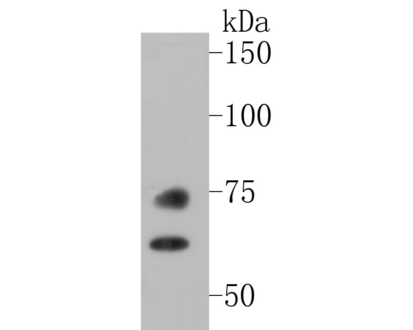
Fig1: Western blot analysis of Lamin A/C on A549 cell lysate. Proteins were transferred to a PVDF membrane and blocked with 5% BSA in PBS for 1 hour at room temperature. The primary antibody (ET7110-12, 1/500) was used in 5% BSA at room temperature for 2 hours. Goat Anti-Rabbit IgG - HRP Secondary Antibody (HA1001) at 1:5,000 dilution was used for 1 hour at room temperature.Application
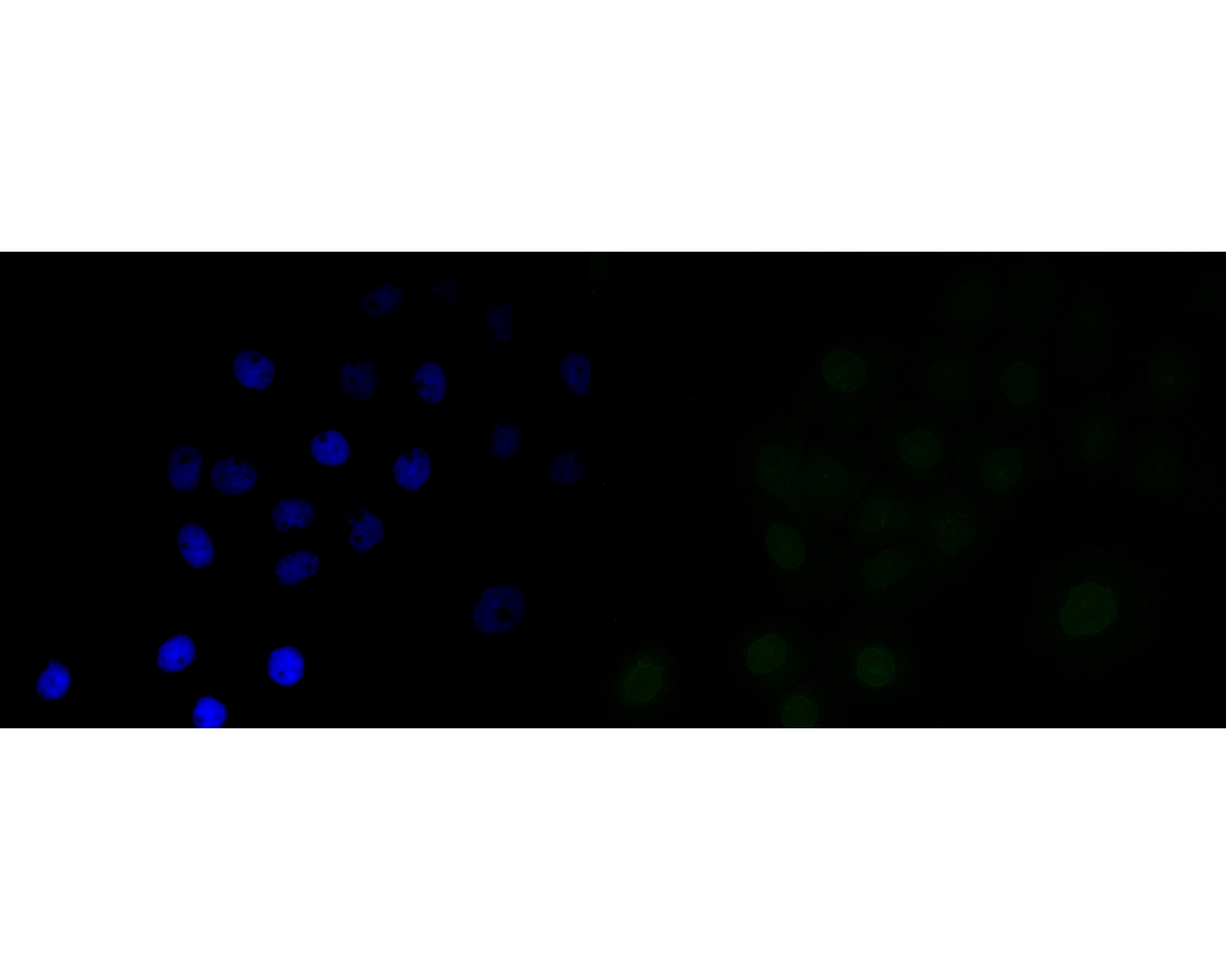
Fig2: ICC staining of Lamin A/C in A431 cells (green). Formalin fixed cells were permeabilized with 0.1% Triton X-100 in TBS for 10 minutes at room temperature and blocked with 1% Blocker BSA for 15 minutes at room temperature. Cells were probed with the primary antibody (ET7110-12, 1/50) for 1 hour at room temperature, washed with PBS. Alexa Fluor®488 Goat anti-Rabbit IgG was used as the secondary antibody at 1/1,000 dilution. The nuclear counter stain is DAPI (blue).Application
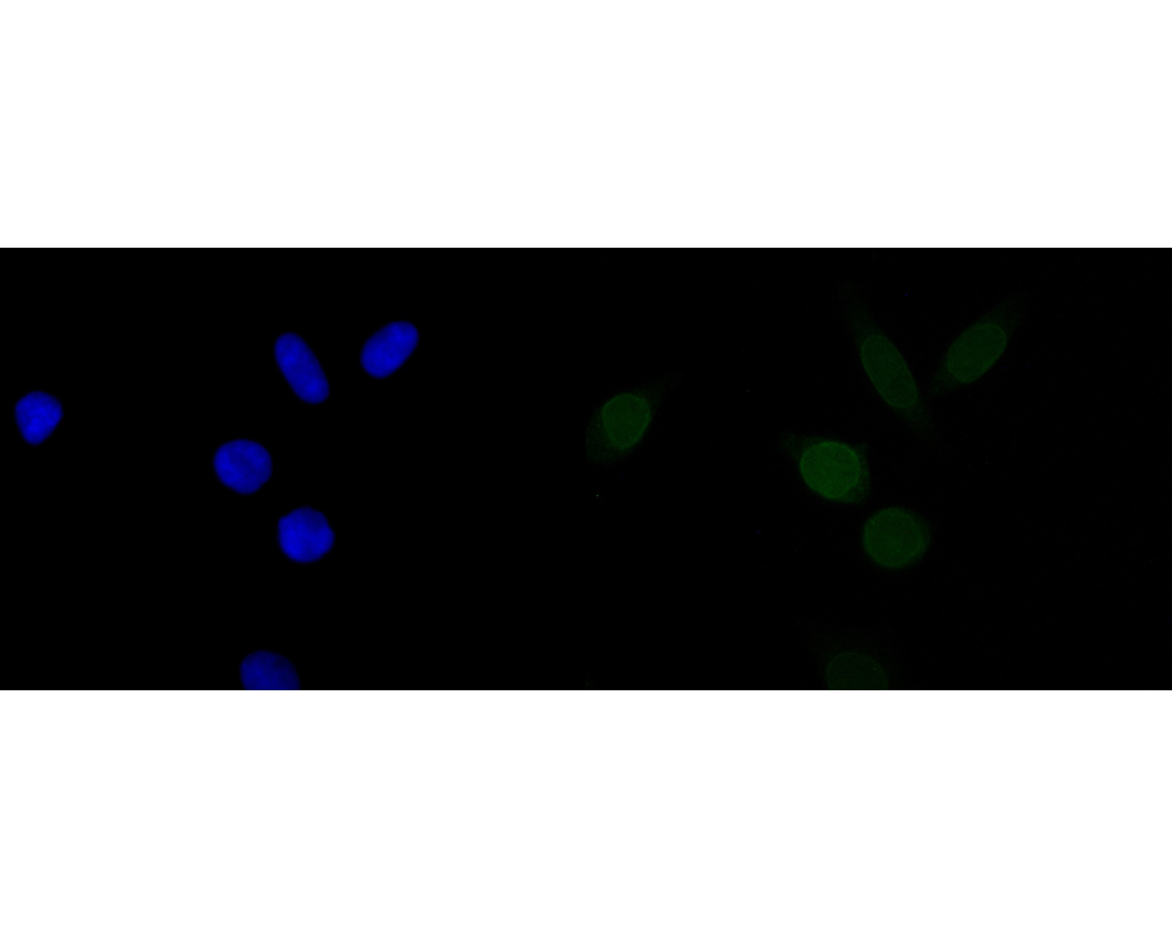
Fig3: ICC staining of Lamin A/C in SiHa cells (green). Formalin fixed cells were permeabilized with 0.1% Triton X-100 in TBS for 10 minutes at room temperature and blocked with 1% Blocker BSA for 15 minutes at room temperature. Cells were probed with the primary antibody (ET7110-12, 1/100) for 1 hour at room temperature, washed with PBS. Alexa Fluor®488 Goat anti-Rabbit IgG was used as the secondary antibody at 1/1,000 dilution. The nuclear counter stain is DAPI (blue).Application
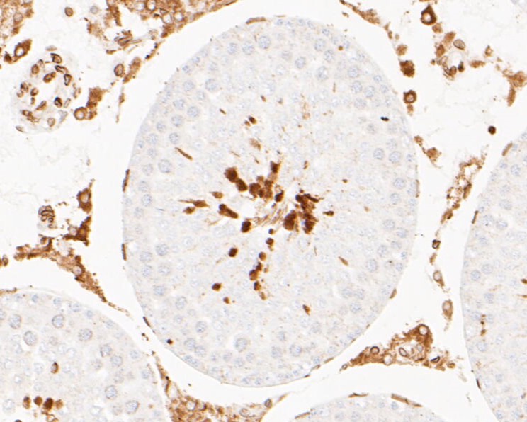
Fig4: Immunohistochemical analysis of paraffin-embedded rat testis tissue using anti-Lamin A/C antibody. The section was pre-treated using heat mediated antigen retrieval with sodium citrate buffer (pH 6.0) for 20 minutes. The tissues were blocked in 5% BSA for 30 minutes at room temperature, washed with ddH2O and PBS, and then probed with the primary antibody (ET7110-12, 1/50) for 30 minutes at room temperature. The detection was performed using an HRP conjugated compact polymer system. DAB was used as the chromogen. Tissues were counterstained with hematoxylin and mounted with DPX.Application
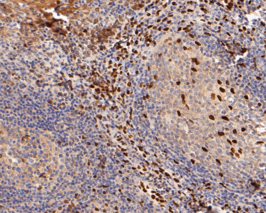
Fig5: Immunohistochemical analysis of paraffin-embedded human tonsil tissue using anti-Lamin A/C antibody. The section was pre-treated using heat mediated antigen retrieval with sodium citrate buffer (pH 6.0) for 20 minutes. The tissues were blocked in 5% BSA for 30 minutes at room temperature, washed with ddH2O and PBS, and then probed with the primary antibody (ET7110-12, 1/50) for 30 minutes at room temperature. The detection was performed using an HRP conjugated compact polymer system. DAB was used as the chromogen. Tissues were counterstained with hematoxylin and mounted with DPX.Application
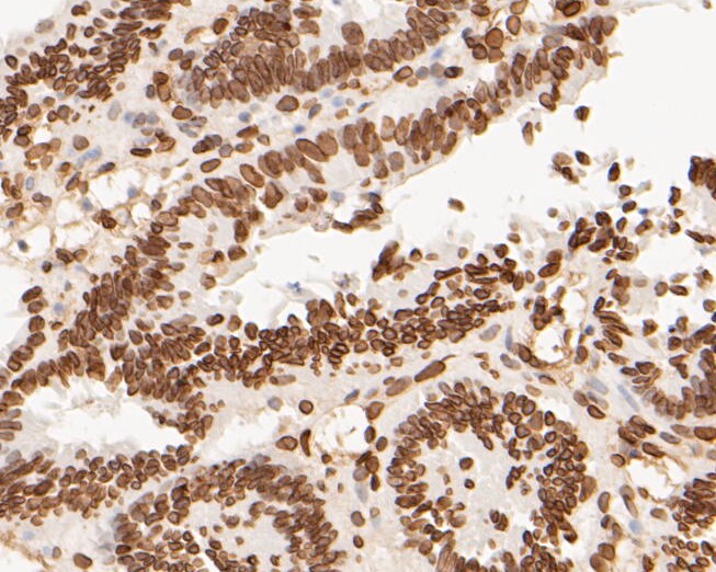
Fig6: Immunohistochemical analysis of paraffin-embedded human colon carcinoma tissue using anti-Lamin A/C antibody. The section was pre-treated using heat mediated antigen retrieval with sodium citrate buffer (pH 6.0) for 20 minutes. The tissues were blocked in 5% BSA for 30 minutes at room temperature, washed with ddH2O and PBS, and then probed with the primary antibody (ET7110-12, 1/50) for 30 minutes at room temperature. The detection was performed using an HRP conjugated compact polymer system. DAB was used as the chromogen. Tissues were counterstained with hematoxylin and mounted with DPX.Application
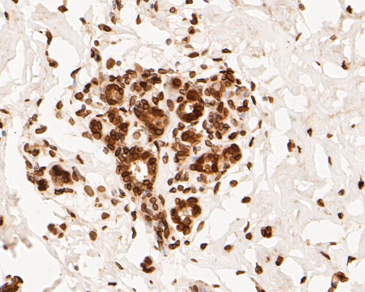
Fig7: Immunohistochemical analysis of paraffin-embedded human breast tissue using anti-Lamin A/C antibody. The section was pre-treated using heat mediated antigen retrieval with sodium citrate buffer (pH 6.0) for 20 minutes. The tissues were blocked in 5% BSA for 30 minutes at room temperature, washed with ddH2O and PBS, and then probed with the primary antibody (ET7110-12, 1/50) for 30 minutes at room temperature. The detection was performed using an HRP conjugated compact polymer system. DAB was used as the chromogen. Tissues were counterstained with hematoxylin and mounted with DPX.Application
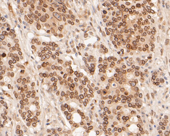
Fig8: Immunohistochemical analysis of paraffin-embedded human gastric carcinoma tissue using anti-Lamin A/C antibody. The section was pre-treated using heat mediated antigen retrieval with sodium citrate buffer (pH 6.0) for 20 minutes. The tissues were blocked in 5% BSA for 30 minutes at room temperature, washed with ddH2O and PBS, and then probed with the primary antibody (ET7110-12, 1/50) for 30 minutes at room temperature. The detection was performed using an HRP conjugated compact polymer system. DAB was used as the chromogen. Tissues were counterstained with hematoxylin and mounted with DPX.Application
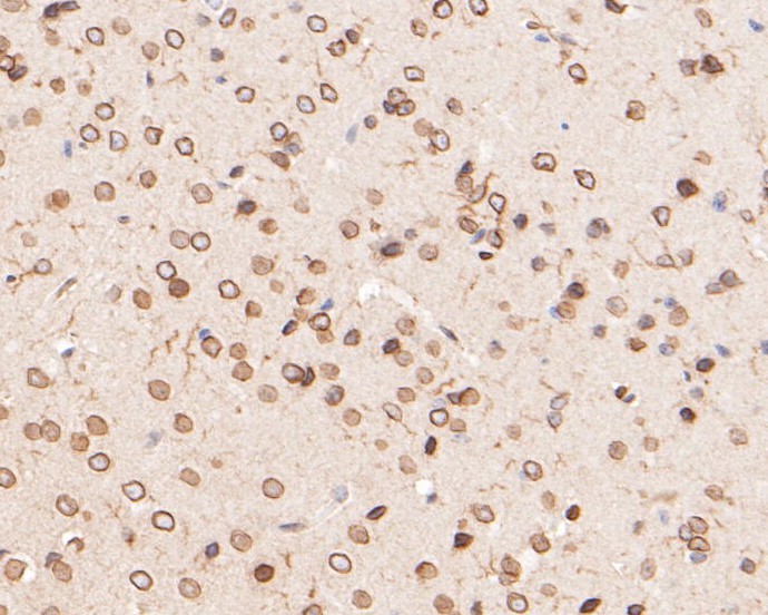
Fig9: Immunohistochemical analysis of paraffin-embedded mouse brain tissue using anti-Lamin A/C antibody. The section was pre-treated using heat mediated antigen retrieval with sodium citrate buffer (pH 6.0) for 20 minutes. The tissues were blocked in 5% BSA for 30 minutes at room temperature, washed with ddH2O and PBS, and then probed with the primary antibody (ET7110-12, 1/200) for 30 minutes at room temperature. The detection was performed using an HRP conjugated compact polymer system. DAB was used as the chromogen. Tissues were counterstained with hematoxylin and mounted with DPX.Application
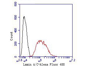
Fig10: Flow cytometric analysis of Lamin A/C was done on Hela cells. The cells were fixed, permeabilized and stained with the primary antibody (ET7110-12, 1/50) (red). After incubation of the primary antibody at room temperature for an hour, the cells were stained with a Alexa Fluor 488-conjugated Goat anti-Rabbit IgG Secondary antibody at 1/1000 dilution for 30 minutes.Unlabelled sample was used as a control (cells without incubation with primary antibody; black).| Product Name | Lamin A/C [JE51-60] |
|---|---|
| Antibody Type | Primary Antibodies |
| Antigen Alias | 70 kDa lamin antibody Cardiomyopathy dilated 1A (autosomal dominant) antibody CDCD1 antibody CDDC antibody CMD1A antibody CMT2B1 antibody EMD2 antibody FPL antibody FPLD antibody FPLD2 antibody HGPS antibody IDC antibody Lamin A antibody Lamin A/C antibody Lamin A/C like 1 antibody Lamin antibody Lamin C antibody Lamin-A/C antibody LDP1 antibody LFP antibody LGMD1B antibody Limb girdle muscular dystrophy 1B (autosomal dominant) antibody LMN 1 antibody LMN A antibody LMN C antibody LMN1 antibody LMNA antibody LMNA_HUMAN antibody LMNC antibody LMNL1 antibody Prelamin A/C antibody PRO1 antibody Renal carcinoma antigen NY REN 32 antibody Renal carcinoma antigen NY-REN-32 antibody Renal carcinoma antigen NYREN32 antibody |
| Product description | A unique family of cysteine proteases has been described that differs in sequence, structure and substrate specificity from any previously described protease family. This family, termed Ced-3/ICE, is comprised of ICE, CPP32, ICH-1/Nedd-2, Tx, Mch2, Mch3 (ICE-LAP3 or CMH-1), Mch4 and ICE-LAP6. Ced-3/ICE family members function as key components of the apoptotic machinery and act to destroy specific target proteins which are critical to cellular longevity. Nuclear lamins are critical to maintaining the integrity of the nuclear envelope and cellular morphology. The nuclear Lamin A is cleaved by Mch2, but not CPP32. Nuclear Lamin B is fragmented as a consequence of apoptosis by an unidentified member of the ICE family. Lamin C is a splice variant of Lamin A, differing only at the carboxy-terminus. Lamins A and C are identical for the first 566 amino acids, with Lamin C differing only in six unique carboxy-terminal amino acids. |
| Immunogen | Synthetic peptide within N-terminal human Lamin A/C |
| Clonality | Monoclonal |
|---|---|
| Isotype | IgG |
| Host Species | Recombinant rabbit |
| Tested Applications | WBICCIHC-PFC |
| WB:1:500-1:2,000 ICC:1:50-1:100 IHC-P:1:50-1:200 FC:1:50-1:100 |
|
| Species Reactivity | HumanMouseRat |
| Concentration | 1mg/mL |
| Alternative Names | 70 kDa lamin antibody
Cardiomyopathy dilated 1A (autosomal dominant) antibody
CDCD1 antibody CDDC antibody CMD1A antibody CMT2B1 antibody EMD2 antibody FPL antibody FPLD antibody FPLD2 antibody HGPS antibody IDC antibody Lamin A antibody Lamin A/C antibody Lamin A/C like 1 antibody Lamin antibody Lamin C antibody Lamin-A/C antibody LDP1 antibody LFP antibody LGMD1B antibody Limb girdle muscular dystrophy 1B (autosomal dominant) antibody LMN 1 antibody LMN A antibody LMN C antibody LMN1 antibody LMNA antibody LMNA_HUMAN antibody LMNC antibody LMNL1 antibody Prelamin A/C antibody PRO1 antibody Renal carcinoma antigen NY REN 32 antibody Renal carcinoma antigen NY-REN-32 antibody Renal carcinoma antigen NYREN32 antibody |
|---|---|
| Molecular Weight(MW) | 74/65 kDa |
| Cellular Localization | Nucleus. |
Application
Fig1: Western blot analysis of Lamin A/C on A549 cell lysate. Proteins were transferred to a PVDF membrane and blocked with 5% BSA in PBS for 1 hour at room temperature. The primary antibody (ET7110-12, 1/500) was used in 5% BSA at room temperature for 2 hours. Goat Anti-Rabbit IgG - HRP Secondary Antibody (HA1001) at 1:5,000 dilution was used for 1 hour at room temperature.
Application
Fig2: ICC staining of Lamin A/C in A431 cells (green). Formalin fixed cells were permeabilized with 0.1% Triton X-100 in TBS for 10 minutes at room temperature and blocked with 1% Blocker BSA for 15 minutes at room temperature. Cells were probed with the primary antibody (ET7110-12, 1/50) for 1 hour at room temperature, washed with PBS. Alexa Fluor®488 Goat anti-Rabbit IgG was used as the secondary antibody at 1/1,000 dilution. The nuclear counter stain is DAPI (blue).
Application
Fig3: ICC staining of Lamin A/C in SiHa cells (green). Formalin fixed cells were permeabilized with 0.1% Triton X-100 in TBS for 10 minutes at room temperature and blocked with 1% Blocker BSA for 15 minutes at room temperature. Cells were probed with the primary antibody (ET7110-12, 1/100) for 1 hour at room temperature, washed with PBS. Alexa Fluor®488 Goat anti-Rabbit IgG was used as the secondary antibody at 1/1,000 dilution. The nuclear counter stain is DAPI (blue).
Application
Fig4: Immunohistochemical analysis of paraffin-embedded rat testis tissue using anti-Lamin A/C antibody. The section was pre-treated using heat mediated antigen retrieval with sodium citrate buffer (pH 6.0) for 20 minutes. The tissues were blocked in 5% BSA for 30 minutes at room temperature, washed with ddH2O and PBS, and then probed with the primary antibody (ET7110-12, 1/50) for 30 minutes at room temperature. The detection was performed using an HRP conjugated compact polymer system. DAB was used as the chromogen. Tissues were counterstained with hematoxylin and mounted with DPX.
Application
Fig5: Immunohistochemical analysis of paraffin-embedded human tonsil tissue using anti-Lamin A/C antibody. The section was pre-treated using heat mediated antigen retrieval with sodium citrate buffer (pH 6.0) for 20 minutes. The tissues were blocked in 5% BSA for 30 minutes at room temperature, washed with ddH2O and PBS, and then probed with the primary antibody (ET7110-12, 1/50) for 30 minutes at room temperature. The detection was performed using an HRP conjugated compact polymer system. DAB was used as the chromogen. Tissues were counterstained with hematoxylin and mounted with DPX.
Application
Fig6: Immunohistochemical analysis of paraffin-embedded human colon carcinoma tissue using anti-Lamin A/C antibody. The section was pre-treated using heat mediated antigen retrieval with sodium citrate buffer (pH 6.0) for 20 minutes. The tissues were blocked in 5% BSA for 30 minutes at room temperature, washed with ddH2O and PBS, and then probed with the primary antibody (ET7110-12, 1/50) for 30 minutes at room temperature. The detection was performed using an HRP conjugated compact polymer system. DAB was used as the chromogen. Tissues were counterstained with hematoxylin and mounted with DPX.
Application
Fig7: Immunohistochemical analysis of paraffin-embedded human breast tissue using anti-Lamin A/C antibody. The section was pre-treated using heat mediated antigen retrieval with sodium citrate buffer (pH 6.0) for 20 minutes. The tissues were blocked in 5% BSA for 30 minutes at room temperature, washed with ddH2O and PBS, and then probed with the primary antibody (ET7110-12, 1/50) for 30 minutes at room temperature. The detection was performed using an HRP conjugated compact polymer system. DAB was used as the chromogen. Tissues were counterstained with hematoxylin and mounted with DPX.
Application
Fig8: Immunohistochemical analysis of paraffin-embedded human gastric carcinoma tissue using anti-Lamin A/C antibody. The section was pre-treated using heat mediated antigen retrieval with sodium citrate buffer (pH 6.0) for 20 minutes. The tissues were blocked in 5% BSA for 30 minutes at room temperature, washed with ddH2O and PBS, and then probed with the primary antibody (ET7110-12, 1/50) for 30 minutes at room temperature. The detection was performed using an HRP conjugated compact polymer system. DAB was used as the chromogen. Tissues were counterstained with hematoxylin and mounted with DPX.
Application
Fig9: Immunohistochemical analysis of paraffin-embedded mouse brain tissue using anti-Lamin A/C antibody. The section was pre-treated using heat mediated antigen retrieval with sodium citrate buffer (pH 6.0) for 20 minutes. The tissues were blocked in 5% BSA for 30 minutes at room temperature, washed with ddH2O and PBS, and then probed with the primary antibody (ET7110-12, 1/200) for 30 minutes at room temperature. The detection was performed using an HRP conjugated compact polymer system. DAB was used as the chromogen. Tissues were counterstained with hematoxylin and mounted with DPX.
Application
Fig10: Flow cytometric analysis of Lamin A/C was done on Hela cells. The cells were fixed, permeabilized and stained with the primary antibody (ET7110-12, 1/50) (red). After incubation of the primary antibody at room temperature for an hour, the cells were stained with a Alexa Fluor 488-conjugated Goat anti-Rabbit IgG Secondary antibody at 1/1000 dilution for 30 minutes.Unlabelled sample was used as a control (cells without incubation with primary antibody; black).| Positive Control | A549 cell, A431, SiHa, rat testis tissue, human tonsil tissue, human colon carcinoma tissue, human breast tissue, human gastric carcinoma tissue, mouse brain tissue, Hela. |
|---|---|
| Application Notes | WB:1:500-1:2,000 ICC:1:50-1:100 IHC-P:1:50-1:200 FC:1:50-1:100 |
| Form | Liquid |
|---|---|
| Storage Instructions | Store at +4℃ after thawing. Aliquot store at -20℃. Avoid repeated freeze / thaw cycles. |
| Storage Buffer | 1*TBS (pH7.4), 1%BSA, 50%Glycerol. Preservative: 0.05% Sodium Azide. |


