Application
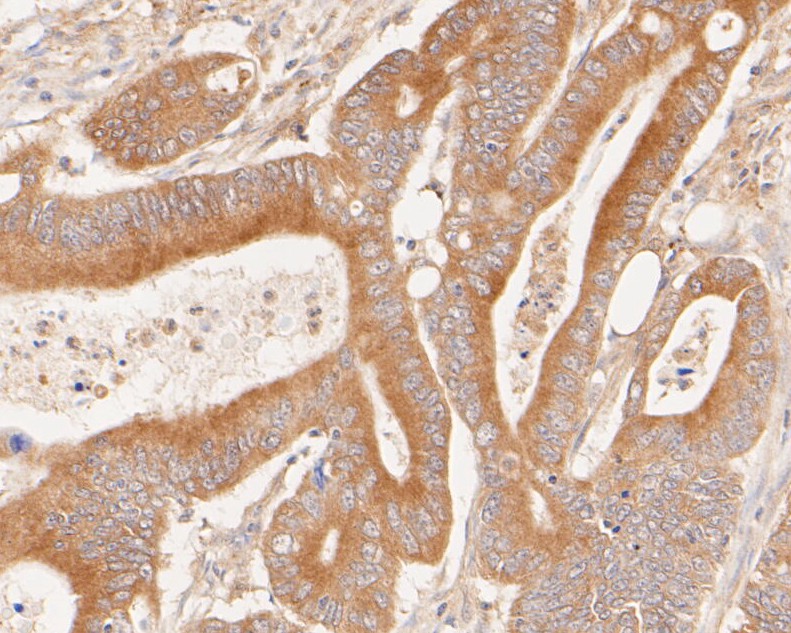
Fig1: Immunohistochemical analysis of paraffin-embedded human colon carcinoma tissue using anti-Phospho-Met (pY1349) antibody. The section was pre-treated using heat mediated antigen retrieval with Tris-EDTA buffer (pH 8.0-8.4) for 20 minutes.The tissues were blocked in 5% BSA for 30 minutes at room temperature, washed with ddH2O and PBS, and then probed with the primary antibody (ET7110-14, 1/50) for 30 minutes at room temperature. The detection was performed using an HRP conjugated compact polymer system. DAB was used as the chromogen. Tissues were counterstained with hematoxylin and mounted with DPX.Application
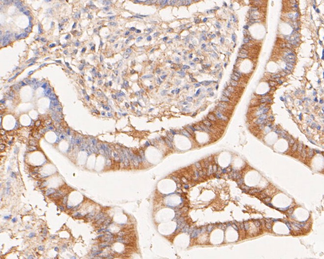
Fig2: Immunohistochemical analysis of paraffin-embedded human small intestine tissue using anti-Phospho-Met (pY1349) antibody. The section was pre-treated using heat mediated antigen retrieval with Tris-EDTA buffer (pH 8.0-8.4) for 20 minutes.The tissues were blocked in 5% BSA for 30 minutes at room temperature, washed with ddH2O and PBS, and then probed with the primary antibody (ET7110-14, 1/50) for 30 minutes at room temperature. The detection was performed using an HRP conjugated compact polymer system. DAB was used as the chromogen. Tissues were counterstained with hematoxylin and mounted with DPX.Application
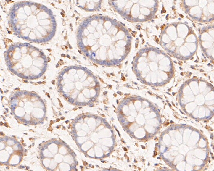
Fig3: Immunohistochemical analysis of paraffin-embedded human rectum tissue using anti-Phospho-Met (pY1349) antibody. The section was pre-treated using heat mediated antigen retrieval with Tris-EDTA buffer (pH 8.0-8.4) for 20 minutes.The tissues were blocked in 5% BSA for 30 minutes at room temperature, washed with ddH2O and PBS, and then probed with the primary antibody (ET7110-14, 1/50) for 30 minutes at room temperature. The detection was performed using an HRP conjugated compact polymer system. DAB was used as the chromogen. Tissues were counterstained with hematoxylin and mounted with DPX.| Product Name | Phospho-Met (pY1349) [JE51-68] |
|---|---|
| Antibody Type | Primary Antibodies |
| Antigen Alias | AUTS9 antibody c met antibody D249 antibody Hepatocyte growth factor receptor antibody HGF antibody HGF receptor antibody HGF/SF receptor antibody HGFR antibody MET antibody Met proto oncogene tyrosine kinase antibody MET proto oncogene, receptor tyrosine kinase antibody Met proto-oncogene (hepatocyte growth factor receptor) antibody Met proto-oncogene antibody Met protooncogene antibody MET_HUMAN antibody Oncogene MET antibody Par4 antibody Proto-oncogene c-Met antibody RCCP2 antibody Scatter factor receptor antibody SF receptor antibody Tyrosine-protein kinase Met antibody |
| Product description | Receptor tyrosine kinase that transduces signals from the extracellular matrix into the cytoplasm by binding to hepatocyte growth factor/HGF ligand. Regulates many physiological processes including proliferation, scattering, morphogenesis and survival. Ligand binding at the cell surface induces autophosphorylation of MET on its intracellular domain that provides docking sites for downstream signaling molecules. Following activation by ligand, interacts with the PI3-kinase subunit PIK3R1, PLCG1, SRC, GRB2, STAT3 or the adapter GAB1. Recruitment of these downstream effectors by MET leads to the activation of several signaling cascades including the RAS-ERK, PI3 kinase-AKT, or PLCgamma-PKC. The RAS-ERK activation is associated with the morphogenetic effects while PI3K/AKT coordinates prosurvival effects. During embryonic development, MET signaling plays a role in gastrulation, development and migration of muscles and neuronal precursors, angiogenesis and kidney formation. In adults, participates in wound healing as well as organ regeneration and tissue remodeling. Promotes also differentiation and proliferation of hematopoietic cells. May regulate cortical bone osteogenesis (By similarity). |
| Immunogen | Synthetic peptide surrounding human Met Y1349. |
| Modification | p-pY1349 |
| Clonality | Monoclonal |
|---|---|
| Isotype | IgG |
| Host Species | Recombinant rabbit |
| Tested Applications | WBIHC |
| WB:1:500 IHC:1:50-2:00 |
|
| Species Reactivity | Human |
| Concentration | 1mg/mL |
| Alternative Names | AUTS9 antibody
c met antibody
D249 antibody Hepatocyte growth factor receptor antibody HGF antibody HGF receptor antibody HGF/SF receptor antibody HGFR antibody MET antibody Met proto oncogene tyrosine kinase antibody MET proto oncogene receptor tyrosine kinase antibody Met proto-oncogene (hepatocyte growth factor receptor) antibody Met proto-oncogene antibody Met protooncogene antibody MET_HUMAN antibody Oncogene MET antibody Par4 antibody Proto-oncogene c-Met antibody RCCP2 antibody Scatter factor receptor antibody SF receptor antibody Tyrosine-protein kinase Met antibody |
|---|---|
| Molecular Weight(MW) | 156 kDa |
| Cellular Localization | Membrane, secreted. |
| SwissProt ID | P08581 |
|---|
Application
Fig1: Immunohistochemical analysis of paraffin-embedded human colon carcinoma tissue using anti-Phospho-Met (pY1349) antibody. The section was pre-treated using heat mediated antigen retrieval with Tris-EDTA buffer (pH 8.0-8.4) for 20 minutes.The tissues were blocked in 5% BSA for 30 minutes at room temperature, washed with ddH2O and PBS, and then probed with the primary antibody (ET7110-14, 1/50) for 30 minutes at room temperature. The detection was performed using an HRP conjugated compact polymer system. DAB was used as the chromogen. Tissues were counterstained with hematoxylin and mounted with DPX.
Application
Fig2: Immunohistochemical analysis of paraffin-embedded human small intestine tissue using anti-Phospho-Met (pY1349) antibody. The section was pre-treated using heat mediated antigen retrieval with Tris-EDTA buffer (pH 8.0-8.4) for 20 minutes.The tissues were blocked in 5% BSA for 30 minutes at room temperature, washed with ddH2O and PBS, and then probed with the primary antibody (ET7110-14, 1/50) for 30 minutes at room temperature. The detection was performed using an HRP conjugated compact polymer system. DAB was used as the chromogen. Tissues were counterstained with hematoxylin and mounted with DPX.
Application
Fig3: Immunohistochemical analysis of paraffin-embedded human rectum tissue using anti-Phospho-Met (pY1349) antibody. The section was pre-treated using heat mediated antigen retrieval with Tris-EDTA buffer (pH 8.0-8.4) for 20 minutes.The tissues were blocked in 5% BSA for 30 minutes at room temperature, washed with ddH2O and PBS, and then probed with the primary antibody (ET7110-14, 1/50) for 30 minutes at room temperature. The detection was performed using an HRP conjugated compact polymer system. DAB was used as the chromogen. Tissues were counterstained with hematoxylin and mounted with DPX.| Positive Control | Human colon carcinoma tissue, human small intestine tissue, human rectum tissue. |
|---|---|
| Application Notes | WB:1:500 IHC:1:50-2:00 |
| Form | Liquid |
|---|---|
| Storage Instructions | Store at +4℃ after thawing. Aliquot store at -20℃. Avoid repeated freeze / thaw cycles. |
| Storage Buffer | 1*TBS (pH7.4), 1%BSA, 50%Glycerol. Preservative: 0.05% Sodium Azide. |


