Application
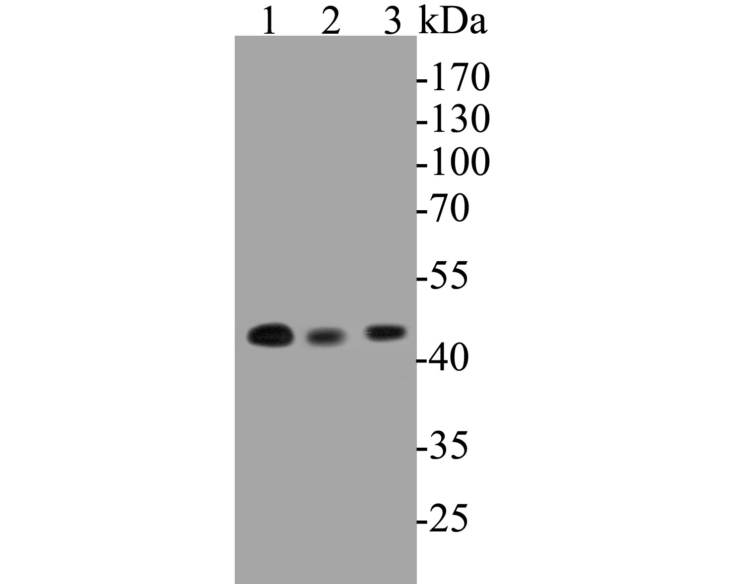
Fig1: Western blot analysis of NUDC on different lysates. Proteins were transferred to a PVDF membrane and blocked with 5% BSA in PBS for 1 hour at room temperature. The primary antibody (ET7110-18, 1/500) was used in 5% BSA at room temperature for 2 hours. Goat Anti-Rabbit IgG - HRP Secondary Antibody (HA1001) at 1:5,000 dilution was used for 1 hour at room temperature. Positive control: Lane 1: K562 cell lysates Lane 2: PC-3M cell lysates Lane 3: U937 cell lysatesApplication
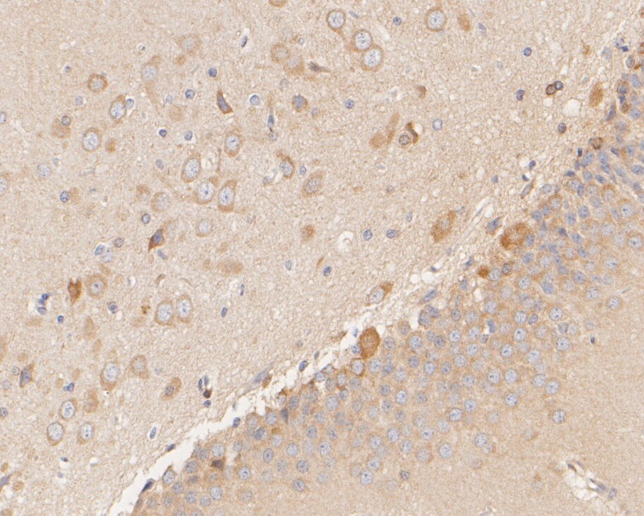
Fig2: Immunohistochemical analysis of paraffin-embedded rat brain tissue using anti-NUDC antibody. The section was pre-treated using heat mediated antigen retrieval with sodium citrate buffer (pH 6.0) for 20 minutes. The tissues were blocked in 5% BSA for 30 minutes at room temperature, washed with ddH2O and PBS, and then probed with the primary antibody (ET7110-18, 1/200) for 30 minutes at room temperature. The detection was performed using an HRP conjugated compact polymer system. DAB was used as the chromogen. Tissues were counterstained with hematoxylin and mounted with DPX.Application
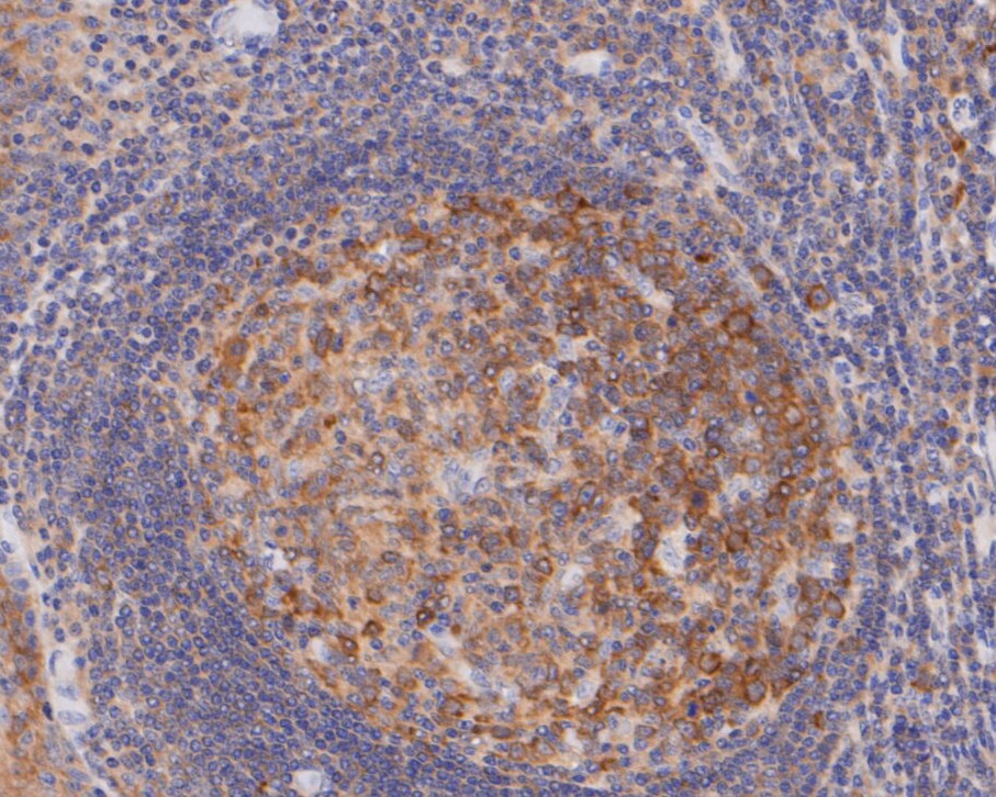
Fig3: Immunohistochemical analysis of paraffin-embedded human tonsil tissue using anti-NUDC antibody. The section was pre-treated using heat mediated antigen retrieval with sodium citrate buffer (pH 6.0) for 20 minutes. The tissues were blocked in 5% BSA for 30 minutes at room temperature, washed with ddH2O and PBS, and then probed with the primary antibody (ET7110-18, 1/200) for 30 minutes at room temperature. The detection was performed using an HRP conjugated compact polymer system. DAB was used as the chromogen. Tissues were counterstained with hematoxylin and mounted with DPX.Application
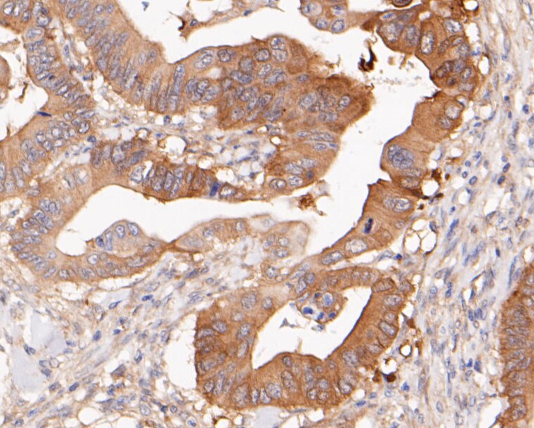
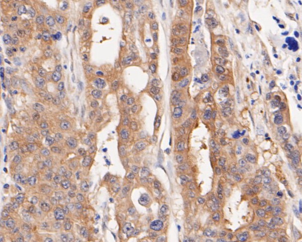
Fig4: Immunohistochemical analysis of paraffin-embedded human colon carcinoma tissue using anti-NUDC antibody. The section was pre-treated using heat mediated antigen retrieval with sodium citrate buffer (pH 6.0) for 20 minutes. The tissues were blocked in 5% BSA for 30 minutes at room temperature, washed with ddH2O and PBS, and then probed with the primary antibody (ET7110-18, 1/200) for 30 minutes at room temperature. The detection was performed using an HRP conjugated compact polymer system. DAB was used as the chromogen. Tissues were counterstained with hematoxylin and mounted with DPX.Application
Fig5: Immunohistochemical analysis of paraffin-embedded human gastric carcinoma tissue using anti-NUDC antibody. The section was pre-treated using heat mediated antigen retrieval with sodium citrate buffer (pH 6.0) for 20 minutes. The tissues were blocked in 5% BSA for 30 minutes at room temperature, washed with ddH2O and PBS, and then probed with the primary antibody (ET7110-18, 1/200) for 30 minutes at room temperature. The detection was performed using an HRP conjugated compact polymer system. DAB was used as the chromogen. Tissues were counterstained with hematoxylin and mounted with DPX.Application
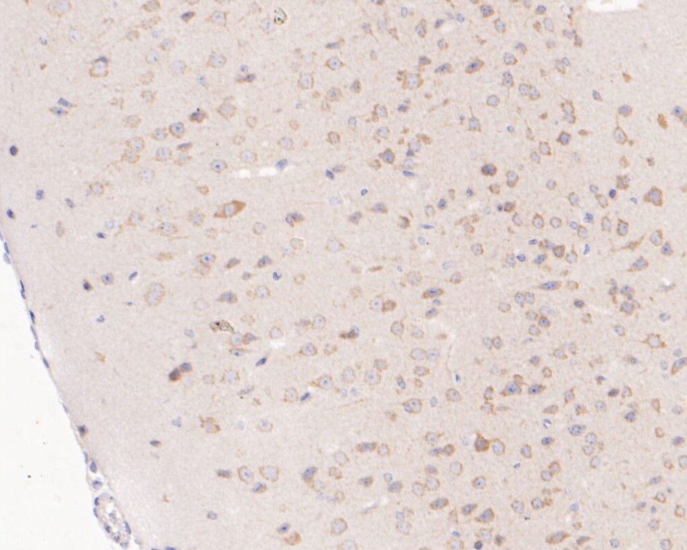
Fig6: Immunohistochemical analysis of paraffin-embedded mouse brain tissue using anti-NUDC antibody. The section was pre-treated using heat mediated antigen retrieval with sodium citrate buffer (pH 6.0) for 20 minutes. The tissues were blocked in 5% BSA for 30 minutes at room temperature, washed with ddH2O and PBS, and then probed with the primary antibody (ET7110-18, 1/200) for 30 minutes at room temperature. The detection was performed using an HRP conjugated compact polymer system. DAB was used as the chromogen. Tissues were counterstained with hematoxylin and mounted with DPX.Application
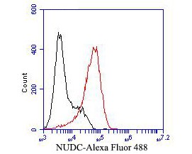
Fig7: Flow cytometric analysis of NUDC was done on F9 cells. The cells were fixed, permeabilized and stained with the primary antibody (ET7110-18, 1/50) (red). After incubation of the primary antibody at room temperature for an hour, the cells were stained with a Alexa Fluor 488-conjugated Goat anti-Rabbit IgG Secondary antibody at 1/1000 dilution for 30 minutes.Unlabelled sample was used as a control (cells without incubation with primary antibody; black).| Product Name | NUDC [JE50-74] |
|---|---|
| Antibody Type | Primary Antibodies |
| Antigen Alias | HNUDC antibody MNUDC antibody MNUDC protein antibody NPD011 antibody Nuclear distribution C homolog antibody Nuclear distribution gene C (A.nidulans) homolog antibody Nuclear distribution gene C homolog antibody Nuclear distribution gene C homolog (A. nidulans) antibody Nuclear distribution protein C homolog antibody Nuclear migration protein nudC antibody nudC antibody NudC nuclear distribution protein antibody NUDC_HUMAN antibody OTTHUMP00000004405 antibody SIG 92 antibody SIG92 antibody |
| Product description | NUDC (nuclear distribution gene C homolog, A. nidulans), also known as HNUDC, MNUDC or NPD011, is a ubiquitously expressed protein that is conserved from fungus to human. Highly expressed in proliferating cells, NUDC localizes to the cytoplasm and nucleus, contains a CS domain and participates in neurogenesis, mitosis, nuclear migration and cytokinesis. At the onset of mitosis, NUDC is phosphorylated by Plk. This modification of NUDC is required for proper mitotic spindle formation, chromosome separation during mitosis, cytokinesis and cell proliferation. In neurons and fibroblasts, NUDC forms a complex with LIS1 that localizes to the microtubule network and microtubule-organizing center and facilitates nuclear movement and transport in migrating neurons. In addition, the NUDC/LIS1 complex can associate with the minus-end directed Dynein/Dynactin motor complex and, together, these complexes cooperate in the regulation of cytokinesis. |
| Immunogen | Synthetic peptide within C-terminal human NUDC |
| Clonality | Monoclonal |
|---|---|
| Isotype | IgG |
| Host Species | Recombinant rabbit |
| Tested Applications | WBIHC-PFC |
| WB:1:500-1:2,000: IHC-P:1:100-1:500: FC:1:50-1:100: |
|
| Species Reactivity | HumanMouseRat |
| Concentration | 1mg/ml |
| Alternative Names | HNUDC antibody
MNUDC antibody
MNUDC protein antibody NPD011 antibody Nuclear distribution C homolog antibody Nuclear distribution gene C (A.nidulans) homolog antibody Nuclear distribution gene C homolog antibody Nuclear distribution gene C homolog (A. nidulans) antibody Nuclear distribution protein C homolog antibody Nuclear migration protein nudC antibody nudC antibody NudC nuclear distribution protein antibody NUDC_HUMAN antibody OTTHUMP00000004405 antibody SIG 92 antibody SIG92 antibody |
|---|---|
| Molecular Weight(MW) | 38 kDa (Predicted band size) |
| Cellular Localization | Nucleus, Cytoskeleton. |
Application
Fig1: Western blot analysis of NUDC on different lysates. Proteins were transferred to a PVDF membrane and blocked with 5% BSA in PBS for 1 hour at room temperature. The primary antibody (ET7110-18, 1/500) was used in 5% BSA at room temperature for 2 hours. Goat Anti-Rabbit IgG - HRP Secondary Antibody (HA1001) at 1:5,000 dilution was used for 1 hour at room temperature. Positive control: Lane 1: K562 cell lysates Lane 2: PC-3M cell lysates Lane 3: U937 cell lysates
Application
Fig2: Immunohistochemical analysis of paraffin-embedded rat brain tissue using anti-NUDC antibody. The section was pre-treated using heat mediated antigen retrieval with sodium citrate buffer (pH 6.0) for 20 minutes. The tissues were blocked in 5% BSA for 30 minutes at room temperature, washed with ddH2O and PBS, and then probed with the primary antibody (ET7110-18, 1/200) for 30 minutes at room temperature. The detection was performed using an HRP conjugated compact polymer system. DAB was used as the chromogen. Tissues were counterstained with hematoxylin and mounted with DPX.
Application
Fig3: Immunohistochemical analysis of paraffin-embedded human tonsil tissue using anti-NUDC antibody. The section was pre-treated using heat mediated antigen retrieval with sodium citrate buffer (pH 6.0) for 20 minutes. The tissues were blocked in 5% BSA for 30 minutes at room temperature, washed with ddH2O and PBS, and then probed with the primary antibody (ET7110-18, 1/200) for 30 minutes at room temperature. The detection was performed using an HRP conjugated compact polymer system. DAB was used as the chromogen. Tissues were counterstained with hematoxylin and mounted with DPX.
Application
Fig4: Immunohistochemical analysis of paraffin-embedded human colon carcinoma tissue using anti-NUDC antibody. The section was pre-treated using heat mediated antigen retrieval with sodium citrate buffer (pH 6.0) for 20 minutes. The tissues were blocked in 5% BSA for 30 minutes at room temperature, washed with ddH2O and PBS, and then probed with the primary antibody (ET7110-18, 1/200) for 30 minutes at room temperature. The detection was performed using an HRP conjugated compact polymer system. DAB was used as the chromogen. Tissues were counterstained with hematoxylin and mounted with DPX.
Application
Fig5: Immunohistochemical analysis of paraffin-embedded human gastric carcinoma tissue using anti-NUDC antibody. The section was pre-treated using heat mediated antigen retrieval with sodium citrate buffer (pH 6.0) for 20 minutes. The tissues were blocked in 5% BSA for 30 minutes at room temperature, washed with ddH2O and PBS, and then probed with the primary antibody (ET7110-18, 1/200) for 30 minutes at room temperature. The detection was performed using an HRP conjugated compact polymer system. DAB was used as the chromogen. Tissues were counterstained with hematoxylin and mounted with DPX.
Application
Fig6: Immunohistochemical analysis of paraffin-embedded mouse brain tissue using anti-NUDC antibody. The section was pre-treated using heat mediated antigen retrieval with sodium citrate buffer (pH 6.0) for 20 minutes. The tissues were blocked in 5% BSA for 30 minutes at room temperature, washed with ddH2O and PBS, and then probed with the primary antibody (ET7110-18, 1/200) for 30 minutes at room temperature. The detection was performed using an HRP conjugated compact polymer system. DAB was used as the chromogen. Tissues were counterstained with hematoxylin and mounted with DPX.
Application
Fig7: Flow cytometric analysis of NUDC was done on F9 cells. The cells were fixed, permeabilized and stained with the primary antibody (ET7110-18, 1/50) (red). After incubation of the primary antibody at room temperature for an hour, the cells were stained with a Alexa Fluor 488-conjugated Goat anti-Rabbit IgG Secondary antibody at 1/1000 dilution for 30 minutes.Unlabelled sample was used as a control (cells without incubation with primary antibody; black).| Positive Control | K562 cell lysates, PC-3M cell lysates, U937 cell lysates, rat brain tissue, human tonsil tissue, human colon carcinoma tissue, human gastric carcinoma tissue, mouse brain tissue, F9. |
|---|---|
| Application Notes | WB:1:500-1:2,000: IHC-P:1:100-1:500: FC:1:50-1:100: |
| Form | Liquid |
|---|---|
| Storage Instructions | Store at +4℃ after thawing. Aliquot store at -20℃. Avoid repeated freeze / thaw cycles. |
| Storage Buffer | 1*TBS (pH7.4), 1%BSA, 50%Glycerol. Preservative: 0.05% Sodium Azide. |


