Application
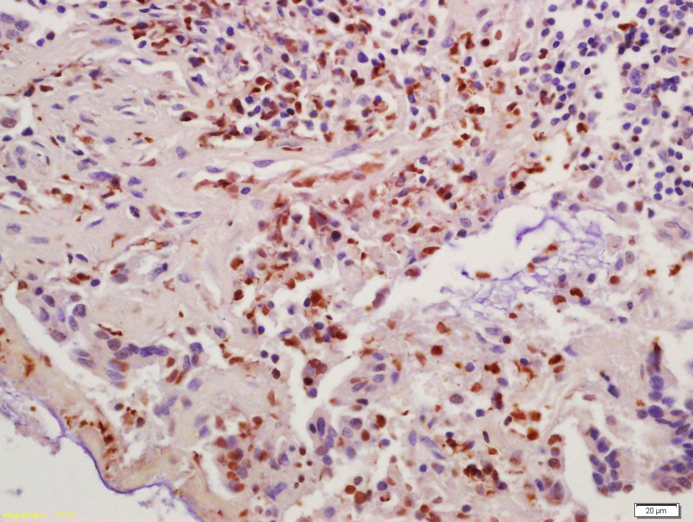
Fig1: Tissue/cell: human colon carcinoma; 4% Paraformaldehyde-fixed and paraffin-embedded; Antigen retrieval: citrate buffer ( 0.01M, pH 6.0 ), Boiling bathing for 15min; Block endogenous peroxidase by 3% Hydrogen peroxide for 30min; Blocking buffer (normal goat serum,C-0005) at 37℃ for 20 min; Incubation: Anti-Cyclin B1 Polyclonal Antibody, Unconjugated(ER1906-93) 1:200, overnight at 4℃, followed by conjugation to the secondary antibody(SP-0023) and DAB(C-0010) stainingApplication
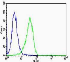
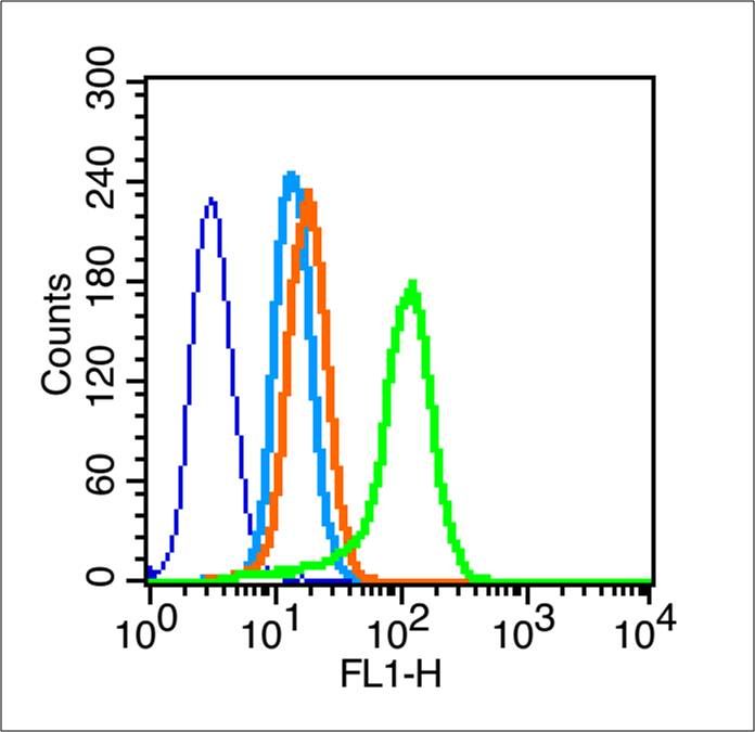
Fig2: Cell: Hela Concentration:1:100 Host/Isotype:Rabbit/IgG Flow cytometric analysis of primary antibody (Cat#: ER1906-93) on Hela(green) compared with isotype control in the absence of primary antibody (blue) followed by Alexa Fluor 488-conjugated goat anti-rabbit IgG(H+L) secondary antibody .Application
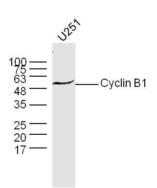
Fig3: Sample: U251 Cell (Human) Lysate at 30 ug Primary: Anti- Cyclin B1 (ER1906-93) at 1/300 dilution Secondary: IRDye800CW Goat Anti-Rabbit IgG at 1/20000 dilution Predicted band size: 48 kD Observed band size: 50 kDApplication
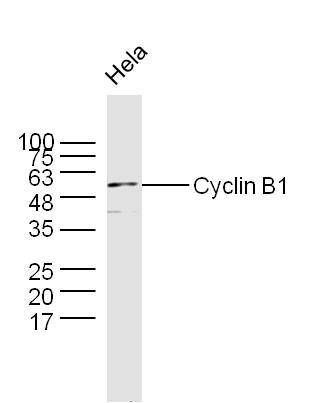
Fig4: Sample: Hela Cell (Human) Lysate at 30 ug Primary: Anti- Cyclin B1 (ER1906-93) at 1/300 dilution Secondary: IRDye800CW Goat Anti-Rabbit IgG at 1/20000 dilution Predicted band size: 48 kD Observed band size: 50 kDApplication
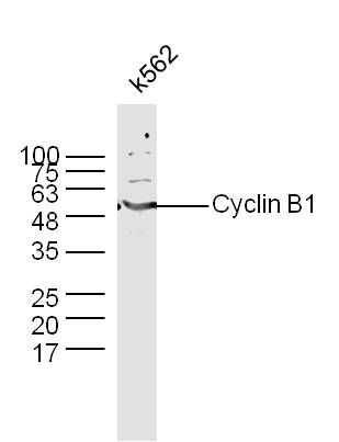
Fig5: Sample: K562 Cell (Human) Lysate at 30 ug Primary: Anti- Cyclin B1 (ER1906-93) at 1/300 dilution Secondary: IRDye800CW Goat Anti-Rabbit IgG at 1/20000 dilution Predicted band size: 48 kD Observed band size: 50 kDApplication
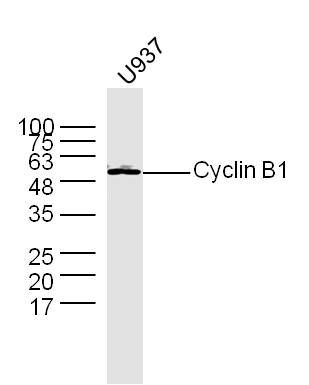
Fig6: Sample: U937 Cell (Human) Lysate at 30 ug Primary: Anti- Cyclin B1 (ER1906-93) at 1/300 dilution Secondary: IRDye800CW Goat Anti-Rabbit IgG at 1/20000 dilution Predicted band size: 48 kD Observed band size: 50 kDApplication
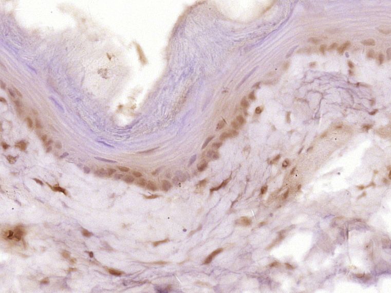
Fig7: Paraformaldehyde-fixed, paraffin embedded (Rat esophageal); Antigen retrieval by boiling in sodium citrate buffer (pH6.0) for 15min; Block endogenous peroxidase by 3% hydrogen peroxide for 20 minutes; Blocking buffer (normal goat serum) at 37℃ for 30min; Antibody incubation with (Cyclin B1) Polyclonal Antibody, Unconjugated (ER1906-93) at 1:400 overnight at 4℃, followed by operating according to SP Kit(Rabbit) (sp-0023) instructionsand DAB staining.Application
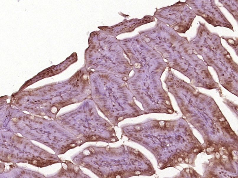
Fig8: Paraformaldehyde-fixed, paraffin embedded (Mouse small intestine); Antigen retrieval by boiling in sodium citrate buffer (pH6.0) for 15min; Block endogenous peroxidase by 3% hydrogen peroxide for 20 minutes; Blocking buffer (normal goat serum) at 37℃ for 30min; Antibody incubation with (Cyclin B1) Polyclonal Antibody, Unconjugated (ER1906-93) at 1:400 overnight at 4℃, followed by operating according to SP Kit(Rabbit) (sp-0023) instructionsand DAB staining.Application
Fig9: Blank control (blue line): A549 (blue). Primary Antibody (green line): Rabbit Anti-Cyclin B1 antibody(ER1906-93). Dilution: 1μg /10^6 cells; Isotype Control Antibody (orange line): Rabbit IgG . Secondary Antibody (white blue line): F(ab’)2 fragment goat anti-rabbit IgG-FITC. Dilution: 1μg /test. Protocol The cells were fixed with 2% paraformaldehyde (10 min) and then permeabilized with 0.1% PBS-Tween for 20 min at room temperature.Cells stained with Primary Antibody for 30 min at room temperature. The cells were then incubated in 1 X PBS/2%BSA/10% goat serum to block non-specific protein-protein interactions followed by the antibody for 15 min at room temperature. The secondary antibody used for 40 min at room temperature. Acquisition of 20,000 events was performed.| Product Name | Cyclin B1 |
|---|---|
| Antibody Type | Primary Antibodies |
| Antigen Alias | CCNB 1 CCNB CCNB1 CCNB1_HUMAN G2 mitotic specific cyclin B1 G2/mitotic-specific cyclin-B1. |
| Product description | Cyclin B1 is a regulatory protein involved in mitosis. The gene product complexes with p34 (Cdk1) to form the maturation-promoting factor (MPF). Two alternative transcripts have been found, a constitutively expressed transcript and a cell cycle-regulated transcript that is expressed predominantly during G2/M phase of the cell cycle. The different transcripts result from the use of alternate transcription initiation sites. Cyclin B1 contributes to the switch-like all or none behavior of the cell in deciding to commit to mitosis. Its activation is well-regulated, and positive feedback loops ensure that once the cyclin B1-Cdk1 complex is activated, it is not deactivated. Cyclin B1-Cdk1 is involved in the early events of mitosis, such as chromosome condensation, nuclear envelope breakdown, and spindle pole assembly. Once activated, cyclin B1-Cdk1 promotes several of the events of early mitosis. The active complex phosphorylates and activates 13S condensin, which helps to condense chromosomes. |
| Immunogen | KLH conjugated synthetic peptide derived from human Cyclin B1 271-433/433 |
| Clonality | Polyclonal |
|---|---|
| Isotype | IgG |
| Host Species | Rabbit |
| Tested Applications | WBIHC-PFC |
| WB:1:500-2000: IHC-P:1:400-800: FC:1μg/Test: |
|
| Species Reactivity | HumanMouseRatCow |
| Concentration | 1mg/ml |
| Alternative Names | CCNB 1 CCNB CCNB1 CCNB1_HUMAN G2 mitotic specific cyclin B1 G2/mitotic-specific cyclin-B1. |
|---|---|
| Molecular Weight(MW) | 48 KDa |
| Cellular Localization | Cytoplasm. Nucleus. Cytoplasm, cytoskeleton, centrosome. |
| SwissProt ID | P24860 |
|---|
Application
Fig1: Tissue/cell: human colon carcinoma; 4% Paraformaldehyde-fixed and paraffin-embedded; Antigen retrieval: citrate buffer ( 0.01M, pH 6.0 ), Boiling bathing for 15min; Block endogenous peroxidase by 3% Hydrogen peroxide for 30min; Blocking buffer (normal goat serum,C-0005) at 37℃ for 20 min; Incubation: Anti-Cyclin B1 Polyclonal Antibody, Unconjugated(ER1906-93) 1:200, overnight at 4℃, followed by conjugation to the secondary antibody(SP-0023) and DAB(C-0010) staining
Application
Fig2: Cell: Hela Concentration:1:100 Host/Isotype:Rabbit/IgG Flow cytometric analysis of primary antibody (Cat#: ER1906-93) on Hela(green) compared with isotype control in the absence of primary antibody (blue) followed by Alexa Fluor 488-conjugated goat anti-rabbit IgG(H+L) secondary antibody .
Application
Fig3: Sample: U251 Cell (Human) Lysate at 30 ug Primary: Anti- Cyclin B1 (ER1906-93) at 1/300 dilution Secondary: IRDye800CW Goat Anti-Rabbit IgG at 1/20000 dilution Predicted band size: 48 kD Observed band size: 50 kD
Application
Fig4: Sample: Hela Cell (Human) Lysate at 30 ug Primary: Anti- Cyclin B1 (ER1906-93) at 1/300 dilution Secondary: IRDye800CW Goat Anti-Rabbit IgG at 1/20000 dilution Predicted band size: 48 kD Observed band size: 50 kD
Application
Fig5: Sample: K562 Cell (Human) Lysate at 30 ug Primary: Anti- Cyclin B1 (ER1906-93) at 1/300 dilution Secondary: IRDye800CW Goat Anti-Rabbit IgG at 1/20000 dilution Predicted band size: 48 kD Observed band size: 50 kD
Application
Fig6: Sample: U937 Cell (Human) Lysate at 30 ug Primary: Anti- Cyclin B1 (ER1906-93) at 1/300 dilution Secondary: IRDye800CW Goat Anti-Rabbit IgG at 1/20000 dilution Predicted band size: 48 kD Observed band size: 50 kD
Application
Fig7: Paraformaldehyde-fixed, paraffin embedded (Rat esophageal); Antigen retrieval by boiling in sodium citrate buffer (pH6.0) for 15min; Block endogenous peroxidase by 3% hydrogen peroxide for 20 minutes; Blocking buffer (normal goat serum) at 37℃ for 30min; Antibody incubation with (Cyclin B1) Polyclonal Antibody, Unconjugated (ER1906-93) at 1:400 overnight at 4℃, followed by operating according to SP Kit(Rabbit) (sp-0023) instructionsand DAB staining.
Application
Fig8: Paraformaldehyde-fixed, paraffin embedded (Mouse small intestine); Antigen retrieval by boiling in sodium citrate buffer (pH6.0) for 15min; Block endogenous peroxidase by 3% hydrogen peroxide for 20 minutes; Blocking buffer (normal goat serum) at 37℃ for 30min; Antibody incubation with (Cyclin B1) Polyclonal Antibody, Unconjugated (ER1906-93) at 1:400 overnight at 4℃, followed by operating according to SP Kit(Rabbit) (sp-0023) instructionsand DAB staining.
Application
Fig9: Blank control (blue line): A549 (blue). Primary Antibody (green line): Rabbit Anti-Cyclin B1 antibody(ER1906-93). Dilution: 1μg /10^6 cells; Isotype Control Antibody (orange line): Rabbit IgG . Secondary Antibody (white blue line): F(ab’)2 fragment goat anti-rabbit IgG-FITC. Dilution: 1μg /test. Protocol The cells were fixed with 2% paraformaldehyde (10 min) and then permeabilized with 0.1% PBS-Tween for 20 min at room temperature.Cells stained with Primary Antibody for 30 min at room temperature. The cells were then incubated in 1 X PBS/2%BSA/10% goat serum to block non-specific protein-protein interactions followed by the antibody for 15 min at room temperature. The secondary antibody used for 40 min at room temperature. Acquisition of 20,000 events was performed.| Positive Control | Tuman colon carcinoma tissue, Hela cell, U251 Cell (Human) Lysate, K562 Cell (Human) Lysate, U937 Cell (Human) Lysate, Rat esophageal tissue, Mouse small intestine tissue, A549 cell. |
|---|---|
| Application Notes | WB:1:500-2000: IHC-P:1:400-800: FC:1μg/Test: |
| Form | Liquid |
|---|---|
| Storage Instructions | Store at -20℃ for one year. Avoid repeated freeze/thaw cycles. The lyophilized antibody is stable at room temperature for at least one month and for greater than a year when kept at -20℃. When reconstituted in sterile pH 7.4 0.01M PBS or diluent of antibody the antibody is stable for at least two weeks at 2-4℃. |
| Storage Buffer | 0.01M TBS(pH7.4) with 1% BSA, 0.03% Proclin300 and 50% Glycerol. |


