Application
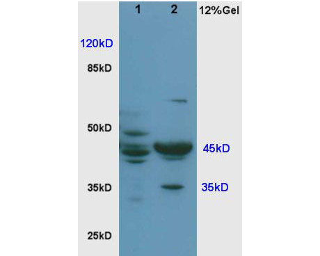
Fig1: Sample: Brain(Rat) lysate at30ug; Lung(Rat) lysate at 30ug; Primary: Anti-Cyclin E (ER1906-94) at 1:200; Secondary: HRP conjugated Goat-Anti-Rabbit IgG(bse-0295G) at 1: 3000; Predicted band size : 45kD Observed band size : 45kDApplication
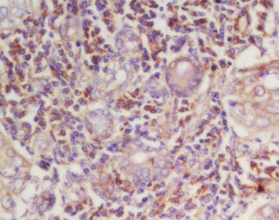
Fig2: Tissue/cell: human laryngocarcinoma; 4% Paraformaldehyde-fixed and paraffin-embedded; Antigen retrieval: citrate buffer ( 0.01M, pH 6.0 ), Boiling bathing for 15min; Block endogenous peroxidase by 3% Hydrogen peroxide for 30min; Blocking buffer (normal goat serum,C-0005) at 37℃ for 20 min; Incubation: Anti-Cyclin-E Polyclonal Antibody, Unconjugated(ER1906-94) 1:200, overnight at 4℃, followed by conjugation to the secondary antibody(SP-0023) and DAB(C-0010) stainingApplication
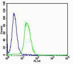
Fig3: Cell: NIH/3T3 Concentration:1:100 Host/Isotype:Rabbit/IgG Flow cytometric analysis of primary antibody (Cat#: ER1906-94) on NIH/3T3(green) compared with Rabbit IgG isotype control in the absence of primary antibody (blue) followed by Alexa Fluor 488-conjugated goat anti-rabbit IgG(H+L) secondary antibody .Application
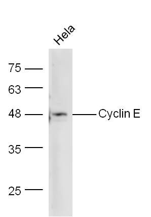
Fig4: Sample: Hela Lysate at 40 ug Primary: Anti-Cyclin E (ER1906-94) at 1/300 dilution Secondary: IRDye800CW Goat Anti-Rabbit IgG at 1/10000 dilution Predicted band size: 45 kD Observed band size: 48 kDApplication
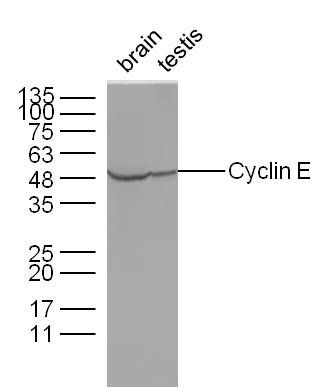
Fig5: Sample: Brain(Mouse) Lysate at 40 ug Testis(Mouse) Lysate at 40 ug Primary: Anti-Cyclin E (ER1906-94) at 1/300 dilution Secondary: IRDye800CW Goat Anti-Rabbit IgG at 1/10000 dilution Predicted band size: 45 kD Observed band size: 48 kDApplication
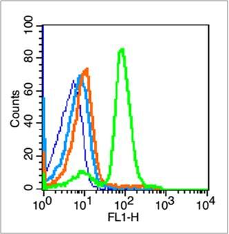
Fig6: Blank control (blue line): Mouse spleen cells (blue). Primary Antibody (green line): Rabbit Anti-Cyclin E1 antibody (ER1906-94) Dilution: 1μg /10^6 cells; Isotype Control Antibody (orange line): Rabbit IgG . Secondary Antibody (white blue line): Goat anti-rabbit IgG-FITC Dilution: 1μg /test. Protocol The cells were fixed with 70% ethanol (overninght at 4℃) and then permeabilized with 0.1% PBS-Tween for 20 min at room temperature. Cells stained with Primary Antibody for 30 min at room temperature. The cells were then incubated in 1 X PBS/2%BSA/10% goat serum to block non-specific protein-protein interactions followed by the antibody for 15 min at room temperature. The secondary antibody used for 40 min at room temperature. Acquisition of 20,000 events was performed.Application
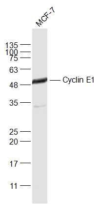
Fig7: Sample: MCF-7(Human) Cell Lysate at 30 ug Primary: Anti-Cyclin E1 (ER1906-94) at 1/1000 dilution Secondary: IRDye800CW Goat Anti-Rabbit IgG at 1/20000 dilution Predicted band size: 45 kD Observed band size: 48 kDApplication
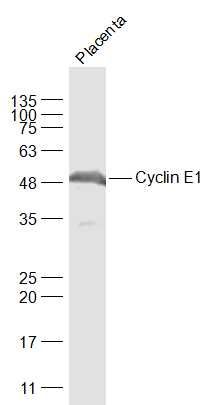
Fig8: Sample: Placenta (Mouse) Lysate at 40 ug Primary: Anti-Cyclin E1 (ER1906-94) at 1/1000 dilution Secondary: IRDye800CW Goat Anti-Rabbit IgG at 1/20000 dilution Predicted band size: 45 kD Observed band size: 48 kD| Product Name | Cyclin E1 |
|---|---|
| Antibody Type | Primary Antibodies |
| Antigen Alias | CCNE 1 CCNE CCNE1 Cyclin Es Cyclin Et CyclinE G1/S specific cyclin E G1/S-specific cyclin-E1 CCNE1_HUMAN pCCNE1. |
| Product description | The protein encoded by this gene belongs to the highly conserved cyclin family, whose members are characterized by a dramatic periodicity in protein abundance through the cell cycle. Cyclins function as regulators of CDK. Different cyclins exhibit distinct expression and degradation patterns which contribute to the temporal coordination of each mitotic event. This cyclin forms a complex with and functions as a regulatory subunit of CDK2, whose activity is required for cell cycle G1/S transition. This protein accumulates at the G1-S phase boundary and is degraded as cells progress through S phase. Overexpression of this gene has been observed in many tumors, which results in chromosome instability, and thus may contribute to tumorigenesis. This protein was found to associate with, and be involved in, the phosphorylation of NPAT protein (nuclear protein mapped to the ATM locus), which participates in cell-cycle regulated histone gene expression and plays a critical role in promoting cell-cycle progression in the absence of pRB. |
| Immunogen | KLH conjugated synthetic peptide derived from rat Cyclin E 375-411/411 |
| Clonality | Polyclonal |
|---|---|
| Isotype | IgG |
| Host Species | Rabbit |
| Tested Applications | WBIHC-PICC/IFFC |
| WB:1:500-2000: IHC-P:1:400-800: FC:1μg/Test: IF:1:100-500: |
|
| Species Reactivity | HumanMouseRat |
| Concentration | 1mg/ml |
| Alternative Names | CCNE 1
CCNE
CCNE1 Cyclin Es Cyclin Et CyclinE G1/S specific cyclin E G1/S-specific cyclin-E1 CCNE1_HUMAN pCCNE1. |
|---|---|
| Molecular Weight(MW) | 45 KDa |
| Cellular Localization | Nucleus. |
| SwissProt ID | P39949 |
|---|
Application
Fig1: Sample: Brain(Rat) lysate at30ug; Lung(Rat) lysate at 30ug; Primary: Anti-Cyclin E (ER1906-94) at 1:200; Secondary: HRP conjugated Goat-Anti-Rabbit IgG(bse-0295G) at 1: 3000; Predicted band size : 45kD Observed band size : 45kD
Application
Fig2: Tissue/cell: human laryngocarcinoma; 4% Paraformaldehyde-fixed and paraffin-embedded; Antigen retrieval: citrate buffer ( 0.01M, pH 6.0 ), Boiling bathing for 15min; Block endogenous peroxidase by 3% Hydrogen peroxide for 30min; Blocking buffer (normal goat serum,C-0005) at 37℃ for 20 min; Incubation: Anti-Cyclin-E Polyclonal Antibody, Unconjugated(ER1906-94) 1:200, overnight at 4℃, followed by conjugation to the secondary antibody(SP-0023) and DAB(C-0010) staining
Application
Fig3: Cell: NIH/3T3 Concentration:1:100 Host/Isotype:Rabbit/IgG Flow cytometric analysis of primary antibody (Cat#: ER1906-94) on NIH/3T3(green) compared with Rabbit IgG isotype control in the absence of primary antibody (blue) followed by Alexa Fluor 488-conjugated goat anti-rabbit IgG(H+L) secondary antibody .
Application
Fig4: Sample: Hela Lysate at 40 ug Primary: Anti-Cyclin E (ER1906-94) at 1/300 dilution Secondary: IRDye800CW Goat Anti-Rabbit IgG at 1/10000 dilution Predicted band size: 45 kD Observed band size: 48 kD
Application
Fig5: Sample: Brain(Mouse) Lysate at 40 ug Testis(Mouse) Lysate at 40 ug Primary: Anti-Cyclin E (ER1906-94) at 1/300 dilution Secondary: IRDye800CW Goat Anti-Rabbit IgG at 1/10000 dilution Predicted band size: 45 kD Observed band size: 48 kD
Application
Fig6: Blank control (blue line): Mouse spleen cells (blue). Primary Antibody (green line): Rabbit Anti-Cyclin E1 antibody (ER1906-94) Dilution: 1μg /10^6 cells; Isotype Control Antibody (orange line): Rabbit IgG . Secondary Antibody (white blue line): Goat anti-rabbit IgG-FITC Dilution: 1μg /test. Protocol The cells were fixed with 70% ethanol (overninght at 4℃) and then permeabilized with 0.1% PBS-Tween for 20 min at room temperature. Cells stained with Primary Antibody for 30 min at room temperature. The cells were then incubated in 1 X PBS/2%BSA/10% goat serum to block non-specific protein-protein interactions followed by the antibody for 15 min at room temperature. The secondary antibody used for 40 min at room temperature. Acquisition of 20,000 events was performed.
Application
Fig7: Sample: MCF-7(Human) Cell Lysate at 30 ug Primary: Anti-Cyclin E1 (ER1906-94) at 1/1000 dilution Secondary: IRDye800CW Goat Anti-Rabbit IgG at 1/20000 dilution Predicted band size: 45 kD Observed band size: 48 kD
Application
Fig8: Sample: Placenta (Mouse) Lysate at 40 ug Primary: Anti-Cyclin E1 (ER1906-94) at 1/1000 dilution Secondary: IRDye800CW Goat Anti-Rabbit IgG at 1/20000 dilution Predicted band size: 45 kD Observed band size: 48 kD| Positive Control | Highly expressed in testis and placenta. Low levels in bronchial epithelial cells. |
|---|---|
| Application Notes | WB:1:500-2000: IHC-P:1:400-800: FC:1μg/Test: IF:1:100-500: |
| Form | Liquid |
|---|---|
| Storage Instructions | Store at -20℃ for one year. Avoid repeated freeze/thaw cycles. The lyophilized antibody is stable at room temperature for at least one month and for greater than a year when kept at -20℃. When reconstituted in sterile pH 7.4 0.01M PBS or diluent of antibody the antibody is stable for at least two weeks at 2-4℃. |
| Storage Buffer | 0.01M TBS(pH7.4) with 1% BSA, 0.03% Proclin300 and 50% Glycerol. |


