Application
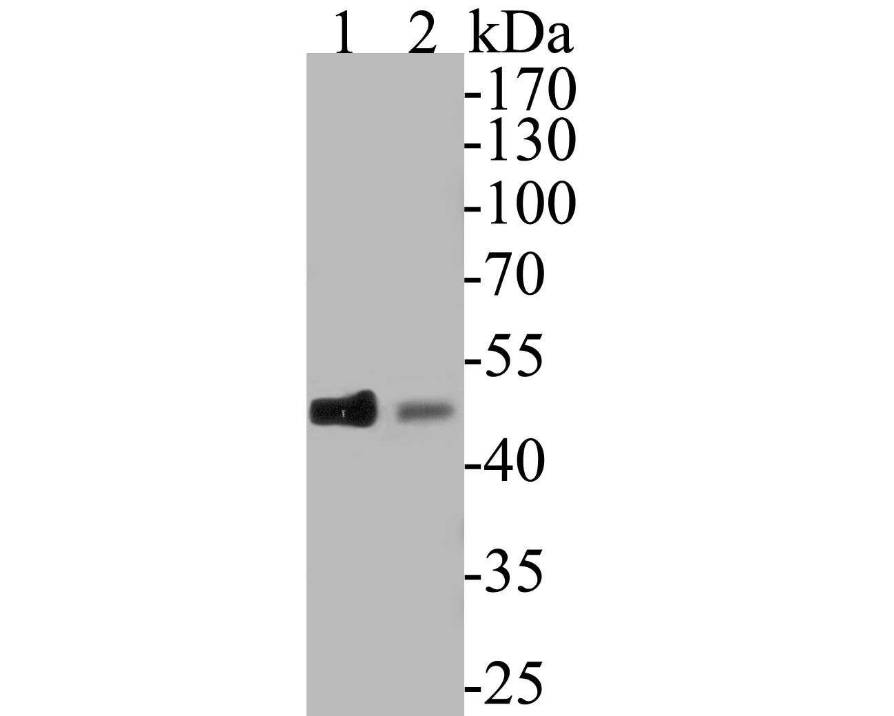
Fig1: Western blot analysis of CDC123 on different lysates. Proteins were transferred to a PVDF membrane and blocked with 5% BSA in PBS for 1 hour at room temperature. The primary antibody (ET7110-32, 1/500) was used in 5% BSA at room temperature for 2 hours. Goat Anti-Rabbit IgG - HRP Secondary Antibody (HA1001) at 1:5,000 dilution was used for 1 hour at room temperature. Positive control: Lane 1: K562 cell lysate Lane 2: A549 cell lysateApplication
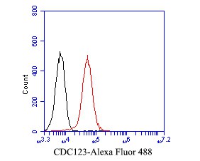
Fig2: Flow cytometric analysis of CDC123 was done on F9 cells. The cells were fixed, permeabilized and stained with the primary antibody (ET7110-32, 1/50) (red). After incubation of the primary antibody at room temperature for an hour, the cells were stained with a Alexa Fluor 488-conjugated Goat anti-Rabbit IgG Secondary antibody at 1/1000 dilution for 30 minutes.Unlabelled sample was used as a control (cells without incubation with primary antibody; black).| Product Name | CDC123 [JE53-14] |
|---|---|
| Antibody Type | Primary Antibodies |
| Antigen Alias | C10orf7 antibody CD123_HUMAN antibody CDC123 antibody Cell division cycle 123 antibody Cell division cycle 123 homolog antibody Cell division cycle protein 123 homolog antibody D123 antibody HT 1080 antibody HT-1080 antibody Protein D123 antibody PZ32 antibody |
| Product description | The eukaryotic cell division cycle consists of a number of gene-controlled sequences that involve cyclin dependent kinases (Cdks) and cell division control (Cdc) proteins. Cdc123 (Cell division cycle protein 123), also known as D123, is a 336 amino acid cytoplasmic protein that is involved in cell cycle control. Widely expressed with high expression in thymus, spleen, ovary, testis, small intestine and leukocytes, Cdc123 functions to destabilize Chfr (checkpoint with forkhead and ring finger domain) proteins which, when accumulated, block the G to S phase transition. Cdc123 prevents the Chfr proteins from collecting in the cell, thereby allowing the cell to enter the S phase. Due to its role in cell cycle control, Cdc123 is thought to be a basal marker for luminal breast cancers. |
| Immunogen | Synthetic peptide within C-terminal human CDC123. |
| Clonality | Monoclonal |
|---|---|
| Isotype | IgG |
| Host Species | Recombinant rabbit |
| Tested Applications | WBFC |
| WB:1:500-1:2,000 FC:1:50-1:100 |
|
| Species Reactivity | HumanMouse |
| Concentration | 1mg/ml |
| Alternative Names | C10orf7 antibody
CD123_HUMAN antibody
CDC123 antibody Cell division cycle 123 antibody Cell division cycle 123 homolog antibody Cell division cycle protein 123 homolog antibody D123 antibody HT 1080 antibody HT-1080 antibody Protein D123 antibody PZ32 antibody |
|---|---|
| Molecular Weight(MW) | 39 kDa (Predicted band size) |
| Cellular Localization | Cytoplasm. |
Application
Fig1: Western blot analysis of CDC123 on different lysates. Proteins were transferred to a PVDF membrane and blocked with 5% BSA in PBS for 1 hour at room temperature. The primary antibody (ET7110-32, 1/500) was used in 5% BSA at room temperature for 2 hours. Goat Anti-Rabbit IgG - HRP Secondary Antibody (HA1001) at 1:5,000 dilution was used for 1 hour at room temperature. Positive control: Lane 1: K562 cell lysate Lane 2: A549 cell lysate
Application
Fig2: Flow cytometric analysis of CDC123 was done on F9 cells. The cells were fixed, permeabilized and stained with the primary antibody (ET7110-32, 1/50) (red). After incubation of the primary antibody at room temperature for an hour, the cells were stained with a Alexa Fluor 488-conjugated Goat anti-Rabbit IgG Secondary antibody at 1/1000 dilution for 30 minutes.Unlabelled sample was used as a control (cells without incubation with primary antibody; black).| Positive Control | K562 cell lysate, A549 cell lysate, F9. |
|---|---|
| Application Notes | WB:1:500-1:2,000 FC:1:50-1:100 |
| Form | Liquid |
|---|---|
| Storage Instructions | Store at +4℃ after thawing. Aliquot store at -20℃. Avoid repeated freeze / thaw cycles. |
| Storage Buffer | 1*TBS (pH7.4), 1%BSA, 50%Glycerol. Preservative: 0.05% Sodium Azide. |


