Application
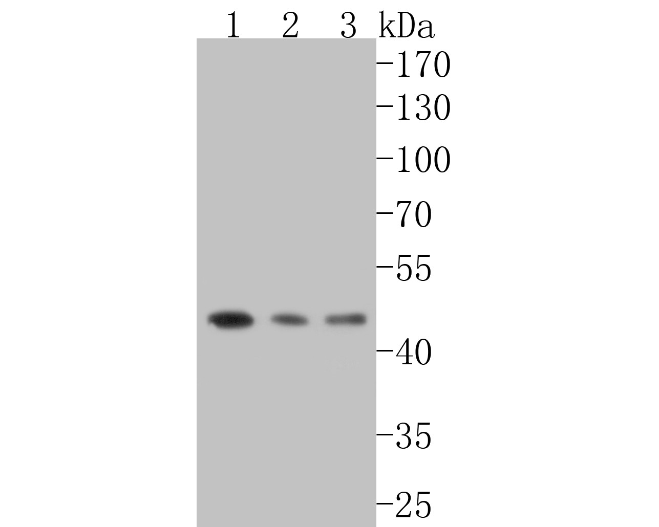
Fig1: Western blot analysis of DP1 on different lysates. Proteins were transferred to a PVDF membrane and blocked with 5% BSA in PBS for 1 hour at room temperature. The primary antibody (ET7110-43, 1/500) was used in 5% BSA at room temperature for 2 hours. Goat Anti-Rabbit IgG - HRP Secondary Antibody (HA1001) at 1:5,000 dilution was used for 1 hour at room temperature. Positive control: Lane 1: SiHa cell lysate Lane 2: SH-SY5Y cell lysate Lane 3: mouse thymus tissue lysateApplication
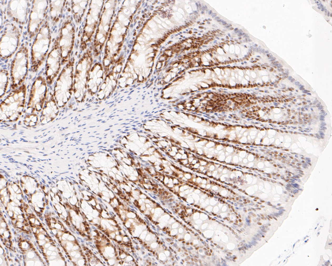
Fig2: Immunohistochemical analysis of paraffin-embedded rat large intestine tissue using anti-DP1 antibody. The section was pre-treated using heat mediated antigen retrieval with sodium citrate buffer (pH 6.0) for 20 minutes. The tissues were blocked in 5% BSA for 30 minutes at room temperature, washed with ddH2O and PBS, and then probed with the primary antibody (ET7110-43, 1/200) for 30 minutes at room temperature. The detection was performed using an HRP conjugated compact polymer system. DAB was used as the chromogen. Tissues were counterstained with hematoxylin and mounted with DPX.Application
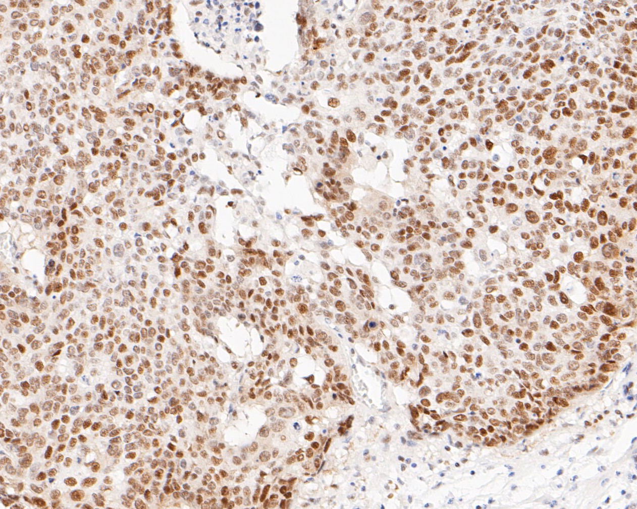
Fig3: Immunohistochemical analysis of paraffin-embedded human lung carcinoma tissue using anti-DP1 antibody. The section was pre-treated using heat mediated antigen retrieval with sodium citrate buffer (pH 6.0) for 20 minutes. The tissues were blocked in 5% BSA for 30 minutes at room temperature, washed with ddH2O and PBS, and then probed with the primary antibody (ET7110-43, 1/200) for 30 minutes at room temperature. The detection was performed using an HRP conjugated compact polymer system. DAB was used as the chromogen. Tissues were counterstained with hematoxylin and mounted with DPX.Application
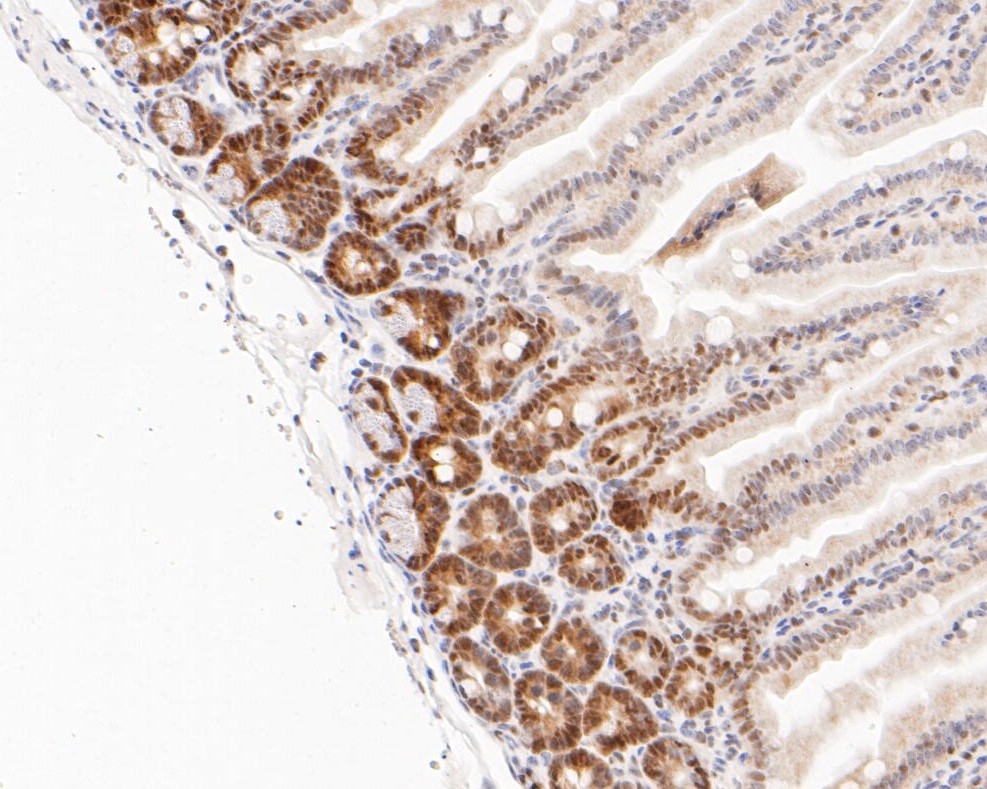
Fig4: Immunohistochemical analysis of paraffin-embedded mouse colon tissue using anti-DP1 antibody. The section was pre-treated using heat mediated antigen retrieval with sodium citrate buffer (pH 6.0) for 20 minutes. The tissues were blocked in 5% BSA for 30 minutes at room temperature, washed with ddH2O and PBS, and then probed with the primary antibody (ET7110-43, 1/200) for 30 minutes at room temperature. The detection was performed using an HRP conjugated compact polymer system. DAB was used as the chromogen. Tissues were counterstained with hematoxylin and mounted with DPX.Application
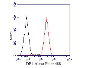
Fig5: Flow cytometric analysis of DP1 was done on THP-1 cells. The cells were fixed, permeabilized and stained with the primary antibody (ET7110-43, 1/50) (red). After incubation of the primary antibody at room temperature for an hour, the cells were stained with a Alexa Fluor 488-conjugated Goat anti-Rabbit IgG Secondary antibody at 1/1000 dilution for 30 minutes.Unlabelled sample was used as a control (cells without incubation with primary antibody; black).| Product Name | DP1 [JE52-55] |
|---|---|
| Antibody Type | Primary Antibodies |
| Antigen Alias | DP 1 antibody DP1 antibody DRTF1 antibody DRTF1-polypeptide 1 antibody E2F dimerization partner 1 antibody E2F related transcription factor antibody TFDP1 antibody TFDP1_HUMAN antibody Transcription factor Dp-1 antibody Transcription factor sequence-specific, DRTF1 antibody |
| Product description | This gene encodes a member of a family of transcription factors that heterodimerize with E2F proteins to enhance their DNA-binding activity and promote transcription from E2F target genes. The encoded protein functions as part of this complex to control the transcriptional activity of numerous genes involved in cell cycle progression from G1 to S phase. Alternative splicing results in multiple transcript variants. Pseudogenes of this gene are found on chromosomes 1, 15, and X. E2F factors bind to DNA as homodimers or heterodimers in association with dimerization partner DP1. TFDP1 may be the first example of a family of related transcription factors. |
| Immunogen | Synthetic peptide within C terminal human DP1. |
| Clonality | Monoclonal |
|---|---|
| Isotype | IgG |
| Host Species | Recombinant rabbit |
| Tested Applications | WBIHC-PFC |
| WB:1:500-1:2,000: IHC-P:1:50-1:200: FC:1:50-1:100: |
|
| Species Reactivity | HumanMouseRat |
| Concentration | 1mg/ml |
| Alternative Names | DP 1 antibody
DP1 antibody
DRTF1 antibody DRTF1-polypeptide 1 antibody E2F dimerization partner 1 antibody E2F related transcription factor antibody TFDP1 antibody TFDP1_HUMAN antibody Transcription factor Dp-1 antibody Transcription factor sequence-specific DRTF1 antibody |
|---|---|
| Molecular Weight(MW) | 45 kDa |
| Cellular Localization | Nucleus, cytoplasm. |
Application
Fig1: Western blot analysis of DP1 on different lysates. Proteins were transferred to a PVDF membrane and blocked with 5% BSA in PBS for 1 hour at room temperature. The primary antibody (ET7110-43, 1/500) was used in 5% BSA at room temperature for 2 hours. Goat Anti-Rabbit IgG - HRP Secondary Antibody (HA1001) at 1:5,000 dilution was used for 1 hour at room temperature. Positive control: Lane 1: SiHa cell lysate Lane 2: SH-SY5Y cell lysate Lane 3: mouse thymus tissue lysate
Application
Fig2: Immunohistochemical analysis of paraffin-embedded rat large intestine tissue using anti-DP1 antibody. The section was pre-treated using heat mediated antigen retrieval with sodium citrate buffer (pH 6.0) for 20 minutes. The tissues were blocked in 5% BSA for 30 minutes at room temperature, washed with ddH2O and PBS, and then probed with the primary antibody (ET7110-43, 1/200) for 30 minutes at room temperature. The detection was performed using an HRP conjugated compact polymer system. DAB was used as the chromogen. Tissues were counterstained with hematoxylin and mounted with DPX.
Application
Fig3: Immunohistochemical analysis of paraffin-embedded human lung carcinoma tissue using anti-DP1 antibody. The section was pre-treated using heat mediated antigen retrieval with sodium citrate buffer (pH 6.0) for 20 minutes. The tissues were blocked in 5% BSA for 30 minutes at room temperature, washed with ddH2O and PBS, and then probed with the primary antibody (ET7110-43, 1/200) for 30 minutes at room temperature. The detection was performed using an HRP conjugated compact polymer system. DAB was used as the chromogen. Tissues were counterstained with hematoxylin and mounted with DPX.
Application
Fig4: Immunohistochemical analysis of paraffin-embedded mouse colon tissue using anti-DP1 antibody. The section was pre-treated using heat mediated antigen retrieval with sodium citrate buffer (pH 6.0) for 20 minutes. The tissues were blocked in 5% BSA for 30 minutes at room temperature, washed with ddH2O and PBS, and then probed with the primary antibody (ET7110-43, 1/200) for 30 minutes at room temperature. The detection was performed using an HRP conjugated compact polymer system. DAB was used as the chromogen. Tissues were counterstained with hematoxylin and mounted with DPX.
Application
Fig5: Flow cytometric analysis of DP1 was done on THP-1 cells. The cells were fixed, permeabilized and stained with the primary antibody (ET7110-43, 1/50) (red). After incubation of the primary antibody at room temperature for an hour, the cells were stained with a Alexa Fluor 488-conjugated Goat anti-Rabbit IgG Secondary antibody at 1/1000 dilution for 30 minutes.Unlabelled sample was used as a control (cells without incubation with primary antibody; black).| Positive Control | SiHa cell lysate, SH-SY5Y cell lysate, mouse thymus tissue lysate, rat large intestine tissue, human lung carcinoma tissue, mouse colon tissue, THP-1. |
|---|---|
| Application Notes | WB:1:500-1:2,000: IHC-P:1:50-1:200: FC:1:50-1:100: |
| Form | Liquid |
|---|---|
| Storage Instructions | Store at +4℃ after thawing. Aliquot store at -20℃. Avoid repeated freeze / thaw cycles. |
| Storage Buffer | 1*TBS (pH7.4), 1%BSA, 50%Glycerol. Preservative: 0.05% Sodium Azide. |


