Application
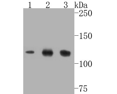
Fig1: Western blot analysis of SIRT1 on different lysates. Proteins were transferred to a PVDF membrane and blocked with 5% BSA in PBS for 1 hour at room temperature. The primary antibody (ET7110-49, 1/500) was used in 5% BSA at room temperature for 2 hours. Goat Anti-Rabbit IgG - HRP Secondary Antibody (HA1001) at 1:5,000 dilution was used for 1 hour at room temperature. Positive control: Lane 1: Jurkat cell lysate Lane 2: HepG2 cell lysate Lane 3: Hela cell lysateApplication
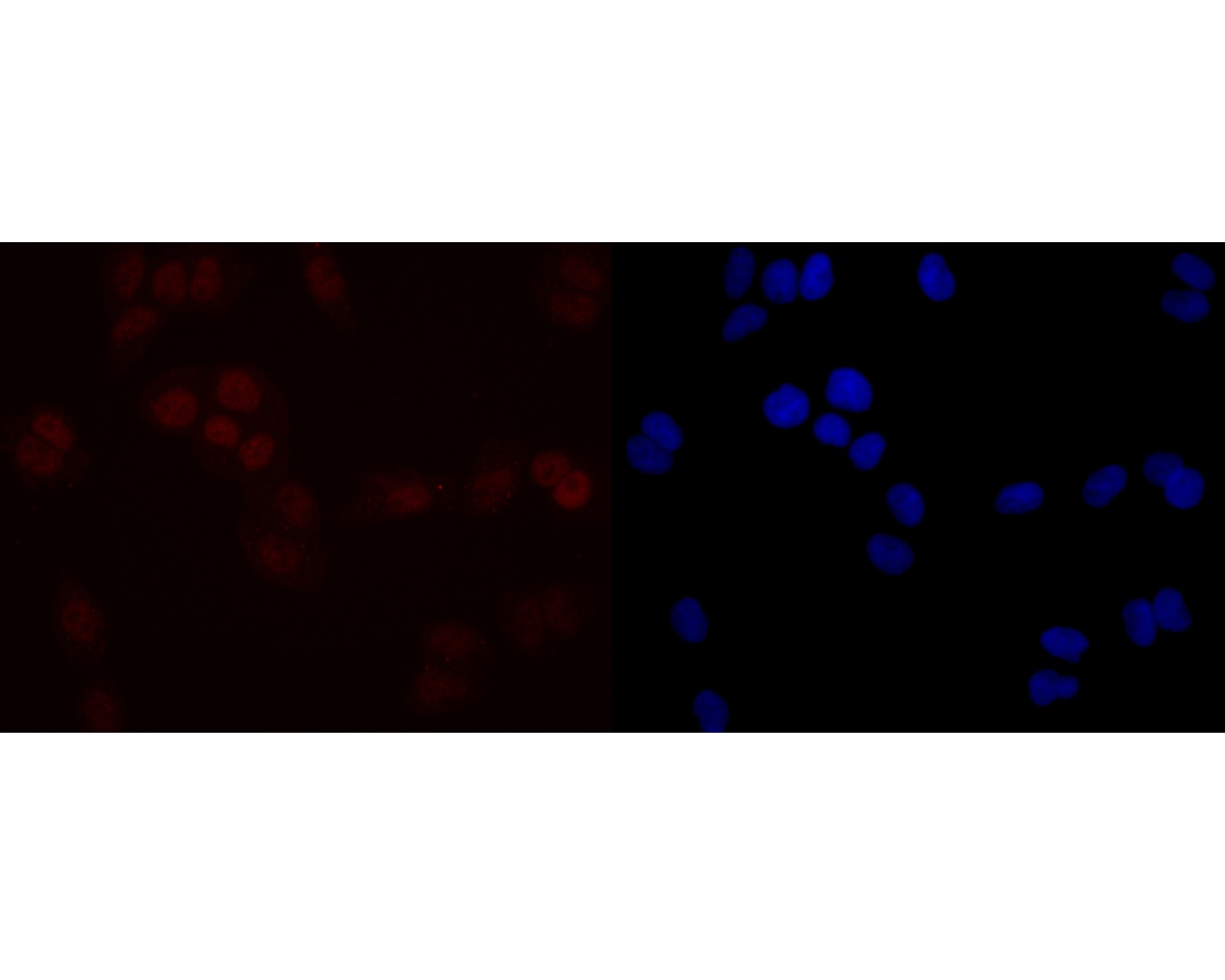
Fig2: ICC staining of SIRT1 in Hela cells (red). Formalin fixed cells were permeabilized with 0.1% Triton X-100 in TBS for 10 minutes at room temperature and blocked with 1% Blocker BSA for 15 minutes at room temperature. Cells were probed with the primary antibody (ET7110-49, 1/50) for 1 hour at room temperature, washed with PBS. Alexa Fluor®594 Goat anti-Rabbit IgG was used as the secondary antibody at 1/1,000 dilution. The nuclear counter stain is DAPI (blue).| Product Name | SIRT1 [JE11-04] |
|---|---|
| Antibody Type | Primary Antibodies |
| Antigen Alias | 75SirT1 antibody hSIR2 antibody hSIRT1 antibody HST2 antibody HST2, S. cerevisiae, homolog of antibody NAD dependent deacetylase sirtuin 1 antibody NAD dependent protein deacetylase sirtuin 1 antibody NAD-dependent deacetylase sirtuin-1 antibody OTTHUMP00000198111 antibody OTTHUMP00000198112 antibody Regulatory protein SIR2 homolog 1 antibody SIR1_HUMAN antibody SIR2 antibody SIR2 like 1 antibody SIR2 like protein 1 antibody SIR2, S.cerevisiae, homolog-like 1 antibody SIR2-like protein 1 antibody SIR2ALPHA antibody SIR2L1 antibody Sirt1 antibody SirtT1 75 kDa fragment antibody Sirtuin (silent mating type information regulation 2 homolog) 1 (S. cerevisiae) antibody Sirtuin 1 antibody Sirtuin type 1 antibody |
| Product description | The silent information regulator (SIR2) family of genes are highly-conserved from prokaryotes to eukaryotes and are involved in diverse processes, including transcriptional regulation, cell cycle progression, DNA-damage repair and aging. In S. cerevisiae, Sir2p deacetylates histones in an NAD-dependent manner, which regulates silencing at the telomeric, rDNA and silent mating-type loci. Sir2p is the founding member of a large family, designated sirtuins, which contain a conserved catalytic domain. The human homologs, which include SIRT1-7, are divided into four main branches: SIRT1-3 are class I, SIRT4 is class II, SIRT5 is class III and SIRT6-7 are class IV. SIRT1 has the closest homology to the yeast Sir2p and is widely expressed in fetal and adult tissues. SIRT1 is highly expressed in heart, brain and skeletal muscle, with low expression in lung and placenta. SIRT1 regulates the p53-dependent DNA damage response pathway by binding to and deacetylating p53, specifically at Lys 382. |
| Immunogen | Synthetic peptide within C terminal human SIRT1. |
| Clonality | Monoclonal |
|---|---|
| Isotype | IgG |
| Host Species | Recombinant rabbit |
| Tested Applications | WBICC |
| WB:1:500-1:2,000: ICC:1:50-1:100: |
|
| Species Reactivity | Human |
| Concentration | 1mg/mL |
| Alternative Names | 75SirT1 antibody
hSIR2 antibody
hSIRT1 antibody HST2 antibody HST2 S. cerevisiae homolog of antibody NAD dependent deacetylase sirtuin 1 antibody NAD dependent protein deacetylase sirtuin 1 antibody NAD-dependent deacetylase sirtuin-1 antibody OTTHUMP00000198111 antibody OTTHUMP00000198112 antibody Regulatory protein SIR2 homolog 1 antibody SIR1_HUMAN antibody SIR2 antibody SIR2 like 1 antibody SIR2 like protein 1 antibody SIR2 S.cerevisiae homolog-like 1 antibody SIR2-like protein 1 antibody SIR2ALPHA antibody SIR2L1 antibody Sirt1 antibody SirtT1 75 kDa fragment antibody Sirtuin (silent mating type information regulation 2 homolog) 1 (S. cerevisiae) antibody Sirtuin 1 antibody Sirtuin type 1 antibody |
|---|---|
| Molecular Weight(MW) | Predicted band size 82 kDa. |
| Cellular Localization | PML body, nucleus, cytoplasm, mitochondrion. |
| SwissProt ID | Q96EB6 |
|---|
Application
Fig1: Western blot analysis of SIRT1 on different lysates. Proteins were transferred to a PVDF membrane and blocked with 5% BSA in PBS for 1 hour at room temperature. The primary antibody (ET7110-49, 1/500) was used in 5% BSA at room temperature for 2 hours. Goat Anti-Rabbit IgG - HRP Secondary Antibody (HA1001) at 1:5,000 dilution was used for 1 hour at room temperature. Positive control: Lane 1: Jurkat cell lysate Lane 2: HepG2 cell lysate Lane 3: Hela cell lysate
Application
Fig2: ICC staining of SIRT1 in Hela cells (red). Formalin fixed cells were permeabilized with 0.1% Triton X-100 in TBS for 10 minutes at room temperature and blocked with 1% Blocker BSA for 15 minutes at room temperature. Cells were probed with the primary antibody (ET7110-49, 1/50) for 1 hour at room temperature, washed with PBS. Alexa Fluor®594 Goat anti-Rabbit IgG was used as the secondary antibody at 1/1,000 dilution. The nuclear counter stain is DAPI (blue).| Positive Control | Jurkat cell lysate, HepG2 cell lysate, Hela cell lysate, Hela. |
|---|---|
| Application Notes | WB:1:500-1:2,000: ICC:1:50-1:100: |
| Form | Liquid |
|---|---|
| Storage Instructions | Store at +4℃ after thawing. Aliquot store at -20℃. Avoid repeated freeze / thaw cycles. |
| Storage Buffer | 1*TBS (pH7.4), 1%BSA, 50%Glycerol. Preservative: 0.05% Sodium Azide. |


