Application
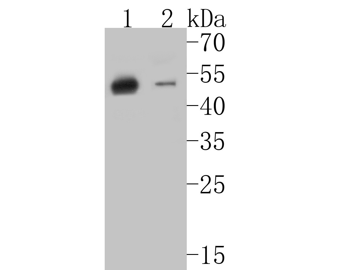
Fig1: Western blot analysis of Cytokeratin 20 on different lysates. Proteins were transferred to a PVDF membrane and blocked with 5% BSA in PBS for 1 hour at room temperature. The primary antibody (ET7110-54, 1/500) was used in 5% BSA at room temperature for 2 hours. Goat Anti-Rabbit IgG - HRP Secondary Antibody (HA1001) at 1:5,000 dilution was used for 1 hour at room temperature. Positive control: Lane 1: human stomach tissue lysate Lane 2: SK-Br-3 cell lysateApplication
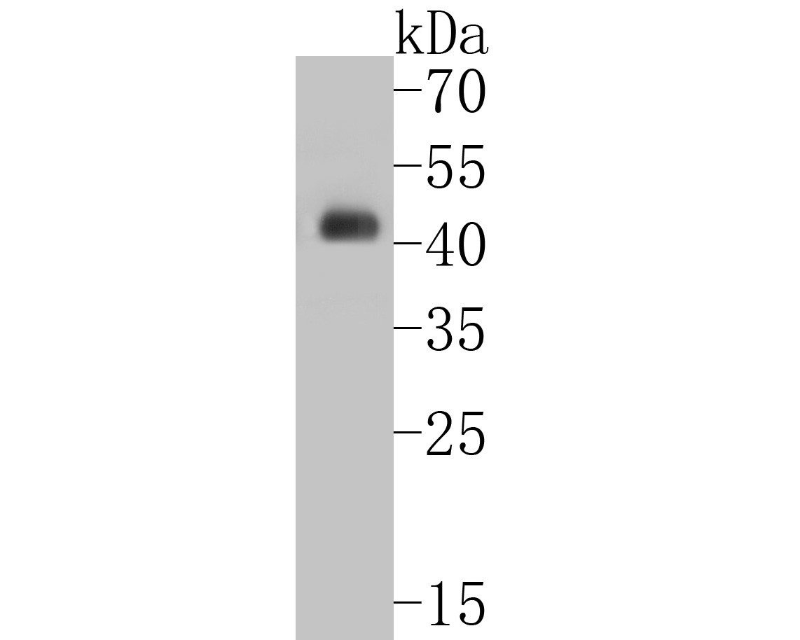
Fig2: Western blot analysis of Cytokeratin 20 on human small intestine tissue lysates. Proteins were transferred to a PVDF membrane and blocked with 5% BSA in PBS for 1 hour at room temperature. The primary antibody (ET7110-54, 1/5000) was used in 5% BSA at room temperature for 2 hours. Goat Anti-Rabbit IgG - HRP Secondary Antibody (HA1001) at 1:5,000 dilution was used for 1 hour at room temperature.Application
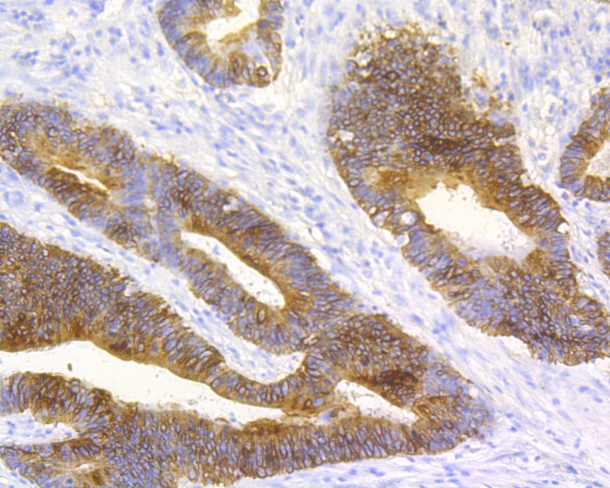
Fig3: Immunohistochemical analysis of paraffin-embedded human colon carcinoma tissue using anti-Cytokeratin 20 antibody. The section was pre-treated using heat mediated antigen retrieval with Tris-EDTA buffer (pH 8.0-8.4) for 20 minutes.The tissues were blocked in 5% BSA for 30 minutes at room temperature, washed with ddH2O and PBS, and then probed with the primary antibody (ET7110-54, 1/50) for 30 minutes at room temperature. The detection was performed using an HRP conjugated compact polymer system. DAB was used as the chromogen. Tissues were counterstained with hematoxylin and mounted with DPX.Application
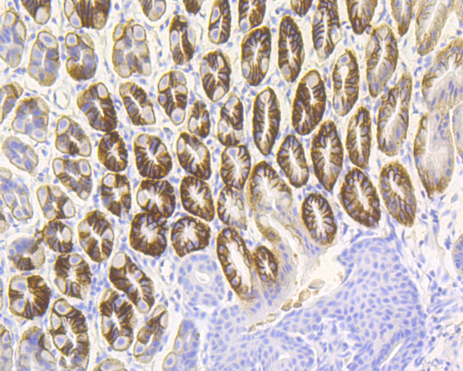
Fig4: Immunohistochemical analysis of paraffin-embedded mouse stomach tissue using anti-Cytokeratin 20 antibody. The section was pre-treated using heat mediated antigen retrieval with Tris-EDTA buffer (pH 8.0-8.4) for 20 minutes.The tissues were blocked in 5% BSA for 30 minutes at room temperature, washed with ddH2O and PBS, and then probed with the primary antibody (ET7110-54, 1/50) for 30 minutes at room temperature. The detection was performed using an HRP conjugated compact polymer system. DAB was used as the chromogen. Tissues were counterstained with hematoxylin and mounted with DPX.| Product Name | Cytokeratin 20 [JE20-39] |
|---|---|
| Antibody Type | Primary Antibodies |
| Antigen Alias | CD20 antibody CK 20 antibody CK-20 antibody CK20 antibody Cytokeratin-20 antibody Cytokeratin20 antibody K1C20_HUMAN antibody K20 antibody KA20 antibody Keratin 20 antibody keratin 20, type I antibody keratin 21, rat, homolog of antibody Keratin antibody Keratin type I cytoskeletal 20 antibody Keratin-20 antibody Keratin20 antibody KRT 20 antibody KRT 21 antibody KRT20 antibody KRT21 antibody MGC35423 antibody OTTHUMP00000164518 antibody Protein IT antibody type I cytoskeletal 20 antibody |
| Product description | The protein encoded by this gene is a member of the keratin family. The keratins are intermediate filament proteins responsible for the structural integrity of epithelial cells and are subdivided into cytokeratins and hair keratins. The type I cytokeratins consist of acidic proteins which are arranged in pairs of heterotypic keratin chains. This cytokeratin is a major cellular protein of mature enterocytes and goblet cells and is specifically expressed in the gastric and intestinal mucosa. Keratin 20 is a type I cytokeratin. It is a major cellular protein of mature enterocytes and goblet cells and is specifically found in the gastric and intestinal mucosa. In immunohistochemistry, antibodies to CK20 can be used to identify a range of adenocarcinoma arising from epithelia that normally contain the CK20 protein. For example, the protein is commonly found in colorectal cancer, transitional cell carcinomas and in Merkel cell carcinoma, but is absent in lung cancer, prostate cancer, and non-mucinous ovarian cancer. It is often used in combination with antibodies to CK7 to distinguish different types of glandular tumour. |
| Immunogen | Synthetic peptide within C terminal Human Cytokeratin 20. |
| Clonality | Monoclonal |
|---|---|
| Isotype | IgG |
| Host Species | Recombinant rabbit |
| Tested Applications | WBIHC-P |
| WB:1:500-1:5,000: IHC-P:1:50-1:200: |
|
| Species Reactivity | HumanMouse |
| Concentration | 1mg/mL |
| Alternative Names | CD20 antibody
CK 20 antibody
CK-20 antibody CK20 antibody Cytokeratin-20 antibody Cytokeratin20 antibody K1C20_HUMAN antibody K20 antibody KA20 antibody Keratin 20 antibody keratin 20 type I antibody keratin 21 rat homolog of antibody Keratin antibody Keratin type I cytoskeletal 20 antibody Keratin-20 antibody Keratin20 antibody KRT 20 antibody KRT 21 antibody KRT20 antibody KRT21 antibody MGC35423 antibody OTTHUMP00000164518 antibody Protein IT antibody type I cytoskeletal 20 antibody |
|---|---|
| Molecular Weight(MW) | 48 kDa |
| Cellular Localization | Cytoplasm. |
Application
Fig1: Western blot analysis of Cytokeratin 20 on different lysates. Proteins were transferred to a PVDF membrane and blocked with 5% BSA in PBS for 1 hour at room temperature. The primary antibody (ET7110-54, 1/500) was used in 5% BSA at room temperature for 2 hours. Goat Anti-Rabbit IgG - HRP Secondary Antibody (HA1001) at 1:5,000 dilution was used for 1 hour at room temperature. Positive control: Lane 1: human stomach tissue lysate Lane 2: SK-Br-3 cell lysate
Application
Fig2: Western blot analysis of Cytokeratin 20 on human small intestine tissue lysates. Proteins were transferred to a PVDF membrane and blocked with 5% BSA in PBS for 1 hour at room temperature. The primary antibody (ET7110-54, 1/5000) was used in 5% BSA at room temperature for 2 hours. Goat Anti-Rabbit IgG - HRP Secondary Antibody (HA1001) at 1:5,000 dilution was used for 1 hour at room temperature.
Application
Fig3: Immunohistochemical analysis of paraffin-embedded human colon carcinoma tissue using anti-Cytokeratin 20 antibody. The section was pre-treated using heat mediated antigen retrieval with Tris-EDTA buffer (pH 8.0-8.4) for 20 minutes.The tissues were blocked in 5% BSA for 30 minutes at room temperature, washed with ddH2O and PBS, and then probed with the primary antibody (ET7110-54, 1/50) for 30 minutes at room temperature. The detection was performed using an HRP conjugated compact polymer system. DAB was used as the chromogen. Tissues were counterstained with hematoxylin and mounted with DPX.
Application
Fig4: Immunohistochemical analysis of paraffin-embedded mouse stomach tissue using anti-Cytokeratin 20 antibody. The section was pre-treated using heat mediated antigen retrieval with Tris-EDTA buffer (pH 8.0-8.4) for 20 minutes.The tissues were blocked in 5% BSA for 30 minutes at room temperature, washed with ddH2O and PBS, and then probed with the primary antibody (ET7110-54, 1/50) for 30 minutes at room temperature. The detection was performed using an HRP conjugated compact polymer system. DAB was used as the chromogen. Tissues were counterstained with hematoxylin and mounted with DPX.| Positive Control | Human stomach tissue lysate, SK-Br-3 cell lysate, human small intestine tissue lysates, human colon carcinoma tissue, mouse stomach tissue. |
|---|---|
| Application Notes | WB:1:500-1:5,000: IHC-P:1:50-1:200: |
| Form | Liquid |
|---|---|
| Storage Instructions | Store at +4℃ after thawing. Aliquot store at -20℃. Avoid repeated freeze / thaw cycles. |
| Storage Buffer | 1*TBS (pH7.4), 1%BSA, 50%Glycerol. Preservative: 0.05% Sodium Azide. |


