Application
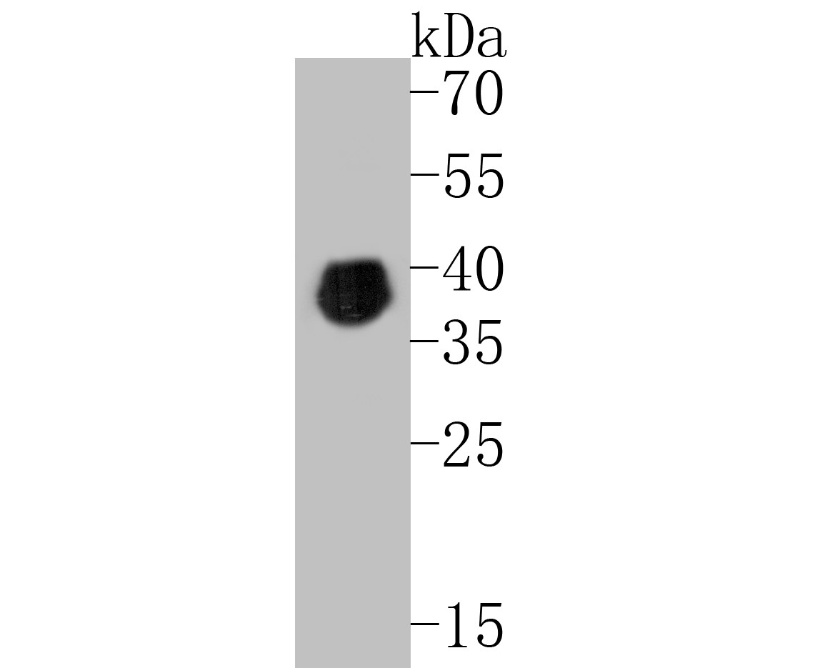
Fig1: Western blot analysis of Pax2 on mouse testis tissue lysates. Proteins were transferred to a PVDF membrane and blocked with 5% BSA in PBS for 1 hour at room temperature. The primary antibody (ET7110-57, 1/500) was used in 5% BSA at room temperature for 2 hours. Goat Anti-Rabbit IgG - HRP Secondary Antibody (HA1001) at 1:5,000 dilution was used for 1 hour at room temperature.Application
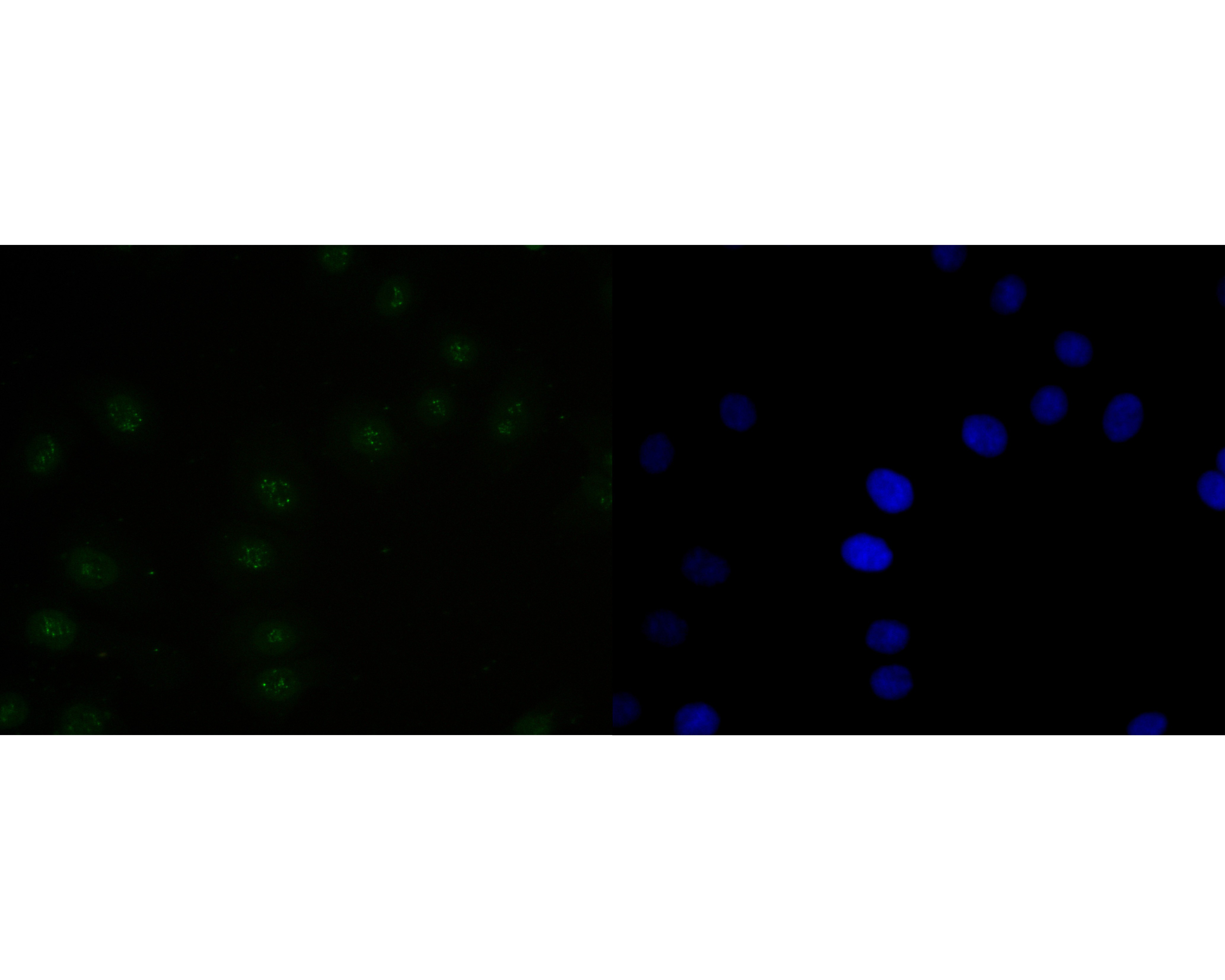
Fig2: ICC staining of Pax2 in SKOV-3 cells (green). Formalin fixed cells were permeabilized with 0.1% Triton X-100 in TBS for 10 minutes at room temperature and blocked with 1% Blocker BSA for 15 minutes at room temperature. Cells were probed with the primary antibody (ET7110-57, 1/100) for 1 hour at room temperature, washed with PBS. Alexa Fluor®488 Goat anti-Rabbit IgG was used as the secondary antibody at 1/1,000 dilution. The nuclear counter stain is DAPI (blue).Application
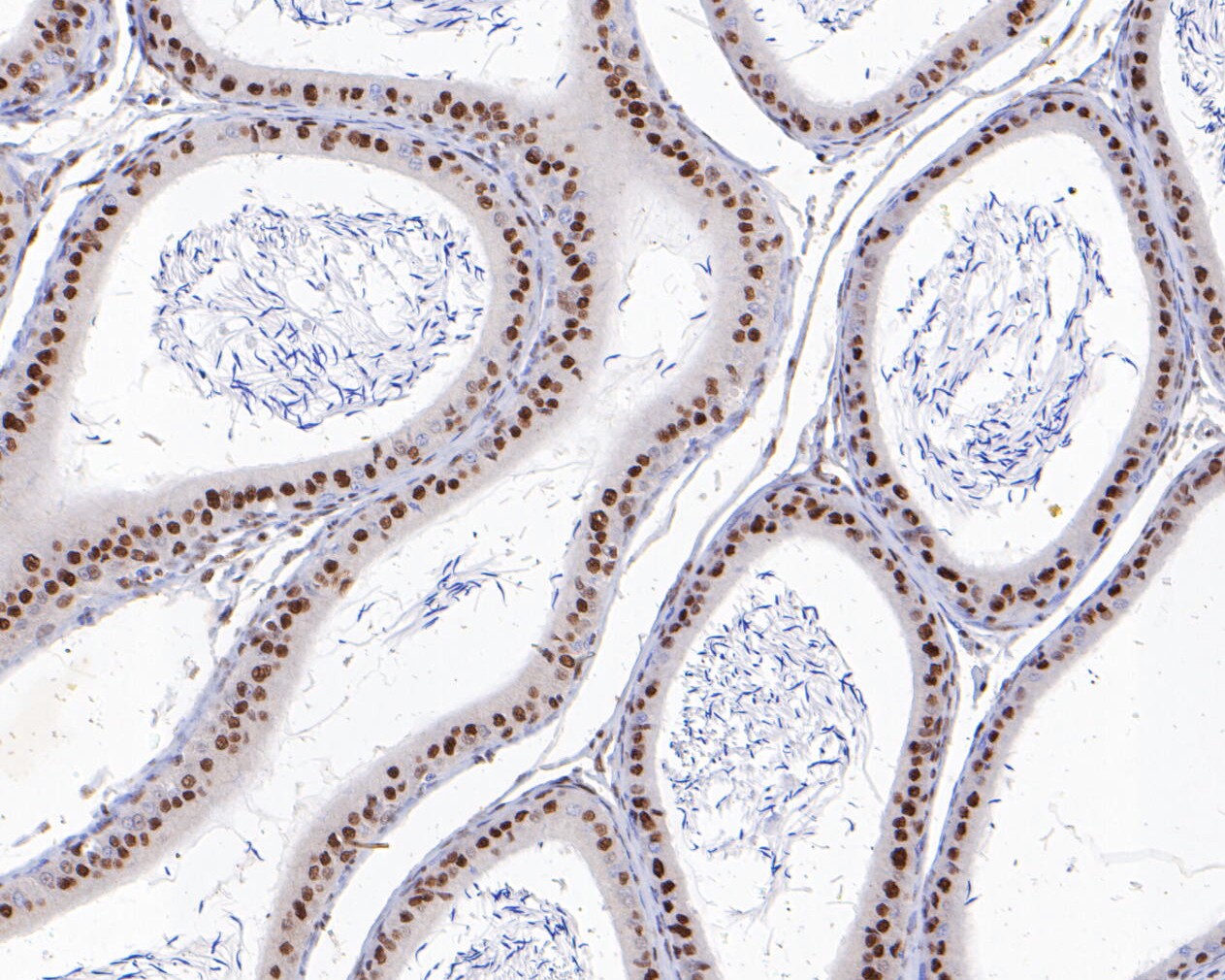
Fig3: Immunohistochemical analysis of paraffin-embedded rat epididymis tissue using anti-Pax2 antibody. The section was pre-treated using heat mediated antigen retrieval with sodium citrate buffer (pH 6.0) for 20 minutes. The tissues were blocked in 5% BSA for 30 minutes at room temperature, washed with ddH2O and PBS, and then probed with the primary antibody (ET7110-57, 1/200) for 30 minutes at room temperature. The detection was performed using an HRP conjugated compact polymer system. DAB was used as the chromogen. Tissues were counterstained with hematoxylin and mounted with DPX.Application
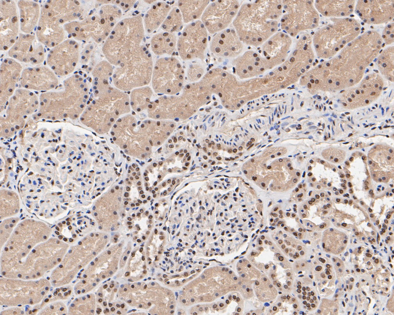
Fig4: Immunohistochemical analysis of paraffin-embedded human kidney tissue using anti-Pax2 antibody. The section was pre-treated using heat mediated antigen retrieval with sodium citrate buffer (pH 6.0) for 20 minutes. The tissues were blocked in 5% BSA for 30 minutes at room temperature, washed with ddH2O and PBS, and then probed with the primary antibody (ET7110-57, 1/200) for 30 minutes at room temperature. The detection was performed using an HRP conjugated compact polymer system. DAB was used as the chromogen. Tissues were counterstained with hematoxylin and mounted with DPX.Application
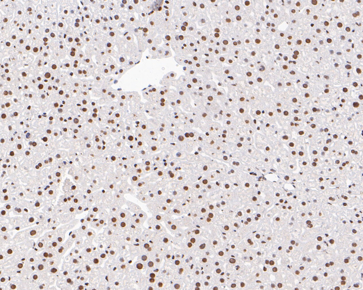
Fig5: Immunohistochemical analysis of paraffin-embedded mouse liver tissue using anti-Pax2 antibody. The section was pre-treated using heat mediated antigen retrieval with sodium citrate buffer (pH 6.0) for 20 minutes. The tissues were blocked in 5% BSA for 30 minutes at room temperature, washed with ddH2O and PBS, and then probed with the primary antibody (ET7110-57, 1/200) for 30 minutes at room temperature. The detection was performed using an HRP conjugated compact polymer system. DAB was used as the chromogen. Tissues were counterstained with hematoxylin and mounted with DPX.Application
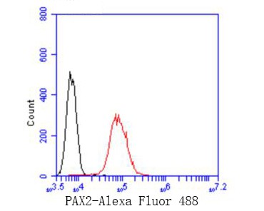
Fig6: Flow cytometric analysis of Pax2 was done on 293T cells. The cells were fixed, permeabilized and stained with the primary antibody (ET7110-57, 1/50) (red). After incubation of the primary antibody at room temperature for an hour, the cells were stained with a Alexa Fluor 488-conjugated Goat anti-Rabbit IgG Secondary antibody at 1/1000 dilution for 30 minutes.Unlabelled sample was used as a control (cells without incubation with primary antibody; black).| Product Name | Pax2 [JE25-49] |
|---|---|
| Antibody Type | Primary Antibodies |
| Antigen Alias | FSGS7 antibody Paired box 2 antibody Paired box gene 2 antibody paired box homeotic gene 2 antibody paired box protein 2 antibody Paired box protein Pax 2 antibody Paired box protein Pax-2 antibody Paired box protein Pax2 antibody PAPRS antibody Pax 2 antibody |
| Product description | PAX2 encodes paired box gene 2, one of many human homologues of the Drosophila melanogaster gene prd. The central feature of this transcription factor gene family is the conserved DNA-binding paired box domain. PAX2 is believed to be a target of transcriptional suppression by the tumor suppressor gene WT1. Pax 2 is a transcription factor controlled by the signaling molecules Wnt1 and Fgf8. Pax2 along with other transcription factors Pax5, Pax8, En1, and En 2 are expressed across the Otx2-Gbx2 boundary in the mid-hindbrain region. These transcription factors work with the signaling molecules Wnt1 and Fgf8 to maintain the MHB organizer. The MHB controls midbrain and cerebellum development. Pax2 is the earliest known gene to be expressed across the Otx2-Gbx2 boundary. It is first expressed in the late primitive streak stage and is expressed in a narrow ring centered at the MHB during somitogenesis. Transgene expression of the mid-hindbrain and developing kidney is directed by Pax2. There are three distinct MHB-specific enhancers in the upstream region of Pax2. Expression at the MHB from the four-somite stage onwards is directed by the two late enhancers in the proximal and distal regions of Pax2. The early enhancer located in the intermediate region activates the mid-hindbrain region of late gastrula embryos. The activation of Pax2, Pax5, and Pax8 is a conserved feature of all vertebrates. |
| Immunogen | Synthetic peptide within N terminal Human Pax2. |
| Clonality | Monoclonal |
|---|---|
| Isotype | IgG |
| Host Species | Recombinant rabbit |
| Tested Applications | WBICCIHC-PFC |
| WB:1:500-1:2,000: ICC:1:50-1:100: IHC-P:1:50-1:200: FC:1:50-1:100: |
|
| Species Reactivity | HumanMouseRat |
| Concentration | 1mg/mL |
| Alternative Names | FSGS7 antibody
Paired box 2 antibody
Paired box gene 2 antibody paired box homeotic gene 2 antibody paired box protein 2 antibody Paired box protein Pax 2 antibody Paired box protein Pax-2 antibody Paired box protein Pax2 antibody PAPRS antibody Pax 2 antibody |
|---|---|
| Molecular Weight(MW) | 45 kDa |
| Cellular Localization | Nucleus. |
Application
Fig1: Western blot analysis of Pax2 on mouse testis tissue lysates. Proteins were transferred to a PVDF membrane and blocked with 5% BSA in PBS for 1 hour at room temperature. The primary antibody (ET7110-57, 1/500) was used in 5% BSA at room temperature for 2 hours. Goat Anti-Rabbit IgG - HRP Secondary Antibody (HA1001) at 1:5,000 dilution was used for 1 hour at room temperature.
Application
Fig2: ICC staining of Pax2 in SKOV-3 cells (green). Formalin fixed cells were permeabilized with 0.1% Triton X-100 in TBS for 10 minutes at room temperature and blocked with 1% Blocker BSA for 15 minutes at room temperature. Cells were probed with the primary antibody (ET7110-57, 1/100) for 1 hour at room temperature, washed with PBS. Alexa Fluor®488 Goat anti-Rabbit IgG was used as the secondary antibody at 1/1,000 dilution. The nuclear counter stain is DAPI (blue).
Application
Fig3: Immunohistochemical analysis of paraffin-embedded rat epididymis tissue using anti-Pax2 antibody. The section was pre-treated using heat mediated antigen retrieval with sodium citrate buffer (pH 6.0) for 20 minutes. The tissues were blocked in 5% BSA for 30 minutes at room temperature, washed with ddH2O and PBS, and then probed with the primary antibody (ET7110-57, 1/200) for 30 minutes at room temperature. The detection was performed using an HRP conjugated compact polymer system. DAB was used as the chromogen. Tissues were counterstained with hematoxylin and mounted with DPX.
Application
Fig4: Immunohistochemical analysis of paraffin-embedded human kidney tissue using anti-Pax2 antibody. The section was pre-treated using heat mediated antigen retrieval with sodium citrate buffer (pH 6.0) for 20 minutes. The tissues were blocked in 5% BSA for 30 minutes at room temperature, washed with ddH2O and PBS, and then probed with the primary antibody (ET7110-57, 1/200) for 30 minutes at room temperature. The detection was performed using an HRP conjugated compact polymer system. DAB was used as the chromogen. Tissues were counterstained with hematoxylin and mounted with DPX.
Application
Fig5: Immunohistochemical analysis of paraffin-embedded mouse liver tissue using anti-Pax2 antibody. The section was pre-treated using heat mediated antigen retrieval with sodium citrate buffer (pH 6.0) for 20 minutes. The tissues were blocked in 5% BSA for 30 minutes at room temperature, washed with ddH2O and PBS, and then probed with the primary antibody (ET7110-57, 1/200) for 30 minutes at room temperature. The detection was performed using an HRP conjugated compact polymer system. DAB was used as the chromogen. Tissues were counterstained with hematoxylin and mounted with DPX.
Application
Fig6: Flow cytometric analysis of Pax2 was done on 293T cells. The cells were fixed, permeabilized and stained with the primary antibody (ET7110-57, 1/50) (red). After incubation of the primary antibody at room temperature for an hour, the cells were stained with a Alexa Fluor 488-conjugated Goat anti-Rabbit IgG Secondary antibody at 1/1000 dilution for 30 minutes.Unlabelled sample was used as a control (cells without incubation with primary antibody; black).| Positive Control | Mouse testis tissue lysates, SKOV-3, rat epididymis tissue, human kidney tissue, mouse liver tissue, 293T. |
|---|---|
| Application Notes | WB:1:500-1:2,000: ICC:1:50-1:100: IHC-P:1:50-1:200: FC:1:50-1:100: |
| Form | Liquid |
|---|---|
| Storage Instructions | Store at +4℃ after thawing. Aliquot store at -20℃. Avoid repeated freeze / thaw cycles. |
| Storage Buffer | 1*TBS (pH7.4), 1%BSA, 50%Glycerol. Preservative: 0.05% Sodium Azide. |


