Application
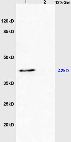
Fig1: Sample: Heart (Mouse) Lysate at 40 ug Liver (Mouse) Lysate at 40 ug Primary: Anti-EDG3 (ER1907-94) at 1/300 dilution Secondary: HRP conjugated Goat-Anti-rabbit IgG (bs-0295G-HRP) at 1/5000 dilution Predicted band size: 42 kD Observed band size: 42 kDApplication
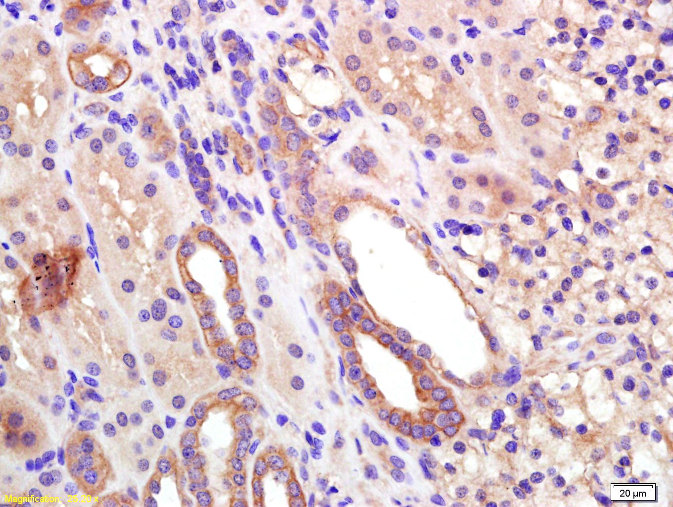
Fig2: Tissue/cell: human kidney tissue; 4% Paraformaldehyde-fixed and paraffin-embedded; Antigen retrieval: citrate buffer ( 0.01M, pH 6.0 ), Boiling bathing for 15min; Block endogenous peroxidase by 3% Hydrogen peroxide for 30min; Blocking buffer (normal goat serum,C-0005) at 37℃ for 20 min; Incubation: Anti-EDG3 Polyclonal Antibody, Unconjugated(ER1907-94) 1:200, overnight at 4℃, followed by conjugation to the secondary antibody(SP-0023) and DAB(C-0010) stainingApplication
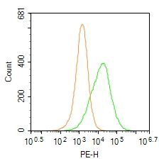
Fig3: Blank control: Hela. Primary Antibody (green line): Rabbit Anti-EDG3 antibody (ER1907-94) Dilution: 0.2μg /10^6 cells; Isotype Control Antibody (orange line): Rabbit IgG . Secondary Antibody : Goat anti-rabbit IgG-PE Dilution: 1μg /test. Protocol The cells were incubated in 5%BSA to block non-specific protein-protein interactions for 30 min at at room temperature .Cells stained with Primary Antibody for 30 min at room temperature. The secondary antibody used for 40 min at room temperature. Acquisition of 20,000 events was performed.WB
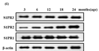
C, Western blot images and their quantification of sphingosine-1-phosphate receptor-3 (S1PR1-3) accumulation in the colon muscle layers from different groups of rats.IF
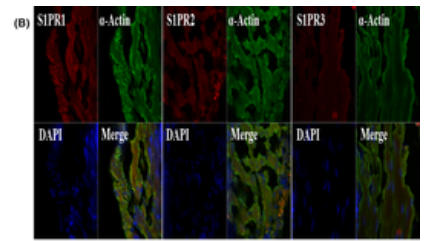
B, Representative double immunofluorescence staining of colon smooth muscle (CSM) for α-actin and sphingosine-1-phosphate receptor-3 (S1PR1-3). Nuclei were identified by DAPI staining.| Product Name | EDG3 |
|---|---|
| Antibody Type | Primary Antibodies |
| Product description | Receptor for the lysosphingolipid sphingosine 1-phosphate (S1P). S1P is a bioactive lysophospholipid that elicits diverse physiological effect on most types of cells and tissues. When expressed in rat HTC4 hepatoma cells, is capable of mediating S1P-induced cell proliferation and suppression of apoptosis. |
| Immunogen | KLH conjugated synthetic peptide derived from human EDG3 145-250/378 |
| Clonality | Polyclonal |
|---|---|
| Isotype | IgG |
| Host Species | Rabbit |
| Tested Applications | FCIHC-PWB |
| WB:1:500-2000: IHC-P:1:400-800: FC:0.2u/test: |
|
| Species Reactivity | CowHumanMouseRabbitRat |
| Concentration | 1mg/ml |
| Alternative Names | EDG 3 EDG3 Endothelial differentiation G protein coupled receptor 3 Endothelial differentiation G-protein coupled receptor 3 Endothelial differentiation gene 3 Endothelial differentiation sphingolipid G protein coupled receptor 3 FLJ37523 FLJ93220 G protein coupled receptor endothelial differentiation gene 3 LPB 3 LPB3 S1P receptor 3 S1P receptor Edg 3 S1P receptor Edg-3 S1P receptor EDG3 S1P3 S1PR3 S1PR3_HUMAN Sphingosine 1 phosphate receptor 3 Sphingosine 1 phosphate receptor Edg 3 Sphingosine 1 phosphate receptor Edg3 Sphingosine 1-phosphate receptor 3 Sphingosine 1-phosphate receptor Edg-3. |
|---|---|
| Molecular Weight(MW) | 42 KDa |
| Cellular Localization | Cell membrane; Multi-pass membrane protein. |
| SwissProt ID | Q99500 |
|---|
Application
Fig1: Sample: Heart (Mouse) Lysate at 40 ug Liver (Mouse) Lysate at 40 ug Primary: Anti-EDG3 (ER1907-94) at 1/300 dilution Secondary: HRP conjugated Goat-Anti-rabbit IgG (bs-0295G-HRP) at 1/5000 dilution Predicted band size: 42 kD Observed band size: 42 kD
Application
Fig2: Tissue/cell: human kidney tissue; 4% Paraformaldehyde-fixed and paraffin-embedded; Antigen retrieval: citrate buffer ( 0.01M, pH 6.0 ), Boiling bathing for 15min; Block endogenous peroxidase by 3% Hydrogen peroxide for 30min; Blocking buffer (normal goat serum,C-0005) at 37℃ for 20 min; Incubation: Anti-EDG3 Polyclonal Antibody, Unconjugated(ER1907-94) 1:200, overnight at 4℃, followed by conjugation to the secondary antibody(SP-0023) and DAB(C-0010) staining
Application
Fig3: Blank control: Hela. Primary Antibody (green line): Rabbit Anti-EDG3 antibody (ER1907-94) Dilution: 0.2μg /10^6 cells; Isotype Control Antibody (orange line): Rabbit IgG . Secondary Antibody : Goat anti-rabbit IgG-PE Dilution: 1μg /test. Protocol The cells were incubated in 5%BSA to block non-specific protein-protein interactions for 30 min at at room temperature .Cells stained with Primary Antibody for 30 min at room temperature. The secondary antibody used for 40 min at room temperature. Acquisition of 20,000 events was performed.
WB
C, Western blot images and their quantification of sphingosine-1-phosphate receptor-3 (S1PR1-3) accumulation in the colon muscle layers from different groups of rats.
IF
B, Representative double immunofluorescence staining of colon smooth muscle (CSM) for α-actin and sphingosine-1-phosphate receptor-3 (S1PR1-3). Nuclei were identified by DAPI staining.| Positive Control | Expressed in all tissues, but most abundantly in heart, placenta, kidney, and liver. |
|---|---|
| Application Notes | WB:1:500-2000: IHC-P:1:400-800: FC:0.2u/test: |
| Form | Liquid |
|---|---|
| Storage Instructions | Store at -20℃ for one year. Avoid repeated freeze/thaw cycles. The lyophilized antibody is stable at room temperature for at least one month and for greater than a year when kept at -20℃. When reconstituted in sterile pH 7.4 0.01M PBS or diluent of antibody the antibody is stable for at least two weeks at 2-4℃. |
| Storage Buffer | 0.01M TBS(pH7.4) with 1% BSA, 0.03% Proclin300 and 50% Glycerol. |


