Application
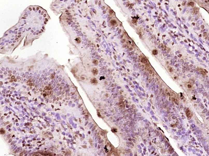
Fig1: Paraformaldehyde-fixed, paraffin embedded (Mouse small intestine); Antigen retrieval by boiling in sodium citrate buffer (pH6.0) for 15min; Block endogenous peroxidase by 3% hydrogen peroxide for 20 minutes; Blocking buffer (normal goat serum) at 37℃ for 30min; Antibody incubation with (Egr1) Polyclonal Antibody, Unconjugated (ER1907-98) at 1:400 overnight at 4℃, followed by operating according to SP Kit(Rabbit) (sp-0023) instructionsand DAB staining.Application
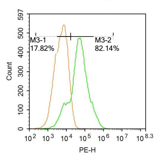
Fig2: U-937 cells were fixed with 4% PFA for 10min at room temperature,permeabilized with 90% ice-cold methanol for 20 min at room temperature,and incubated in 5% BSA blocking buffer for 30 min at room temperature. Cells were then stained with Egr1 Antibody(ER1907-98) at 1:500 dilution in blocking buffer and incubated for 30 min at room temperature, washed twice with 2%BSA in PBS, followed by secondary antibody incubation for 40 min at room temperature. Acquisitions of 20,000 events were performed.Cells stained with primary antibody (green), and isotype control (orange).Application
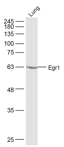
Fig3: Sample: Lung (Mouse) Lysate at 40 ug Primary: Anti-Egr1 (ER1907-98) at 1/300 dilution Secondary: IRDye800CW Goat Anti-Rabbit IgG at 1/20000 dilution Predicted band size: 60 kD Observed band size: 60 kDApplication
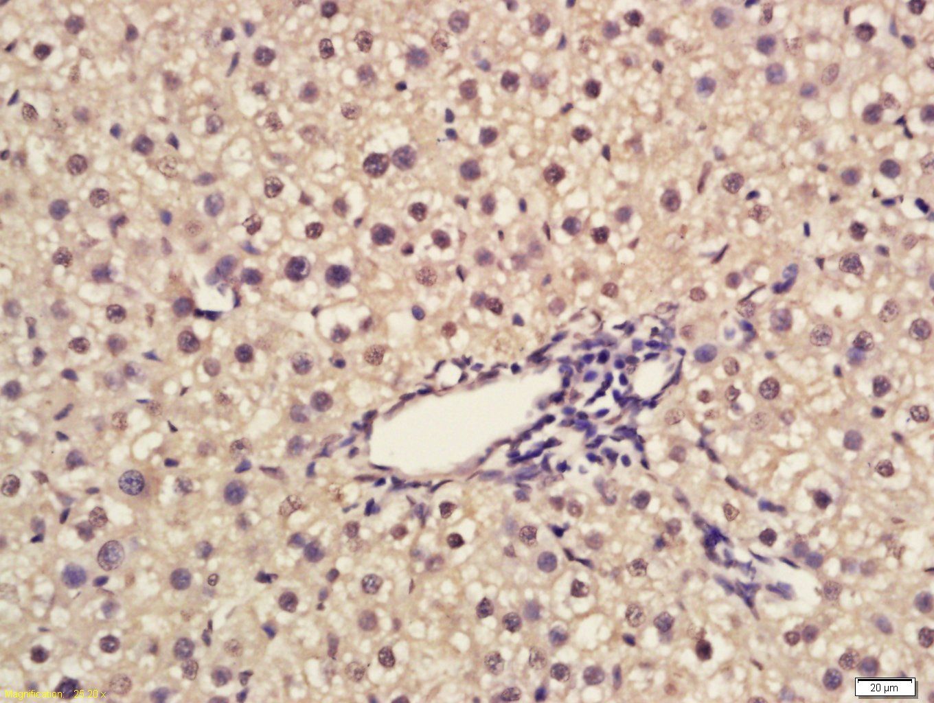
Fig4: Tissue/cell: rat liver tissue; 4% Paraformaldehyde-fixed and paraffin-embedded; Antigen retrieval: citrate buffer ( 0.01M, pH 6.0 ), Boiling bathing for 15min; Block endogenous peroxidase by 3% Hydrogen peroxide for 30min; Blocking buffer (normal goat serum,C-0005) at 37℃ for 20 min; Incubation: Anti-Egr1 Polyclonal Antibody, Unconjugated(ER1907-98) 1:200, overnight at 4℃, followed by conjugation to the secondary antibody(SP-0023) and DAB(C-0010) staining| Product Name | Egr1 |
|---|---|
| Antibody Type | Primary Antibodies |
| Antigen Alias | Egr-1 Early growth response 1 Krox-24 Ngf1 Ngfi NGFI-A zif-268 Egr1 A530045N19Rik egr Egr-1 ETR103 Krox-1 Krox-24 Krox24 NGF1-A NGFI-A NGFIA TIS8 Zenk Zfp-6 AT 225 AT225 Early growth response protein 1 G0S 30 G0S30 KROX 24 Krox 24 protein KROX24 TIS 8 TIS8 Transcription factor ETR 103 Transcription factor ETR103 Transcription factor Zif 268 Transcription factor Zif268 Zfp 6 ZIF268 Zinc finger protein 225 ZNF 225 ZNF225 EGR1_HUMAN. |
| Product description | Transcriptional regulator. Recognizes and binds to the DNA sequence 5'-GCG(T/G)GGGCG-3'(EGR-site) in the promoter region of target genes. Binds double-stranded target DNA, irrespective of the cytosine methylation status. Regulates the transcription of numerous target genes, and thereby plays an important role in regulating the response to growth factors, DNA damage, and ischemia. Plays a role in the regulation of cell survival, proliferation and cell death. Activates expression of p53/TP53 and TGFB1, and thereby helps prevent tumor formation. Required for normal progress through mitosis and normal proliferation of hepatocytes after partial hepatectomy. Mediates responses to ischemia and hypoxia; regulates the expression of proteins such as IL1B and CXCL2 that are involved in inflammatory processes and development of tissue damage after ischemia. Regulates biosynthesis of luteinizing hormone (LHB) in the pituitary. Regulates the amplitude of the expression rhythms of clock genes: ARNTL/BMAL1, PER2 and NR1D1 in the liver via the activation of PER1 (clock repressor) transcription. Regulates the rhythmic expression of core-clock gene ARNTL/BMAL1 in the suprachiasmatic nucleus (SCN). |
| Immunogen | KLH conjugated synthetic peptide derived from human Egr-1 401-500/453 |
| Clonality | Polyclonal |
|---|---|
| Isotype | IgG |
| Host Species | Rabbit |
| Tested Applications | WBIHC-PFC |
| WB:1:500-2000: IHC-P:1:400-800: FC:0.2ug/test: |
|
| Species Reactivity | HumanMouseRat |
| Concentration | 1mg/ml |
| Alternative Names | Egr-1 Early growth response 1 Krox-24 Ngf1 Ngfi NGFI-A zif-268 Egr1 A530045N19Rik egr Egr-1 ETR103 Krox-1 Krox-24 Krox24 NGF1-A NGFI-A NGFIA TIS8 Zenk Zfp-6 AT 225 AT225 Early growth response protein 1 G0S 30 G0S30 KROX 24 Krox 24 protein KROX24 TIS 8 TIS8 Transcription factor ETR 103 Transcription factor ETR103 Transcription factor Zif 268 Transcription factor Zif268 Zfp 6 ZIF268 Zinc finger protein 225 ZNF 225 ZNF225 EGR1_HUMAN. |
|---|---|
| Molecular Weight(MW) | 60 KDa |
| Cellular Localization | Nucleus. |
| SwissProt ID | P18146 |
|---|
Application
Fig1: Paraformaldehyde-fixed, paraffin embedded (Mouse small intestine); Antigen retrieval by boiling in sodium citrate buffer (pH6.0) for 15min; Block endogenous peroxidase by 3% hydrogen peroxide for 20 minutes; Blocking buffer (normal goat serum) at 37℃ for 30min; Antibody incubation with (Egr1) Polyclonal Antibody, Unconjugated (ER1907-98) at 1:400 overnight at 4℃, followed by operating according to SP Kit(Rabbit) (sp-0023) instructionsand DAB staining.
Application
Fig2: U-937 cells were fixed with 4% PFA for 10min at room temperature,permeabilized with 90% ice-cold methanol for 20 min at room temperature,and incubated in 5% BSA blocking buffer for 30 min at room temperature. Cells were then stained with Egr1 Antibody(ER1907-98) at 1:500 dilution in blocking buffer and incubated for 30 min at room temperature, washed twice with 2%BSA in PBS, followed by secondary antibody incubation for 40 min at room temperature. Acquisitions of 20,000 events were performed.Cells stained with primary antibody (green), and isotype control (orange).
Application
Fig3: Sample: Lung (Mouse) Lysate at 40 ug Primary: Anti-Egr1 (ER1907-98) at 1/300 dilution Secondary: IRDye800CW Goat Anti-Rabbit IgG at 1/20000 dilution Predicted band size: 60 kD Observed band size: 60 kD
Application
Fig4: Tissue/cell: rat liver tissue; 4% Paraformaldehyde-fixed and paraffin-embedded; Antigen retrieval: citrate buffer ( 0.01M, pH 6.0 ), Boiling bathing for 15min; Block endogenous peroxidase by 3% Hydrogen peroxide for 30min; Blocking buffer (normal goat serum,C-0005) at 37℃ for 20 min; Incubation: Anti-Egr1 Polyclonal Antibody, Unconjugated(ER1907-98) 1:200, overnight at 4℃, followed by conjugation to the secondary antibody(SP-0023) and DAB(C-0010) staining| Positive Control | Mouse small intestine, U-937 cells, Mouse Lung Lysate, rat small intestine tissue, rat liver tissue. |
|---|---|
| Application Notes | WB:1:500-2000: IHC-P:1:400-800: FC:0.2ug/test: |
| Form | Liquid |
|---|---|
| Storage Instructions | Store at -20℃ for one year. Avoid repeated freeze/thaw cycles. The lyophilized antibody is stable at room temperature for at least one month and for greater than a year when kept at -20℃. When reconstituted in sterile pH 7.4 0.01M PBS or diluent of antibody the antibody is stable for at least two weeks at 2-4℃. |
| Storage Buffer | 0.01M TBS(pH7.4) with 1% BSA, 0.03% Proclin300 and 50% Glycerol. |


