Application
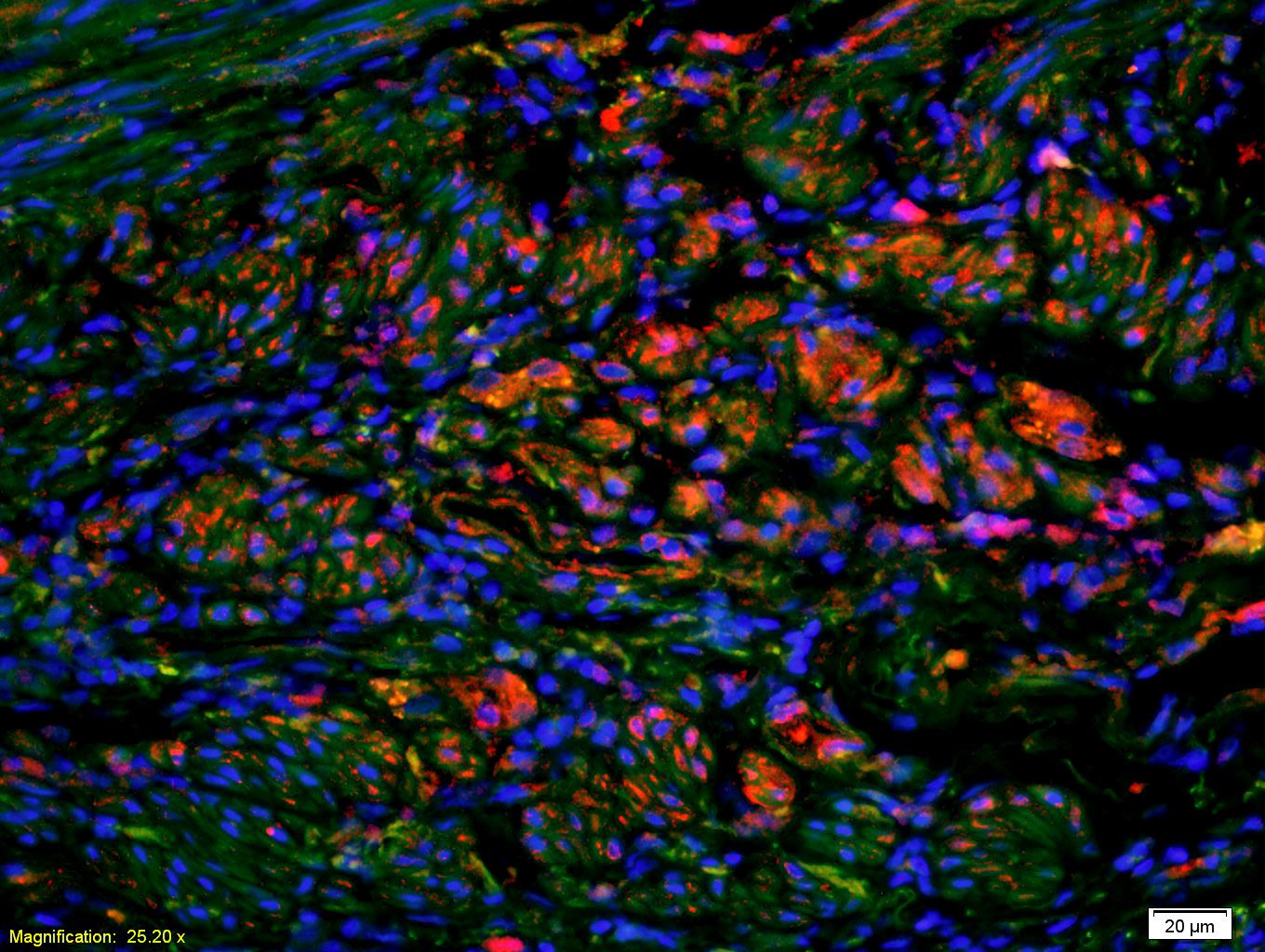
Fig1: Tissue/cell: human rectal carcinoma;4% Paraformaldehyde-fixed and paraffin-embedded; Antigen retrieval: citrate buffer ( 0.01M, pH 6.0 ), Boiling bathing for 15min; Blocking buffer (normal goat serum,C-0005) at 37℃ for 20 min; Incubation: Anti-FasL Polyclonal Antibody, Unconjugated(ER1908-69) 1:200, overnight at 4℃; The secondary antibody was Goat Anti-Rabbit IgG, Cy3 conjugated(bs-0295G-Cy3)used at 1:200 dilution for 40 minutes at 37℃. DAPI(5ug/ml,blue,C-0033) was used to stain the cell nucleiApplication
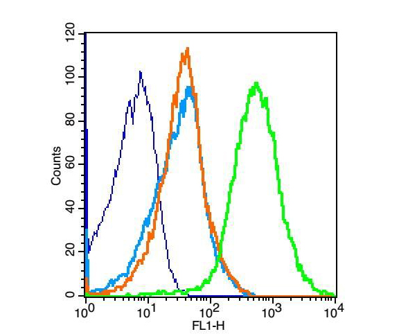
Fig2: Blank control: mouse thymouses(blue) Isotype Control Antibody: Rabbit IgG(orange) ; Secondary Antibody: Goat anti-rabbit IgG-FITC(white blue), Dilution: 1:100 in 1 X PBS containing 0.5% BSA ; Primary Antibody Dilution: 1μl in 100 μL1X PBS containing 0.5% BSA(green).Application
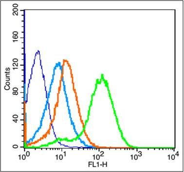
Fig3: Blank control: Mouse Kidney (blue). Primary Antibody:Rabbit Anti-phospho-Fas Ligand antibody (ER1908-69,Green); Dilution: 1μg in 100 μL 1X PBS containing 0.5% BSA; Isotype Control Antibody: Rabbit IgG(orange) ,used under the same conditions; Secondary Antibody: Goat anti-rabbit IgG-FITC(white blue), Dilution: 1:200 in 1 X PBS containing 0.5% BSA. Protocol The cells were fixed with 2% paraformaldehyde for 10 min at 37℃. Primary antibody (ER1908-69, 1μg /1x10^6 cells) were incubated for 30 min at room temperature, followed by 1 X PBS containing 0.5% BSA + 1 0% goat serum (15 min) to block non-specific protein-protein interactions. Then the Goat Anti-rabbit IgG/FITC antibody was added into the blocking buffer mentioned above to react with the primary antibody at 1/200 dilution for 40 min on ice. Acquisition of 20,000 events was performed.Application
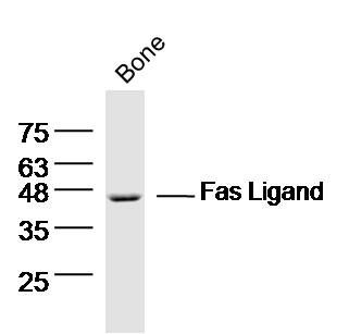
Fig4: Sample: Bone (mouse) Lysate at 40 ug Primary: Anti- Fas ligand (ER1908-69) at 1/300 dilution Secondary: IRDye800CW Goat Anti-Rabbit IgG at 1/20000 dilution Predicted band size: 31 kD Observed band size: 46 kDApplication
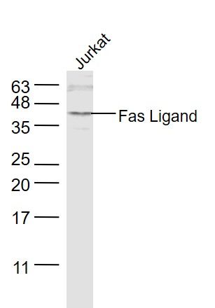
Fig5: Sample: Jurkat(Human) Cell Lysate at 30 ug Primary: Anti-Fas Ligand (ER1908-69) at 1/1000 dilution Secondary: IRDye800CW Goat Anti-Rabbit IgG at 1/20000 dilution Predicted band size: 31 kD Observed band size: 31 kD| Product Name | Fas Ligand |
|---|---|
| Antibody Type | Primary Antibodies |
| Antigen Alias | APO1L Apoptosis (APO 1) antigen ligand 1 Fas-L Apoptosis antigen ligand 1 APTL CD178 CD-178 CD 178 CD95L CD95 ligand CD95-L CD95L protein FAS antigen ligand Fas L Fas Ligand FASL Fasl Fas ligand (TNF superfamily member 6) Generalized lymphoproliferative disease TNF superfamily member 6 TNFSF6 Tumor necrosis factor ligand superfamily member 6 TNFL6_HUMAN Apoptosis antigen ligand Tumor necrosis factor ligand superfamily member 6, membrane form Tumor necrosis factor ligand superfamily member 6, soluble form FASLG. |
| Product description | Cytokine that binds to TNFRSF6/FAS, a receptor that transduces the apoptotic signal into cells. May be involved in cytotoxic T-cell mediated apoptosis and in T-cell development. TNFRSF6/FAS-mediated apoptosis may have a role in the induction of peripheral tolerance, in the antigen-stimulated suicide of mature T-cells, or both. Binding to the decoy receptor TNFRSF6B/DcR3 modulates its effects. |
| Immunogen | KLH conjugated synthetic peptide derived from human Fas Ligand 196-281/281 |
| Clonality | Polyclonal |
|---|---|
| Isotype | IgG |
| Host Species | Rabbit |
| Tested Applications | WBIHC-PICC/IFFC |
| WB:1:500-2000: IHC-P:1:400-800: FC:1μg/Test: IF:1:100-500: ICC:1:100-500: |
|
| Species Reactivity | HumanMouseRatCow |
| Concentration | 1mg/ml |
| Alternative Names | APO1L
Apoptosis (APO 1) antigen ligand 1
Fas-L
Apoptosis antigen ligand 1
APTL
CD178
CD-178
CD 178
CD95L
CD95 ligand
CD95-L
CD95L protein
FAS antigen ligand
Fas L
Fas Ligand
FASL
Fasl
Fas ligand (TNF superfamily member 6)
Generalized lymphoproliferative disease
TNF superfamily member 6
TNFSF6
Tumor necrosis factor ligand superfamily member 6
TNFL6_HUMAN
Apoptosis antigen ligand
Tumor necrosis factor ligand superfamily member 6 membrane form Tumor necrosis factor ligand superfamily member 6 soluble form FASLG. |
|---|---|
| Molecular Weight(MW) | 31 KDa |
| Cellular Localization | Cell membrane; Single-pass type II membrane protein. Secreted. Cytoplasmic vesicle lumen. Lysosome lumen. Note=May be released into the extracellular fluid, probably by cleavage form the cell surface. Is internalized into multivesicular bodies of secretory lysosomes after phosphorylation by FGR and monoubiquitination. |
| SwissProt ID | P48023 |
|---|
Application
Fig1: Tissue/cell: human rectal carcinoma;4% Paraformaldehyde-fixed and paraffin-embedded; Antigen retrieval: citrate buffer ( 0.01M, pH 6.0 ), Boiling bathing for 15min; Blocking buffer (normal goat serum,C-0005) at 37℃ for 20 min; Incubation: Anti-FasL Polyclonal Antibody, Unconjugated(ER1908-69) 1:200, overnight at 4℃; The secondary antibody was Goat Anti-Rabbit IgG, Cy3 conjugated(bs-0295G-Cy3)used at 1:200 dilution for 40 minutes at 37℃. DAPI(5ug/ml,blue,C-0033) was used to stain the cell nuclei
Application
Fig2: Blank control: mouse thymouses(blue) Isotype Control Antibody: Rabbit IgG(orange) ; Secondary Antibody: Goat anti-rabbit IgG-FITC(white blue), Dilution: 1:100 in 1 X PBS containing 0.5% BSA ; Primary Antibody Dilution: 1μl in 100 μL1X PBS containing 0.5% BSA(green).
Application
Fig3: Blank control: Mouse Kidney (blue). Primary Antibody:Rabbit Anti-phospho-Fas Ligand antibody (ER1908-69,Green); Dilution: 1μg in 100 μL 1X PBS containing 0.5% BSA; Isotype Control Antibody: Rabbit IgG(orange) ,used under the same conditions; Secondary Antibody: Goat anti-rabbit IgG-FITC(white blue), Dilution: 1:200 in 1 X PBS containing 0.5% BSA. Protocol The cells were fixed with 2% paraformaldehyde for 10 min at 37℃. Primary antibody (ER1908-69, 1μg /1x10^6 cells) were incubated for 30 min at room temperature, followed by 1 X PBS containing 0.5% BSA + 1 0% goat serum (15 min) to block non-specific protein-protein interactions. Then the Goat Anti-rabbit IgG/FITC antibody was added into the blocking buffer mentioned above to react with the primary antibody at 1/200 dilution for 40 min on ice. Acquisition of 20,000 events was performed.
Application
Fig4: Sample: Bone (mouse) Lysate at 40 ug Primary: Anti- Fas ligand (ER1908-69) at 1/300 dilution Secondary: IRDye800CW Goat Anti-Rabbit IgG at 1/20000 dilution Predicted band size: 31 kD Observed band size: 46 kD
Application
Fig5: Sample: Jurkat(Human) Cell Lysate at 30 ug Primary: Anti-Fas Ligand (ER1908-69) at 1/1000 dilution Secondary: IRDye800CW Goat Anti-Rabbit IgG at 1/20000 dilution Predicted band size: 31 kD Observed band size: 31 kD| Application Notes | WB:1:500-2000: IHC-P:1:400-800: FC:1μg/Test: IF:1:100-500: ICC:1:100-500: |
|---|
| Form | Liquid |
|---|---|
| Storage Instructions | Store at -20℃ for one year. Avoid repeated freeze/thaw cycles. The lyophilized antibody is stable at room temperature for at least one month and for greater than a year when kept at -20℃. When reconstituted in sterile pH 7.4 0.01M PBS or diluent of antibody the antibody is stable for at least two weeks at 2-4℃. |
| Storage Buffer | 0.01M TBS(pH7.4) with 1% BSA, 0.03% Proclin300 and 50% Glycerol. |


