Application
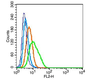
Fig1: Blank control(blue): U937(fixed with 2% paraformaldehyde (10 min)). Primary Antibody: Rabbit Anti-CD64 antibody(ER1908-78), Dilution: 1μg in 100 μL 1X PBS containing 0.5% BSA; Isotype Control Antibody: Rabbit IgG(orange) ,used under the same conditions ); Secondary Antibody: Goat anti-rabbit IgG-PE(white blue), Dilution: 1:200 in 1 X PBS containing 0.5% BSA.Application
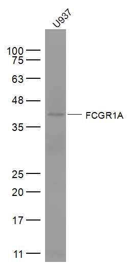
Fig2: Sample: U937(Human) Cell Lysate at 30 ug Primary: Anti-FCGR1A (ER1908-78) at 1/300 dilution Secondary: IRDye800CW Goat Anti-Rabbit IgG at 1/20000 dilution Predicted band size: 41 kD Observed band size: 41 kDApplication
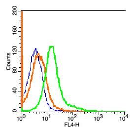
Fig3: Blank control: 293T Cells(blue). Primary Antibody: Rabbit Anti-FCGR1A/CD64/AF647 Conjugated antibody (ER1908-78-AF647), Dilution: 1μg in 100 μL 1X PBS containing 0.5% BSA; Isotype Control Antibody: Rabbit IgG/AF647(orange) ,used under the same conditions.Application
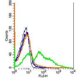
Fig4: Blank control: Mouse spleen cells(blue). Primary Antibody: Rabbit Anti- FCGR1A/CD64 antibody(ER1908-78), Dilution: 5μg in 100 μL 1X PBS containing 0.5% BSA; Isotype Control Antibody: Rabbit IgG (orange) ,used under the same conditions. Secondary Antibody: Goat anti-rabbit IgG-PE(white blue), Dilution: 1:200 in 1 X PBS containing 0.5% BSA. Protocol Primary antibody (ER1908-78, 5μg /1x10^6 cells) were incubated for 30 min on the ice, followed by 1 X PBS containing 0.5% BSA + 1 0% goat serum (15 min) to block non-specific protein-protein interactions. Then the Goat Anti-rabbit IgG/PE antibody was added into the blocking buffer mentioned above to react with the primary antibody at 1/200 dilution for 30 min on ice. Acquisition of 20,000 events was performed.| Product Name | FCGR1A/CD64 |
|---|---|
| Antibody Type | Primary Antibodies |
| Antigen Alias | CD 64 CD64 antigen CD64A Fc fragment of IgG high affinity Ia receptor F Fc fragment of IgG, high affinity Ia, receptor (CD64) Fc fragment of IgG, high affinity Ia, receptor for Fc fragment of IgG, high affinity Ib, receptor (CD64) Fc fragment of IgG, high affinity Ib, receptor for Fc fragment of IgG, high affinity Ic, receptor (CD64) Fc fragment of IgG, high affinity Ic, receptor for Fc gamma receptor Fc gamma receptor I A1 Fc gamma receptor I Fc gamma receptor I B2 Fc gamma RI Fc of IgG high affinity Ia receptor Fc-gamma RIC FCG 1 FCG1 FcgammaRI FCGR 1 FCGR1 FCGR1A FCGR1B FCGR1C FcRI FCRIC IGFR 1 IGFR1 IGFRB IGFRC IgG Fc receptor I IgG Fc receptor IC Immunoglobulin G Fc receptor I Immunoglobulin G Fc receptor IB Immunoglobulin G Fc receptor IC. |
| Immunogen | KLH conjugated synthetic peptide derived from human CD64/IGFR1 101-200/374 |
| Clonality | Polyclonal |
|---|---|
| Isotype | IgG |
| Host Species | Rabbit |
| Tested Applications | WBFC |
| WB:1:500-2000: FC:1μg /test: |
|
| Species Reactivity | HumanMouseRatPigCow |
| Concentration | 1mg/ml |
| Alternative Names | CD 64
CD64 antigen
CD64A
Fc fragment of IgG high affinity Ia receptor
F Fc fragment of IgG high affinity Ia receptor (CD64) Fc fragment of IgG high affinity Ia receptor for Fc fragment of IgG high affinity Ib receptor (CD64) Fc fragment of IgG high affinity Ib receptor for Fc fragment of IgG high affinity Ic receptor (CD64) Fc fragment of IgG high affinity Ic receptor for Fc gamma receptor Fc gamma receptor I A1 Fc gamma receptor I Fc gamma receptor I B2 Fc gamma RI Fc of IgG high affinity Ia receptor Fc-gamma RIC FCG 1 FCG1 FcgammaRI FCGR 1 FCGR1 FCGR1A FCGR1B FCGR1C FcRI FCRIC IGFR 1 IGFR1 IGFRB IGFRC IgG Fc receptor I IgG Fc receptor IC Immunoglobulin G Fc receptor I Immunoglobulin G Fc receptor IB Immunoglobulin G Fc receptor IC. |
|---|---|
| Molecular Weight(MW) | 41 KDa |
| Cellular Localization | Cell membrane; Single-pass type I membrane protein. Note=Stabilized at the cell membrane through interaction with FCER1G. |
| SwissProt ID | P12314 |
|---|
Application
Fig1: Blank control(blue): U937(fixed with 2% paraformaldehyde (10 min)). Primary Antibody: Rabbit Anti-CD64 antibody(ER1908-78), Dilution: 1μg in 100 μL 1X PBS containing 0.5% BSA; Isotype Control Antibody: Rabbit IgG(orange) ,used under the same conditions ); Secondary Antibody: Goat anti-rabbit IgG-PE(white blue), Dilution: 1:200 in 1 X PBS containing 0.5% BSA.
Application
Fig2: Sample: U937(Human) Cell Lysate at 30 ug Primary: Anti-FCGR1A (ER1908-78) at 1/300 dilution Secondary: IRDye800CW Goat Anti-Rabbit IgG at 1/20000 dilution Predicted band size: 41 kD Observed band size: 41 kD
Application
Fig3: Blank control: 293T Cells(blue). Primary Antibody: Rabbit Anti-FCGR1A/CD64/AF647 Conjugated antibody (ER1908-78-AF647), Dilution: 1μg in 100 μL 1X PBS containing 0.5% BSA; Isotype Control Antibody: Rabbit IgG/AF647(orange) ,used under the same conditions.
Application
Fig4: Blank control: Mouse spleen cells(blue). Primary Antibody: Rabbit Anti- FCGR1A/CD64 antibody(ER1908-78), Dilution: 5μg in 100 μL 1X PBS containing 0.5% BSA; Isotype Control Antibody: Rabbit IgG (orange) ,used under the same conditions. Secondary Antibody: Goat anti-rabbit IgG-PE(white blue), Dilution: 1:200 in 1 X PBS containing 0.5% BSA. Protocol Primary antibody (ER1908-78, 5μg /1x10^6 cells) were incubated for 30 min on the ice, followed by 1 X PBS containing 0.5% BSA + 1 0% goat serum (15 min) to block non-specific protein-protein interactions. Then the Goat Anti-rabbit IgG/PE antibody was added into the blocking buffer mentioned above to react with the primary antibody at 1/200 dilution for 30 min on ice. Acquisition of 20,000 events was performed.| Positive Control | Monocyte/macrophage specific. |
|---|---|
| Application Notes | WB:1:500-2000: FC:1μg /test: |
| Form | Liquid |
|---|---|
| Storage Instructions | Store at -20℃ for one year. Avoid repeated freeze/thaw cycles. The lyophilized antibody is stable at room temperature for at least one month and for greater than a year when kept at -20℃. When reconstituted in sterile pH 7.4 0.01M PBS or diluent of antibody the antibody is stable for at least two weeks at 2-4℃. |
| Storage Buffer | 0.01M TBS(pH7.4) with 1% BSA, 0.03% Proclin300 and 50% Glycerol. |


