Application
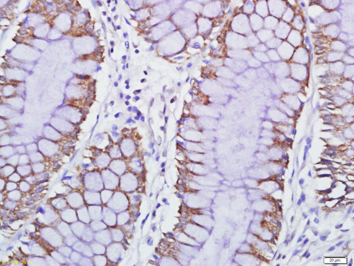
Fig1: Tissue/cell: human colon carcinoma; 4% Paraformaldehyde-fixed and paraffin-embedded; Antigen retrieval: citrate buffer ( 0.01M, pH 6.0 ), Boiling bathing for 15min; Block endogenous peroxidase by 3% Hydrogen peroxide for 30min; Blocking buffer (normal goat serum,C-0005) at 37℃ for 20 min; Incubation: Anti-GPCR EX33 Polyclonal Antibody, Unconjugated(ER1909-85) 1:500, overnight at 4℃, followed by conjugation to the secondary antibody(SP-0023) and DAB(C-0010) stainingApplication
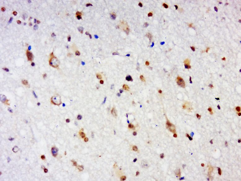
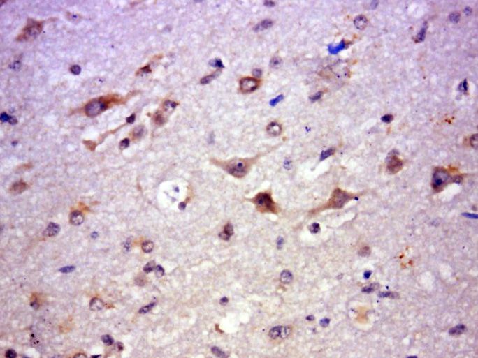
Fig2: Tissue/cell: human brain glioma; 4% Paraformaldehyde-fixed and paraffin-embedded; Antigen retrieval: citrate buffer ( 0.01M, pH 6.0 ), Boiling bathing for 15min; Block endogenous peroxidase by 3% Hydrogen peroxide for 30min; Blocking buffer (normal goat serum,C-0005) at 37℃ for 20 min; Incubation: Anti-EX33 Polyclonal Antibody, Unconjugated(ER1909-85) 1:500, overnight at 4℃, followed by conjugation to the secondary antibody(SP-0023) and DAB(C-0010) stainingApplication
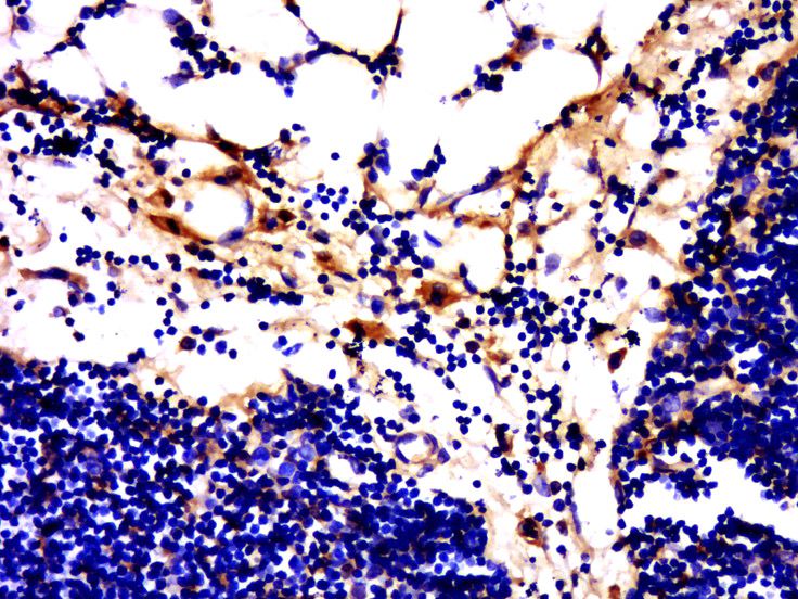
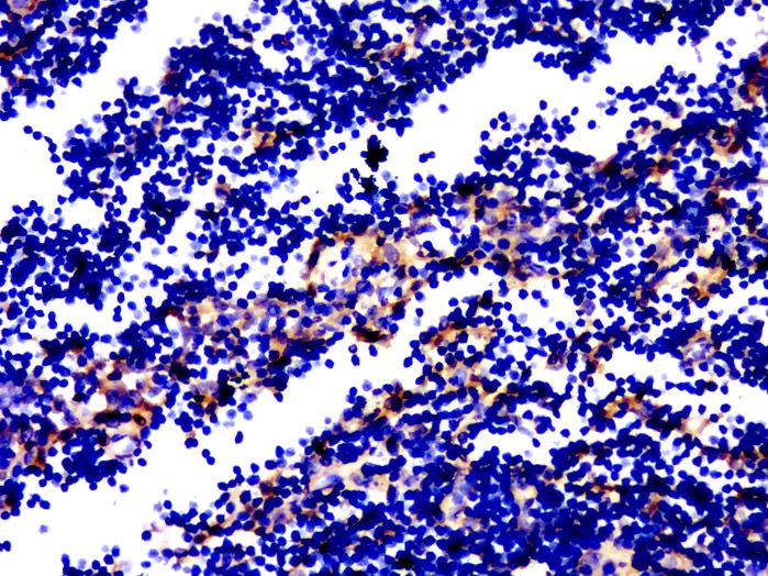
Fig3: Tissue/cell: rabbit thymus tissue; 4% Paraformaldehyde-fixed and paraffin-embedded; Antigen retrieval: citrate buffer ( 0.01M, pH 6.0 ), Boiling bathing for 15min; Block endogenous peroxidase by 3% Hydrogen peroxide for 30min; Blocking buffer (normal goat serum,C-0005) at 37℃ for 20 min; Incubation: Anti-EX33 Polyclonal Antibody, Unconjugated(ER1909-85) 1:500, overnight at 4℃, followed by conjugation to the secondary antibody(SP-0023) and DAB(C-0010) stainingApplication
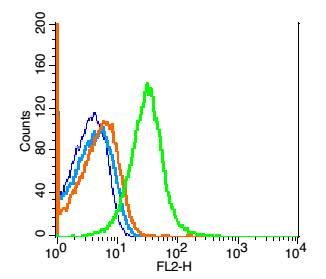
Fig4: Blank control(blue): Mouse thymus cells(fixed with 2% paraformaldehyde (10 min)). Primary Antibody:Rabbit Anti- GPCR EX33 antibody(ER1909-85), Dilution: 1ug in 100 uL 1X PBS containing 0.5% BSA; Isotype Control Antibody: Rabbit IgG(orange) ,used under the same conditions ); Secondary Antibody: Goat anti-rabbit IgG-PE(white blue), Dilution: 1:200 in 1 X PBS containing 0.5% BSA.Application
Fig5: Paraformaldehyde-fixed, paraffin embedded (Human Brain Glioma); Antigen retrieval by boiling in sodium citrate buffer (pH6.0) for 15min; Block endogenous peroxidase by 3% hydrogen peroxide for 20 minutes; Blocking buffer (normal goat serum) at 37℃ for 30min; Antibody incubation with (GPCR EX33) Polyclonal Antibody, Unconjugated (ER1909-85) at 1:500 overnight at 4℃, followed by a conjugated secondary (sp-0023) for 20 minutes and DAB staining.Application
Fig6: araformaldehyde-fixed, paraffin embedded (rabbit thymus); Antigen retrieval by boiling in sodium citrate buffer (pH6.0) for 15min; Block endogenous peroxidase by 3% hydrogen peroxide for 20 minutes; Blocking buffer (normal goat serum) at 37℃ for 30min; Antibody incubation with (GPCR EX33) Polyclonal Antibody, Unconjugated (ER1909-85) at 1:500 overnight at 4℃, followed by a conjugated secondary (sp-0023) for 20 minutes and DAB staining.Application
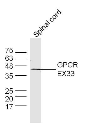
Fig7: Sample: Spinal cord (Mouse) Lysate at 40 ug Primary: Anti-GPCR EX33 (ER1909-85) at 1/300 dilution Secondary: IRDye800CW Goat Anti-Rabbit IgG at 1/20000 dilution Predicted band size: 44 kD Observed band size: 44 kD| Product Name | GPCR EX33 |
|---|---|
| Antibody Type | Primary Antibodies |
| Antigen Alias | EX33 G-protein coupled receptor 84 GPCR4 GPR84 Inflammation-related G-protein coupled receptor EX33 GPR84_HUMAN. |
| Product description | Receptor for medium-chain free fatty acid (FFA) with carbon chain lengths of C9 to C14. Capric acid (C10:0), undecanoic acid (C11:0) and lauric acid (C12:0) are the most potent agonists. Not activated by short-chain and long-chain saturated and unsaturated FFAs. Activation by medium-chain free fatty acid is coupled to a pertussis toxin sensitive G(i/o) protein pathway. May have important roles in processes from fatty acid metabolism to regulation of the immune system. |
| Immunogen | KLH conjugated synthetic peptide derived from human GPCR EX33 protein 21-120/369 |
| Clonality | Polyclonal |
|---|---|
| Isotype | IgG |
| Host Species | Rabbit |
| Tested Applications | WBIHC-PFC |
| WB:1:500-2000: IHC-P:1:400-800: FC:1μg/Test: |
|
| Species Reactivity | HumanMousePigCowRabbit |
| Concentration | 1mg/ml |
| Alternative Names | EX33 G-protein coupled receptor 84 GPCR4 GPR84 Inflammation-related G-protein coupled receptor EX33 GPR84_HUMAN. |
|---|---|
| Molecular Weight(MW) | 44 KDa |
| Cellular Localization | Cell membrane; Multi-pass membrane protein. |
| SwissProt ID | Q9NQS5 |
|---|
Application
Fig1: Tissue/cell: human colon carcinoma; 4% Paraformaldehyde-fixed and paraffin-embedded; Antigen retrieval: citrate buffer ( 0.01M, pH 6.0 ), Boiling bathing for 15min; Block endogenous peroxidase by 3% Hydrogen peroxide for 30min; Blocking buffer (normal goat serum,C-0005) at 37℃ for 20 min; Incubation: Anti-GPCR EX33 Polyclonal Antibody, Unconjugated(ER1909-85) 1:500, overnight at 4℃, followed by conjugation to the secondary antibody(SP-0023) and DAB(C-0010) staining
Application
Fig2: Tissue/cell: human brain glioma; 4% Paraformaldehyde-fixed and paraffin-embedded; Antigen retrieval: citrate buffer ( 0.01M, pH 6.0 ), Boiling bathing for 15min; Block endogenous peroxidase by 3% Hydrogen peroxide for 30min; Blocking buffer (normal goat serum,C-0005) at 37℃ for 20 min; Incubation: Anti-EX33 Polyclonal Antibody, Unconjugated(ER1909-85) 1:500, overnight at 4℃, followed by conjugation to the secondary antibody(SP-0023) and DAB(C-0010) staining
Application
Fig3: Tissue/cell: rabbit thymus tissue; 4% Paraformaldehyde-fixed and paraffin-embedded; Antigen retrieval: citrate buffer ( 0.01M, pH 6.0 ), Boiling bathing for 15min; Block endogenous peroxidase by 3% Hydrogen peroxide for 30min; Blocking buffer (normal goat serum,C-0005) at 37℃ for 20 min; Incubation: Anti-EX33 Polyclonal Antibody, Unconjugated(ER1909-85) 1:500, overnight at 4℃, followed by conjugation to the secondary antibody(SP-0023) and DAB(C-0010) staining
Application
Fig4: Blank control(blue): Mouse thymus cells(fixed with 2% paraformaldehyde (10 min)). Primary Antibody:Rabbit Anti- GPCR EX33 antibody(ER1909-85), Dilution: 1ug in 100 uL 1X PBS containing 0.5% BSA; Isotype Control Antibody: Rabbit IgG(orange) ,used under the same conditions ); Secondary Antibody: Goat anti-rabbit IgG-PE(white blue), Dilution: 1:200 in 1 X PBS containing 0.5% BSA.
Application
Fig5: Paraformaldehyde-fixed, paraffin embedded (Human Brain Glioma); Antigen retrieval by boiling in sodium citrate buffer (pH6.0) for 15min; Block endogenous peroxidase by 3% hydrogen peroxide for 20 minutes; Blocking buffer (normal goat serum) at 37℃ for 30min; Antibody incubation with (GPCR EX33) Polyclonal Antibody, Unconjugated (ER1909-85) at 1:500 overnight at 4℃, followed by a conjugated secondary (sp-0023) for 20 minutes and DAB staining.
Application
Fig6: araformaldehyde-fixed, paraffin embedded (rabbit thymus); Antigen retrieval by boiling in sodium citrate buffer (pH6.0) for 15min; Block endogenous peroxidase by 3% hydrogen peroxide for 20 minutes; Blocking buffer (normal goat serum) at 37℃ for 30min; Antibody incubation with (GPCR EX33) Polyclonal Antibody, Unconjugated (ER1909-85) at 1:500 overnight at 4℃, followed by a conjugated secondary (sp-0023) for 20 minutes and DAB staining.
Application
Fig7: Sample: Spinal cord (Mouse) Lysate at 40 ug Primary: Anti-GPCR EX33 (ER1909-85) at 1/300 dilution Secondary: IRDye800CW Goat Anti-Rabbit IgG at 1/20000 dilution Predicted band size: 44 kD Observed band size: 44 kD| Positive Control | Expressed predominantly in hematopoietic tissues. High levels detected in the bone marrow and lower levels in the peripheral leukocytes and lung. Also expressed in brain, heart, muscle, colon, thymus, spleen, kidney, liver, placenta and intestine. Within the leukocyte population expression is higher in neutrophils and eosinophils relative to T- or B-lymphocytes. |
|---|---|
| Application Notes | WB:1:500-2000: IHC-P:1:400-800: FC:1μg/Test: |
| Form | Liquid |
|---|---|
| Storage Instructions | Store at -20℃ for one year. Avoid repeated freeze/thaw cycles. The lyophilized antibody is stable at room temperature for at least one month and for greater than a year when kept at -20℃. When reconstituted in sterile pH 7.4 0.01M PBS or diluent of antibody the antibody is stable for at least two weeks at 2-4℃. |
| Storage Buffer | 0.01M TBS(pH7.4) with 1% BSA, 0.03% Proclin300 and 50% Glycerol. |


